Gastric Case 1
Prof. Stefan Seewald
GastroZentrum Hirslanden, Zurich
Disclaimer:
- NBI™ and TXI™ Technologies are not intended to replace histopathological sampling as a means of diagnosis
- NBI™ and TXI ™ Technologies are 510(k) cleared in the United State. This case study is being furnished to provide example of NBI™ and TXI™ technology use. The GIF-EZ1500 used in this case is not available in the US market at this time, nor is there an established time for it’s release. The safety and effectiveness of this product and/or the use of these products has not yet been established in the United States market.
- The positions and statements made herein by Prof. Seewald are based on Prof. Seewald’s experiences, thoughts and opinions. As with any product, results may vary, and the techniques, instruments, and settings can vary from facility to facility. The content hereof should not be considered as a substitute for carefully reading all applicable labeling, including the Instructions for Use. Please thoroughly review the relevant user manual(s) for instructions, risks, warnings, and cautions. Techniques, instruments, and setting can vary from facility to facility. It is the clinician’s decision and responsibility in each clinical situation to decide which products, modes, medications, applications, and settings to use.
- The EVIS X1™ endoscopy system is not designed for cardiac applications. Other combinations of equipment may cause ventricular fibrillation or seriously affect the cardiac function of the patient. Improper use of endoscopes may result in patient injury, infection, bleeding, and/or perforation. Complete indications, contraindications, warnings, and cautions are available in the Instructions for Use (IFU)
Scope:GIF-EZ1500
Case: Intramucosal Gastric Carcinoma (AEG Siewert Type III)
Organ: Stomach
Patient information:M, 70
Medical history: Incidental finding
1. WLI
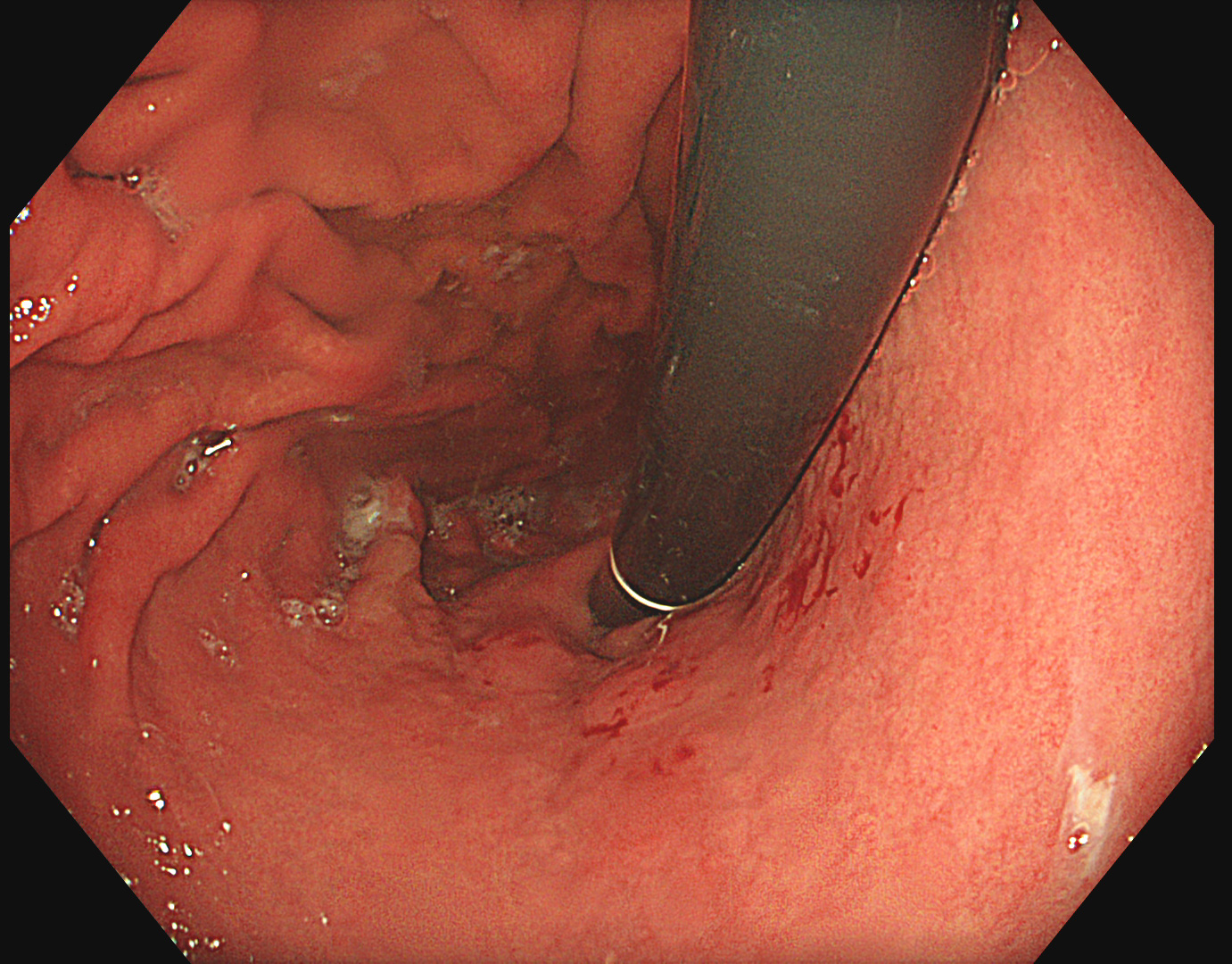
Retroflex assessment of the fundus in white light reveals a suspicious area of patchy appearance spreading from 1-5 o’clock.
2. TXI™ technology
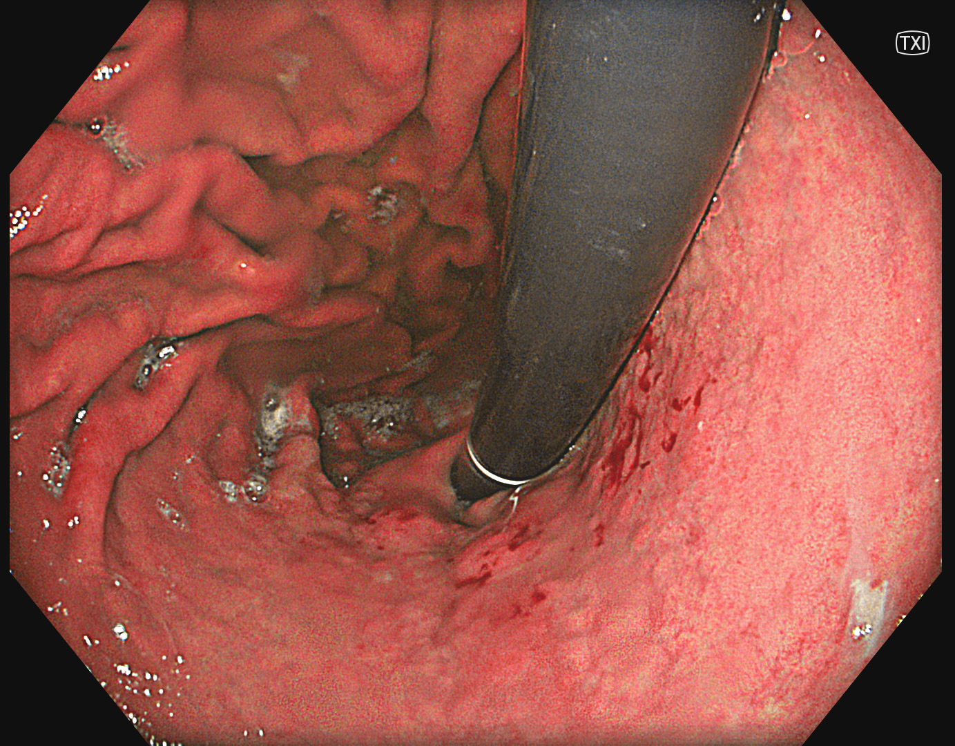
TXI™ technology is enhancing the color contrast and structure between the suspicious area and the surrounding mucosa.
3. NBI™ technology
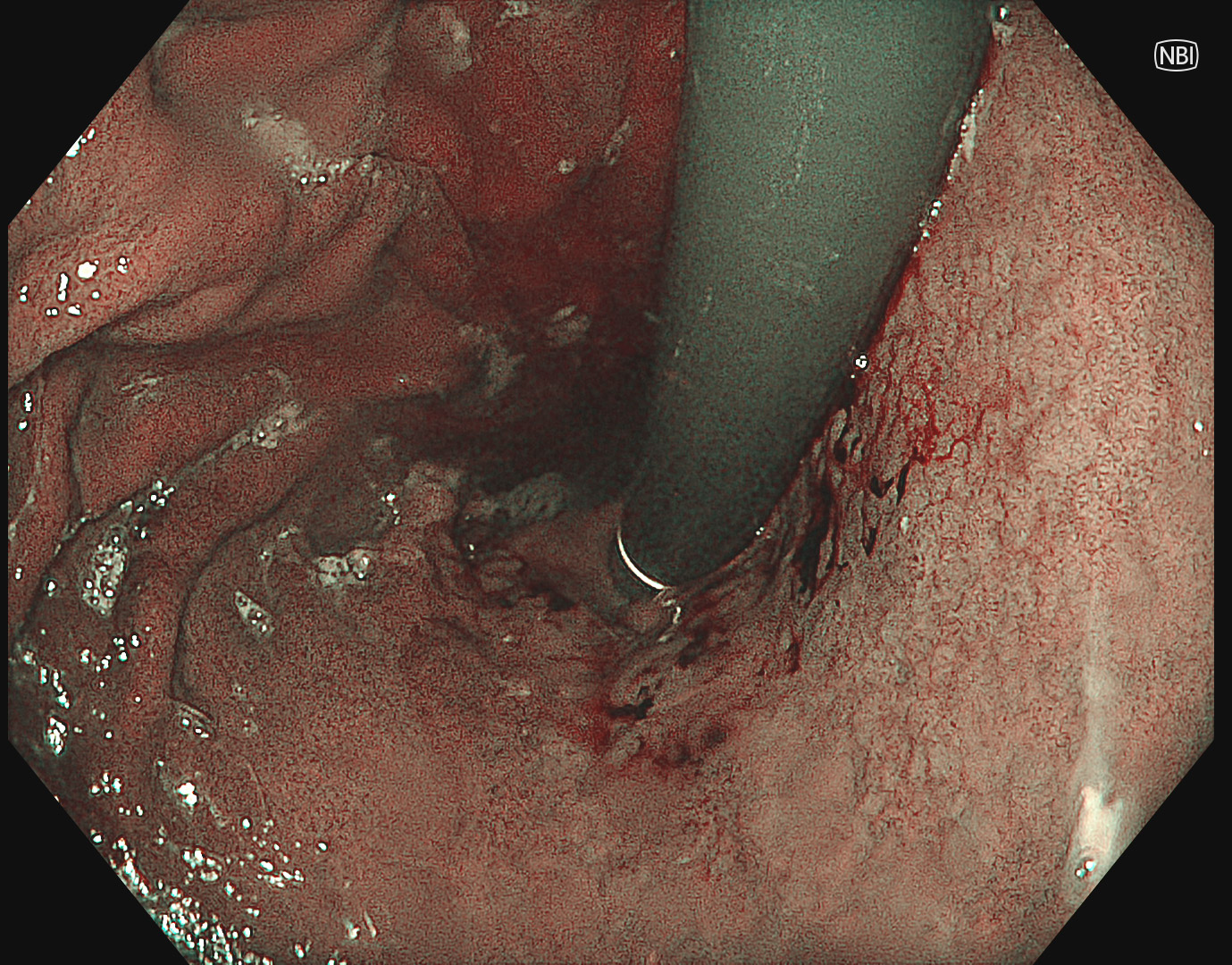
NBI™ technology is bright enough, but for characterization, Near Focus with NBI™ technology is necessary.
4. Near Focus with NBI™ technology
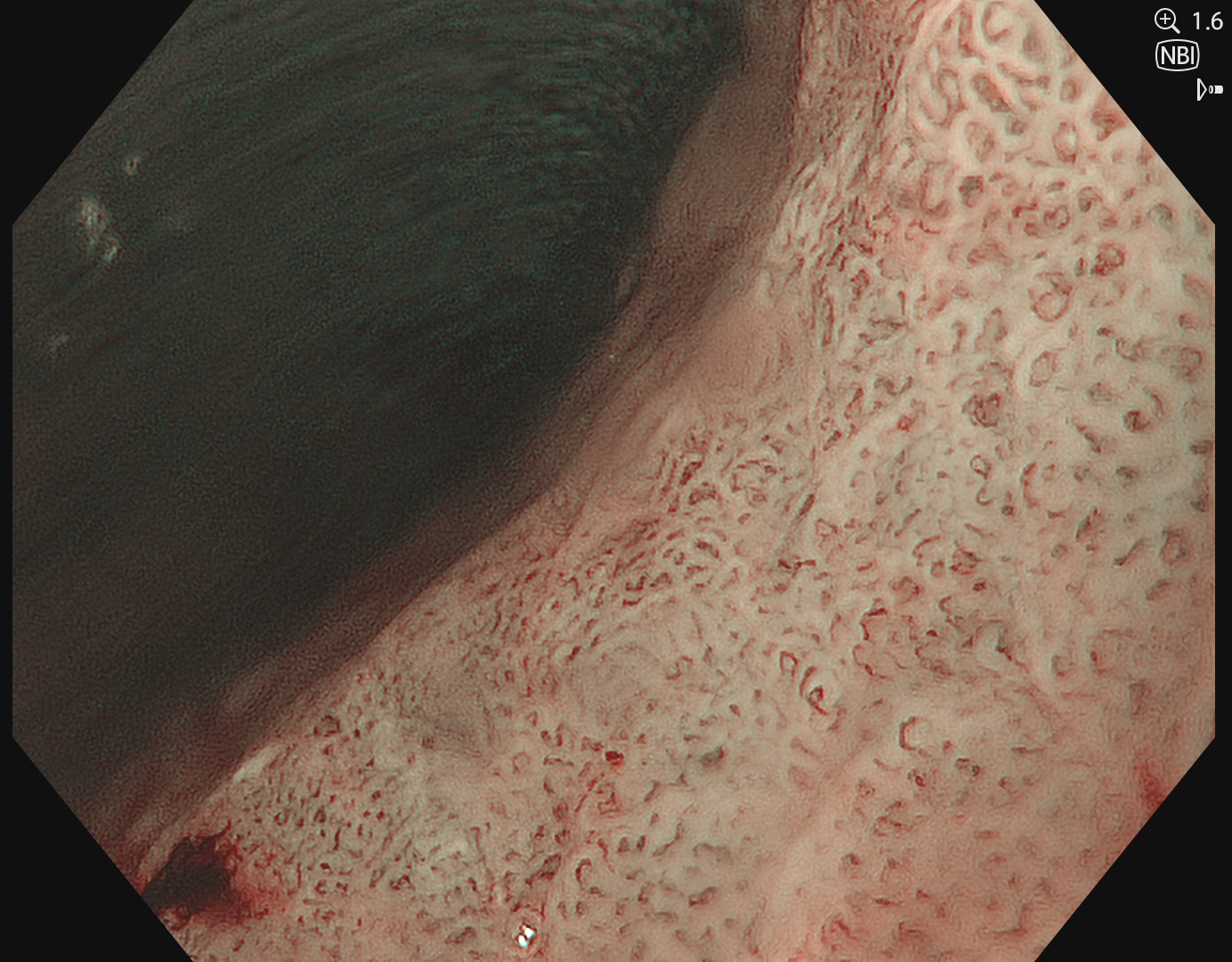
Near Focus with NBI™ technology reveals irregular VS pattern suggesting early gastric carcinoma.
5. TXI™ technology with indigocarmine
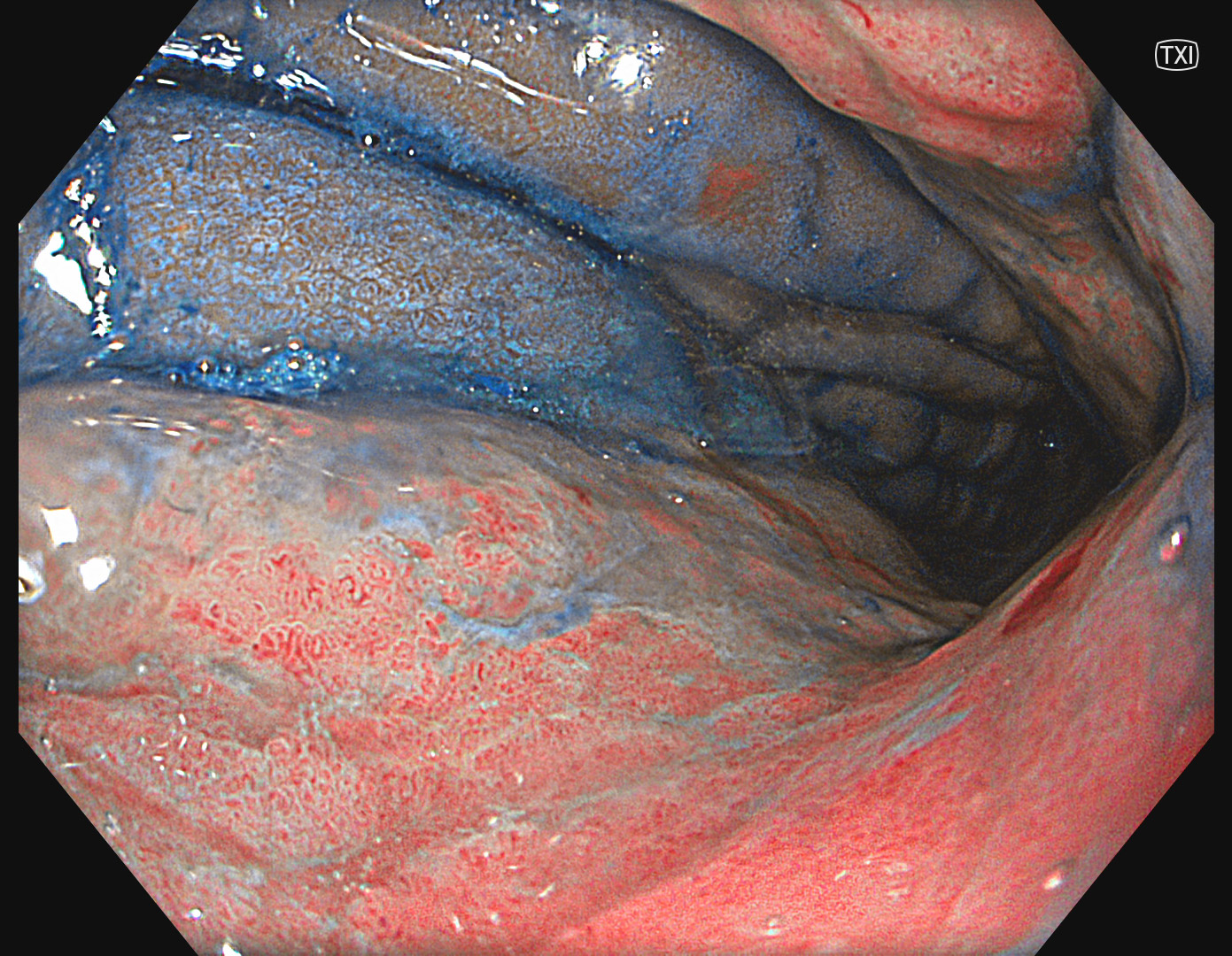
In my opinion, the contrast effect of indigocarmine is enhanced by TXI™ technology and improves the delineation.
6. TXI™ technology with indigocarmine
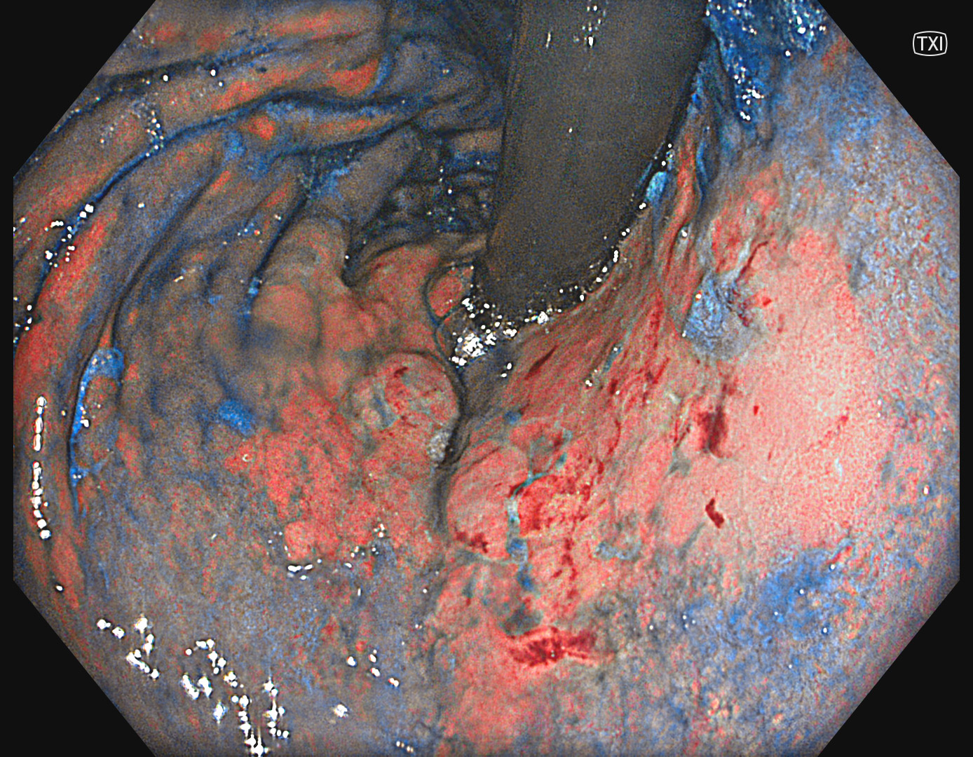
In my opinion, the extent of the lesion can be nicely identified by TXI™ technology in combination with indigocarmine.
Overall Comment
This case illustrates the value of TXI™ technology in detection and delineation of early gastric carcinomas. Especially the delineation was difficult due to indistinct borders. In my opinion, the combination of TXI™ technology and indigocarmine enabled the strong color contrast and delineation of the lesion. NBI™ technology remains my primary modality for optical assessment of histological features.
* Specifications, design and accessories are subject to change without any notice or obligation on the part of the manufacturer.


