Esophageal Case 2
Prof. Stefan Seewald
GastroZentrum Hirslanden, Zurich, Switzerland
Disclaimer:
- NBI™ and TXI™ Technologies are not intended to replace histopathological sampling as a means of diagnosis
- The positions and statements made herein by Prof. Seewald are based on Prof. Seewald’s experiences, thoughts and opinions. As with any product, results may vary, and the techniques, instruments, and settings can vary from facility to facility. The content hereof should not be considered as a substitute for carefully reading all applicable labeling, including the Instructions for Use. Please thoroughly review the relevant user manual(s) for instructions, risks, warnings, and cautions. Techniques, instruments, and setting can vary from facility to facility. It is the clinician’s decision and responsibility in each clinical situation to decide which products, modes, medications, applications, and settings to use.
- The EVIS X1™ endoscopy system is not designed for cardiac applications. Other combinations of equipment may cause ventricular fibrillation or seriously affect the cardiac function of the patient. Improper use of endoscopes may result in patient injury, infection, bleeding, and/or perforation. Complete indications, contraindications, warnings, and cautions are available in the Instructions for Use (IFU)
Scope: GIF-EZ1500
Case: Early Barrett’s cancer (pT1m3 L0 V0 Pn0 G2 R0)
Organ: Esophagus
Patient information: M, 70s
Medical history: Heartburn
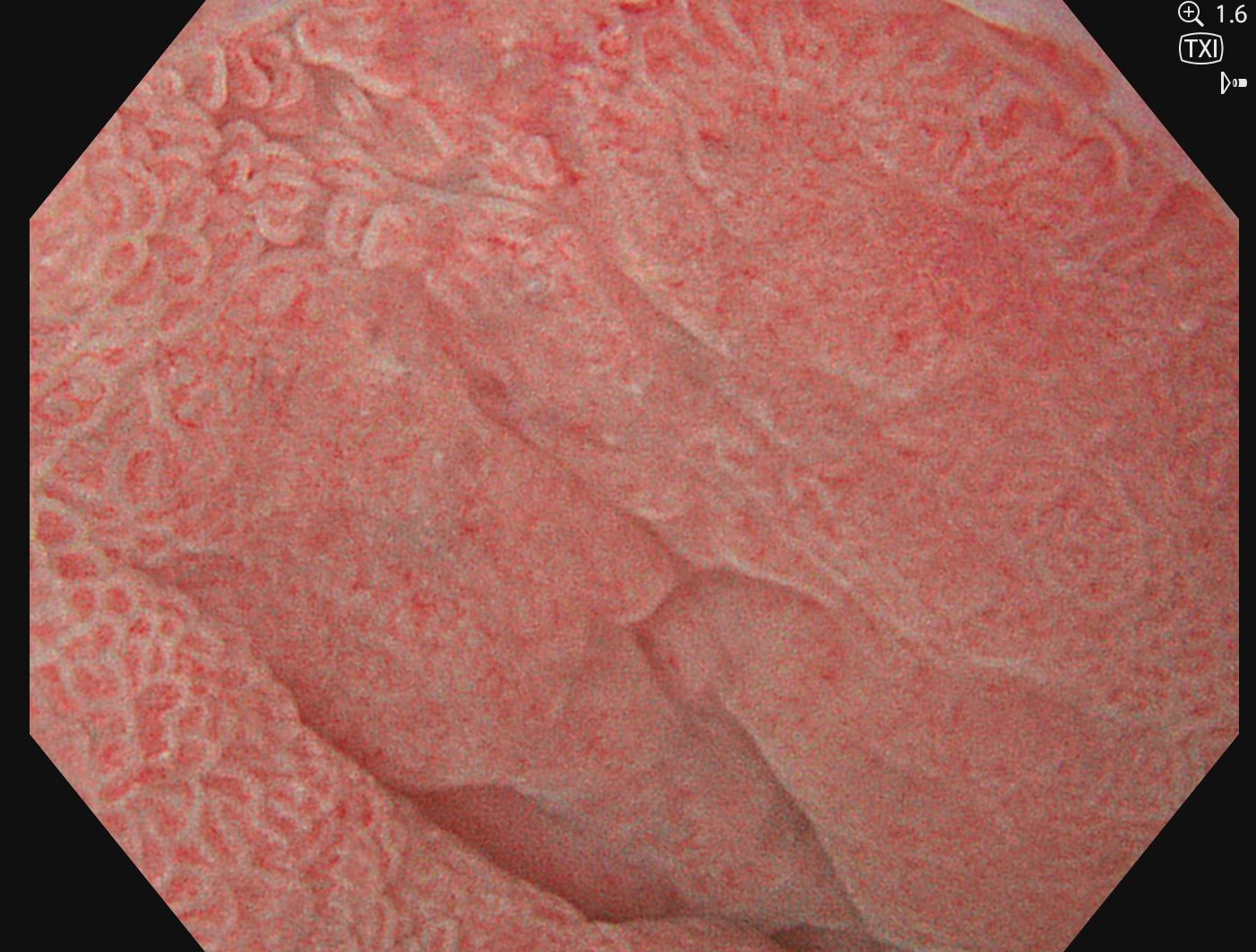
1. TXI™ technology
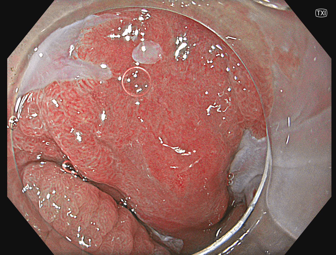
With TXI technology, a broader area of the Barrett appears on the mucosa with an enhanced red color, suggesting a higher vessel density.
2. NBI ™ technology
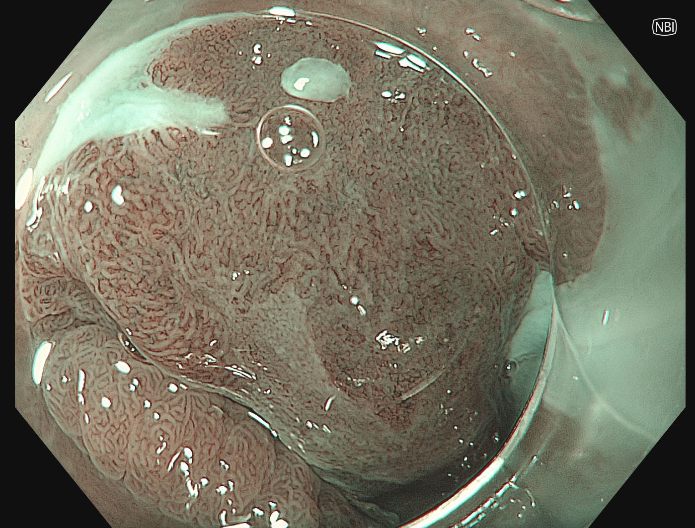
NBI™ technology in normal focus suggests irregularities in mucosal and vascular pattern.
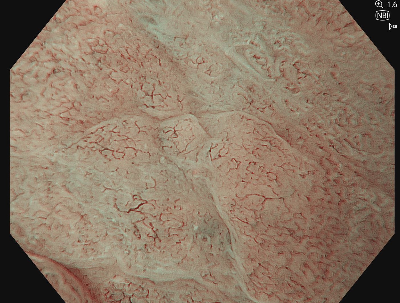
3. Near focus with NBI™ technology
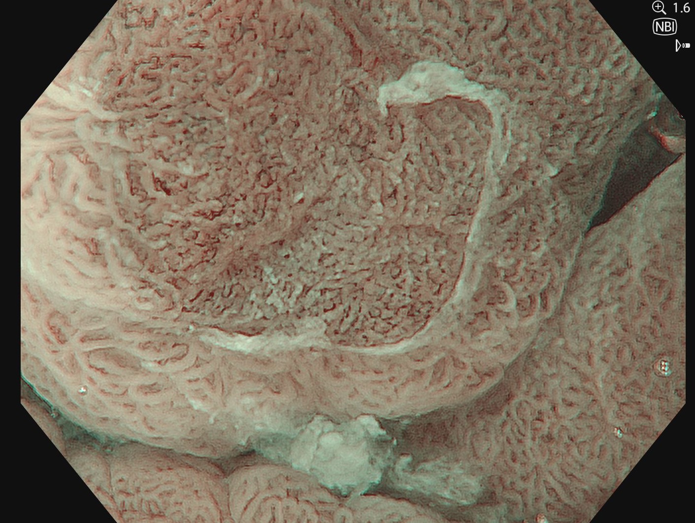
Irregular vascular pattern, characteristic for Barrett‘s carcinoma.
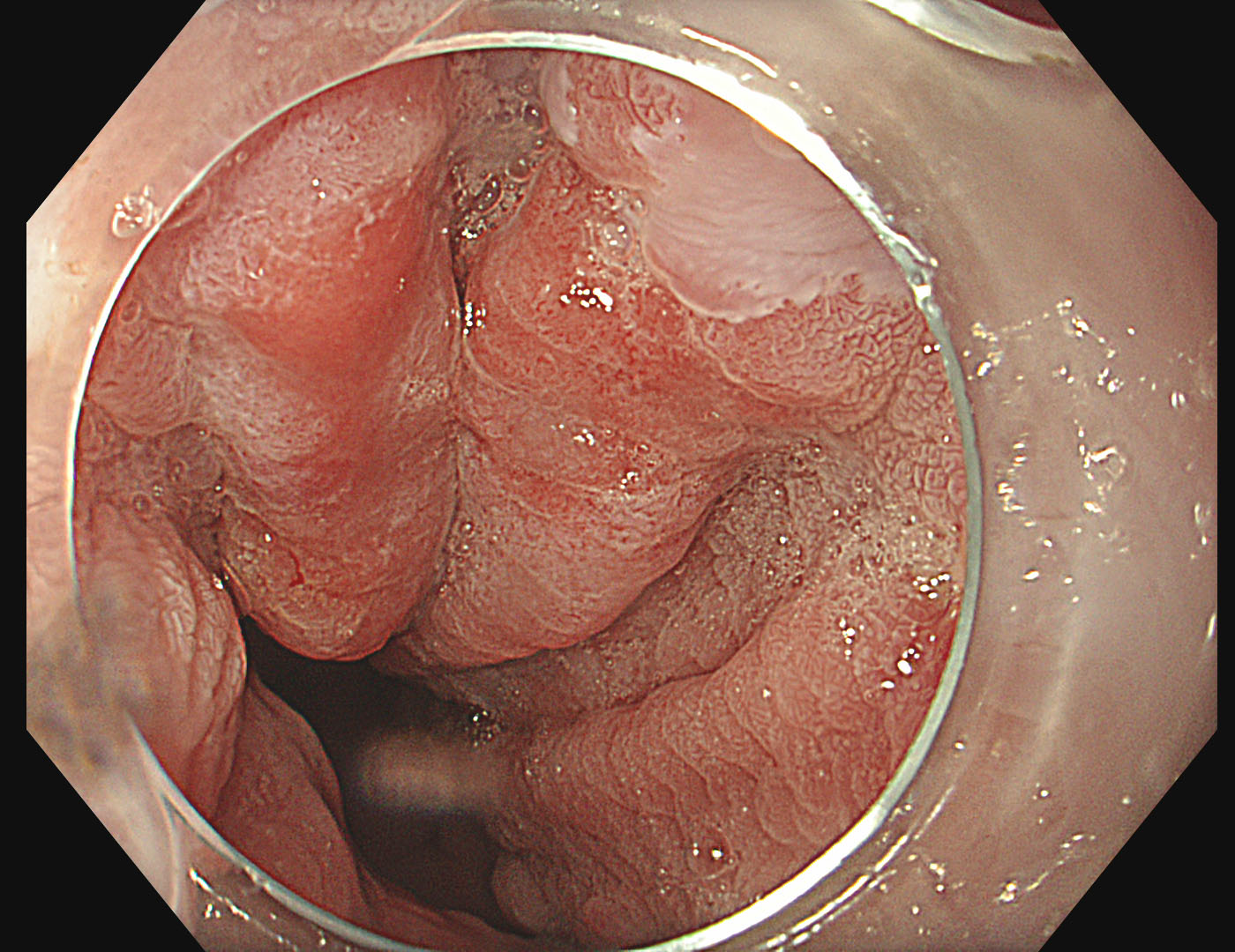
4. Near Focus with TXI™ technology
With TXI™ technology, an irregular surface pattern can be observed.
5. TXI™ technology
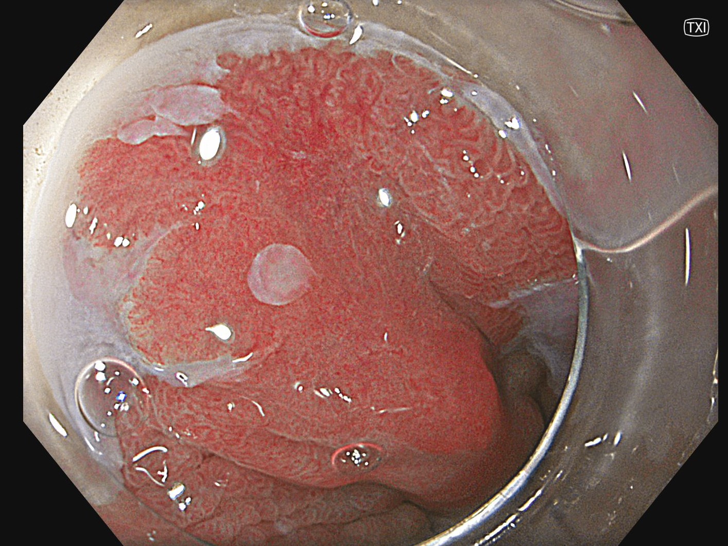
The difference in surface between the upper and lower part of the lesion is easy to observe. An enhanced reddening accompanied by depression at 11 o‘clock indicates the most advanced area.
6. Near focus with NBI™ technology
Near focus with NBI ™ technology and 1.6x electronic zoom under water visualizes irregular vessel and surface structure which are characteristic for Barrett‘s carcinoma.
7. Acetic acid
Acetic acid was applied to estimate the margins of the lesion
8. Acetic acid with TXI™ technology
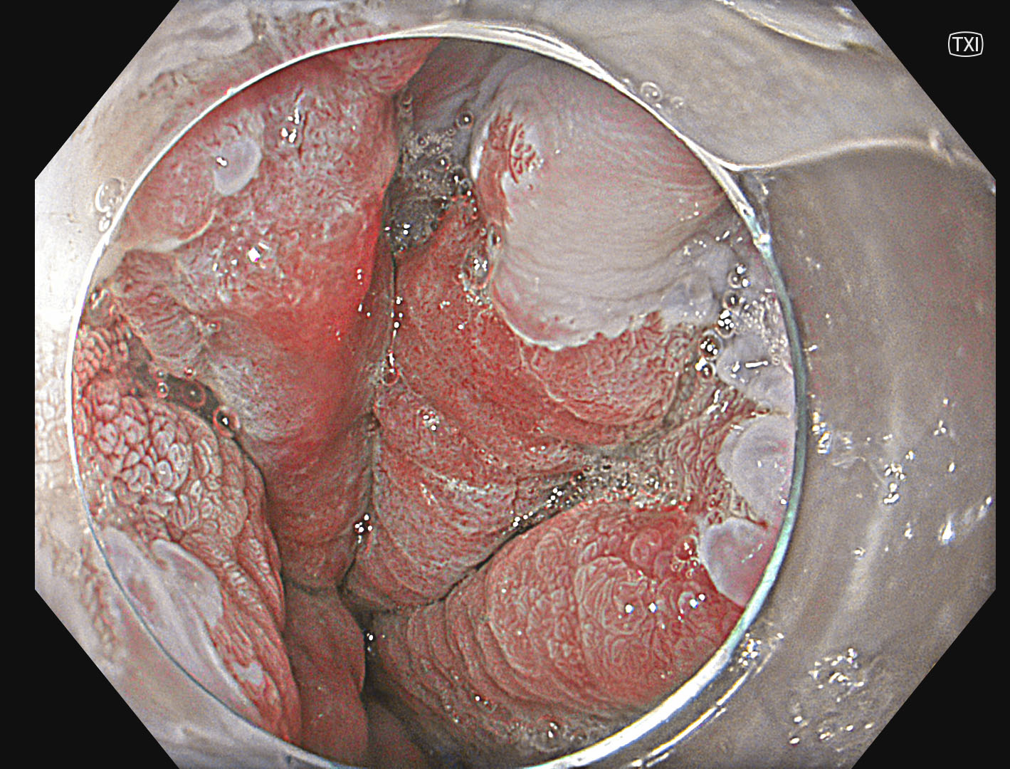
With TXI, a larger extent than anticipated with WLI+acetic acid is suspected.
9. Indigocarmine staining
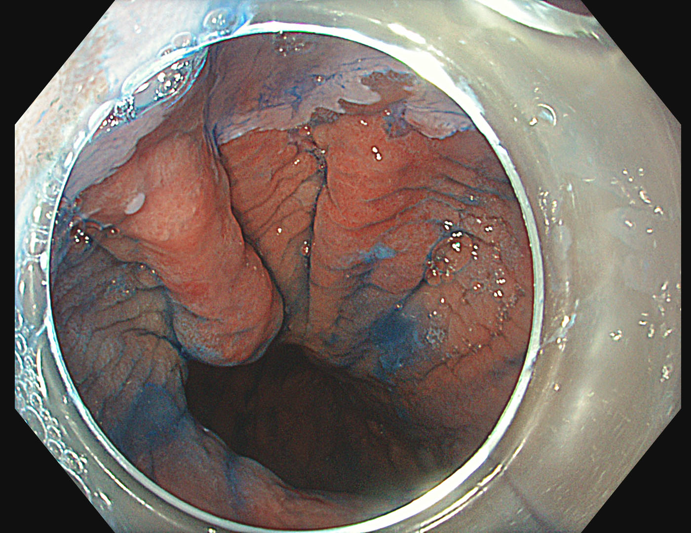
To further delineate the lesion, indigo carmine was applied. Under WLI, an indistinct non-stained area spreads from 10 to 2 o‘clock.
10. Indigocarmine + TXI™ technology
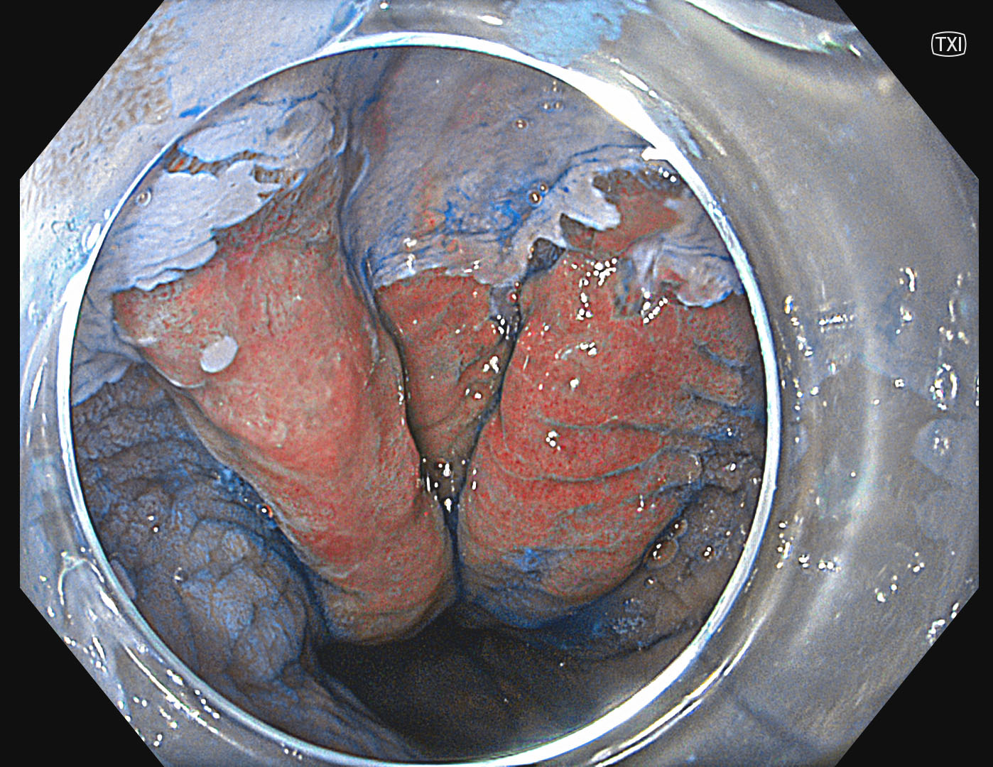
Adding TXI™ technology to indigo carmine is providing an excellent delineation of this lesion.
Case video
Overall Comment
In this complex case, the combination of TXI™ technology, NBI™ technology, and chromoendoscopy was helpful for a proper diagnosis and precise delineation.
* Specifications, design and accessories are subject to change without any notice or obligation on the part of the manufacturer.
Dr. D Nageshwar Reddy Case 3: Squamous Cell Carcinoma
Prof. Stefan Seewald
- Keyword
- Content Type


