Esophageal Case 5

Prof. Rajvinder Singh
University of Adelaide, Australia
Scope:GIF-EZ1500
Case: pT1a esophageal adenocarcinoma
Organ: Lower esophagus
Patient information: M, 82
Medical history: Autoimmune hemolytic anemia, atrial fibrillation, pacemaker, sleep apnea, hypertension, congestive cardiac failure
1. WLI
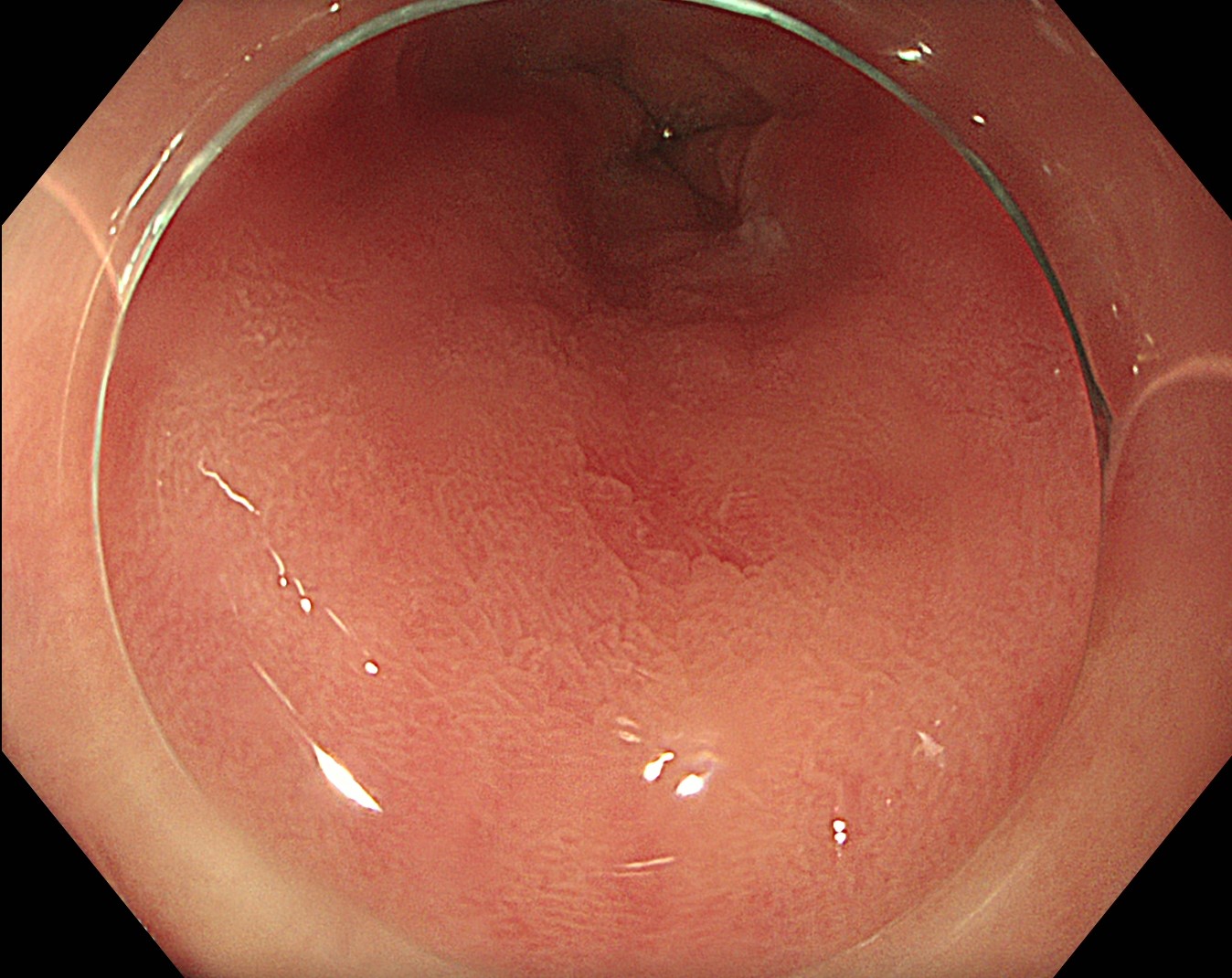
On initial views, a small depressed area is seen at the site of previous biopsy
2. TXI and high-magnification
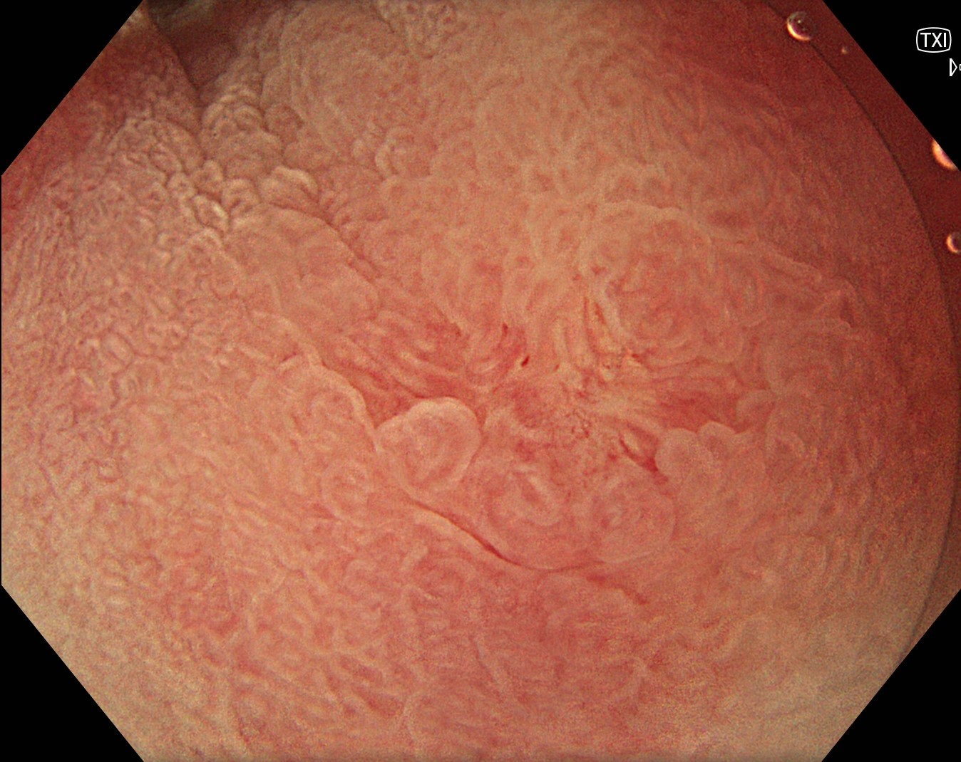
The depressed area is more conspicuous with TXI and magnification
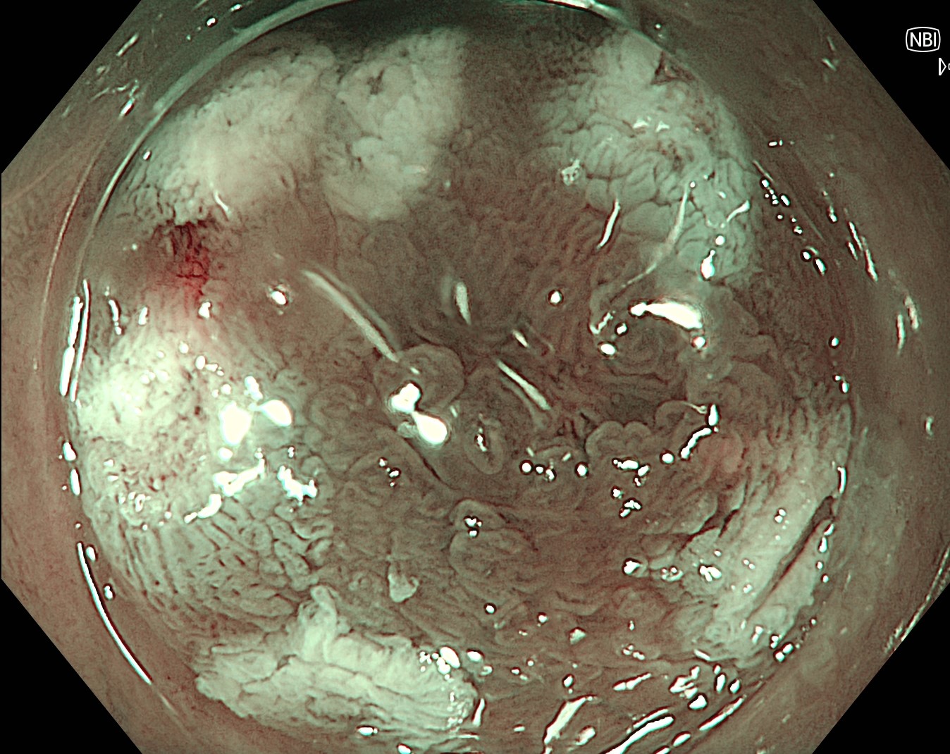
3. NBI
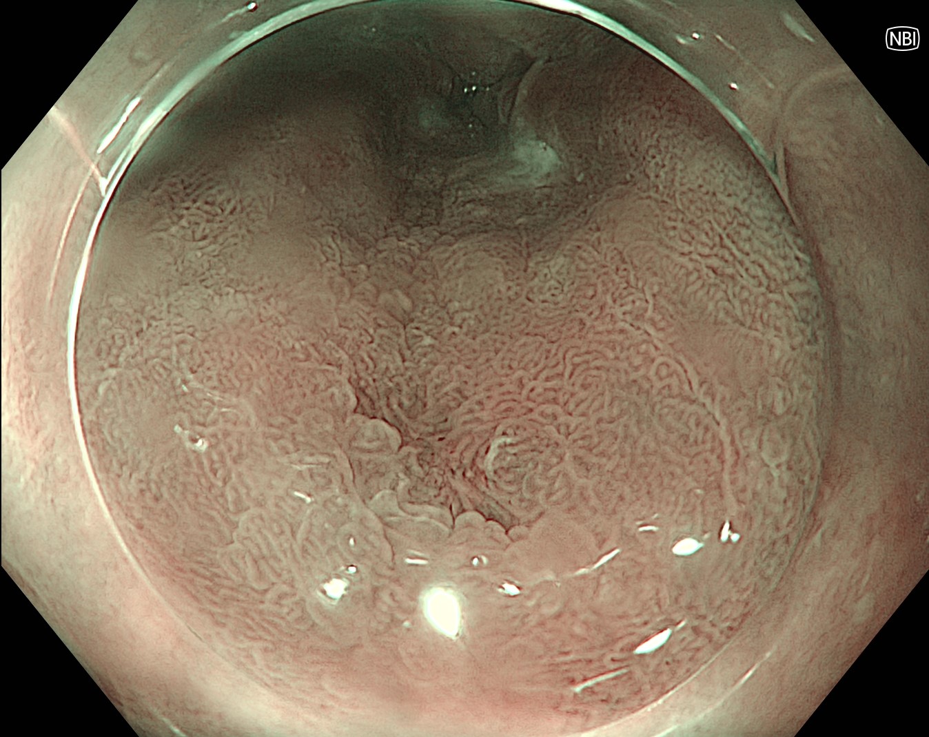
Some tortuous vessels are seen at the base on initial examination with NBI
4. NBI and high-magnification
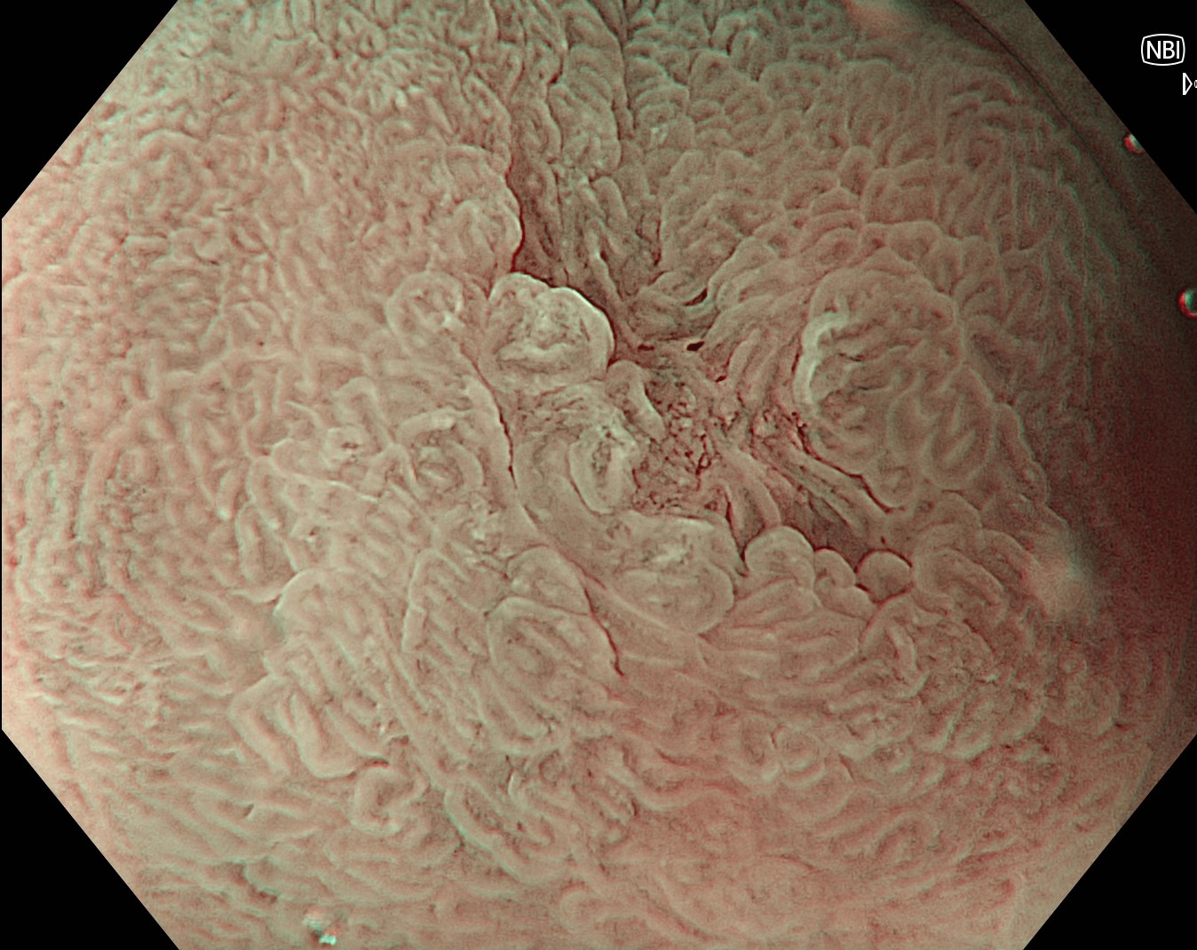
On NBI and high-magnification, tortuous vessels at the base of the depressed area are more clearly seen
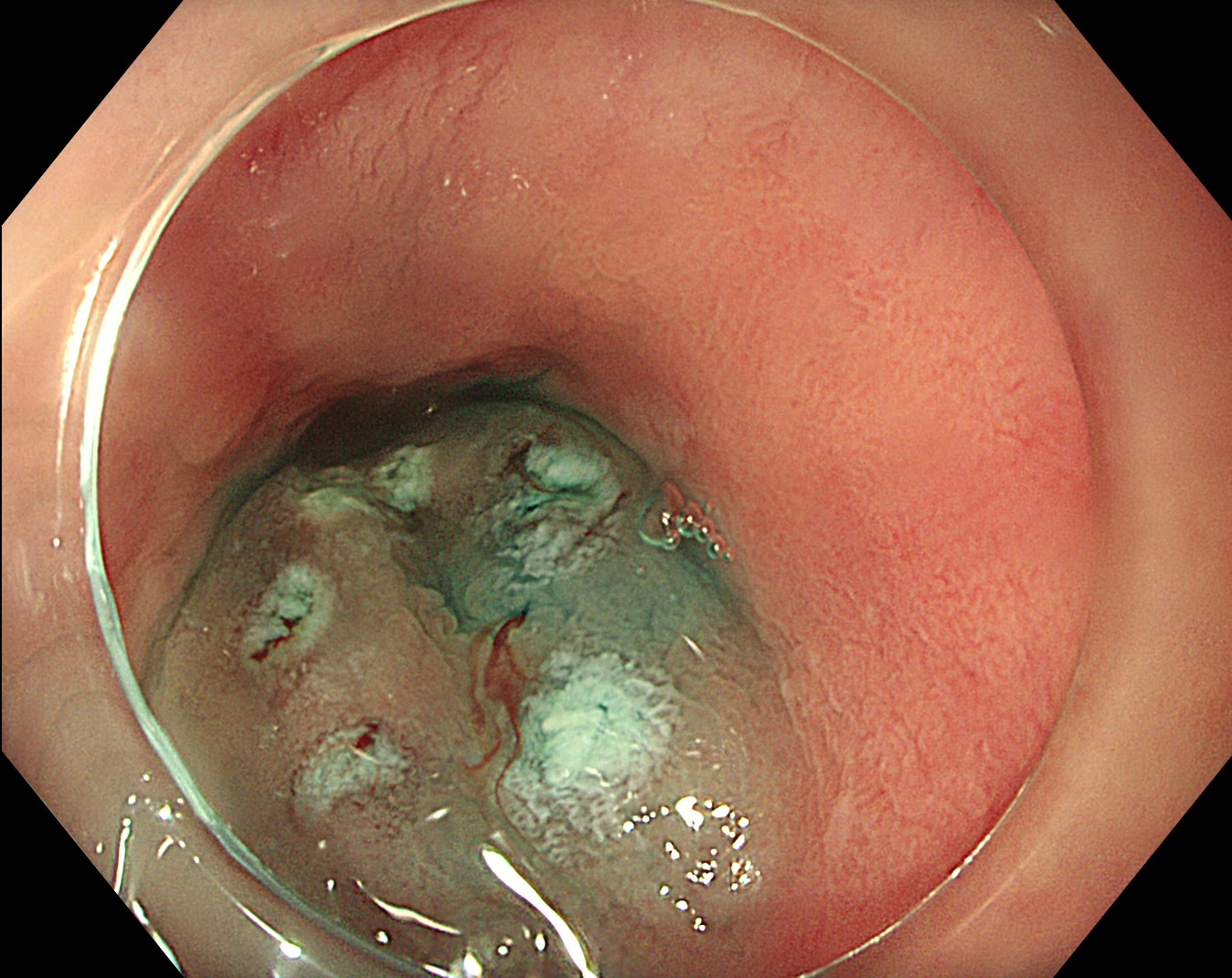
5. Marking
The surroundings are marked while using NBI
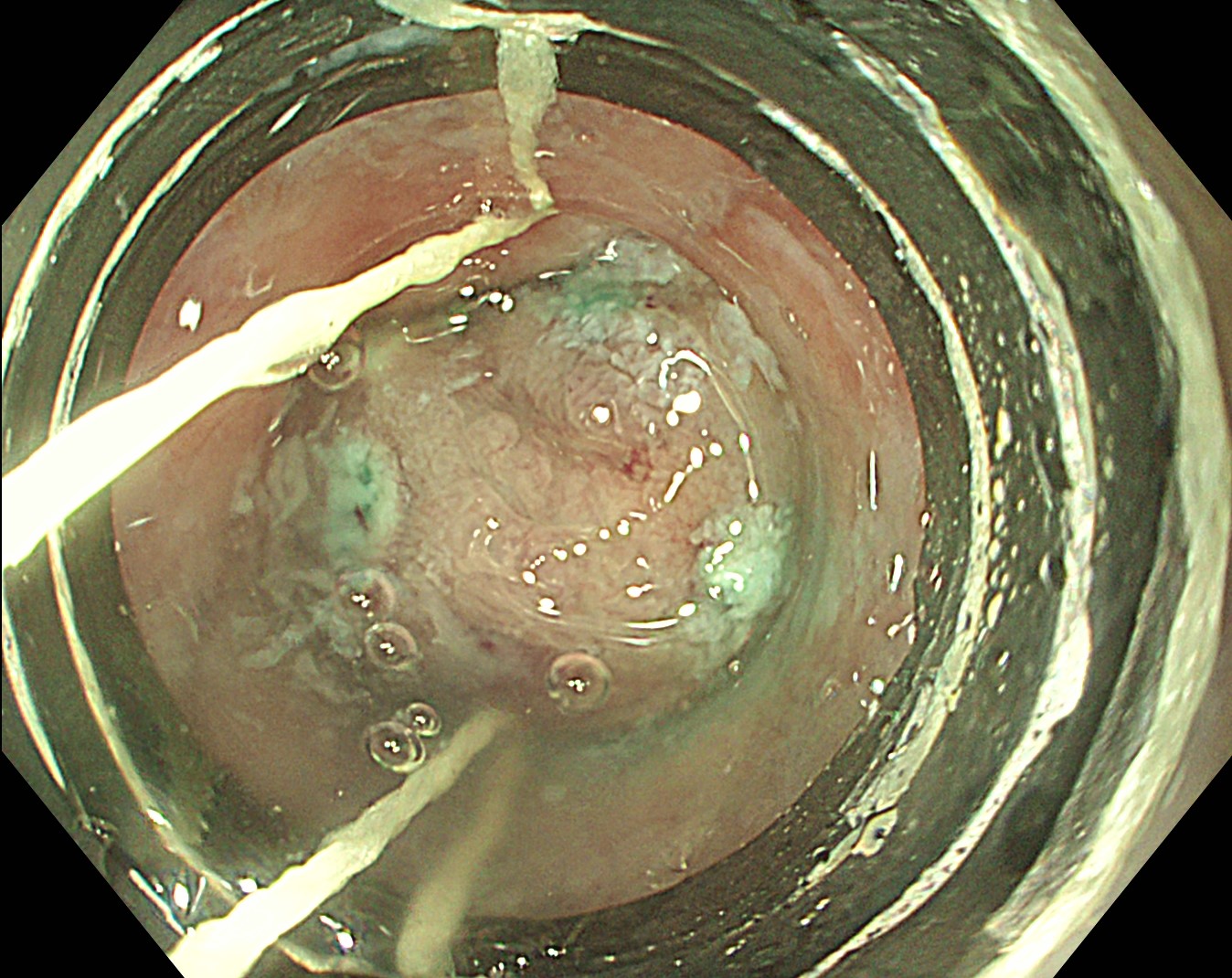
6. Submucosal injection
The area lifted well with submucosal injection
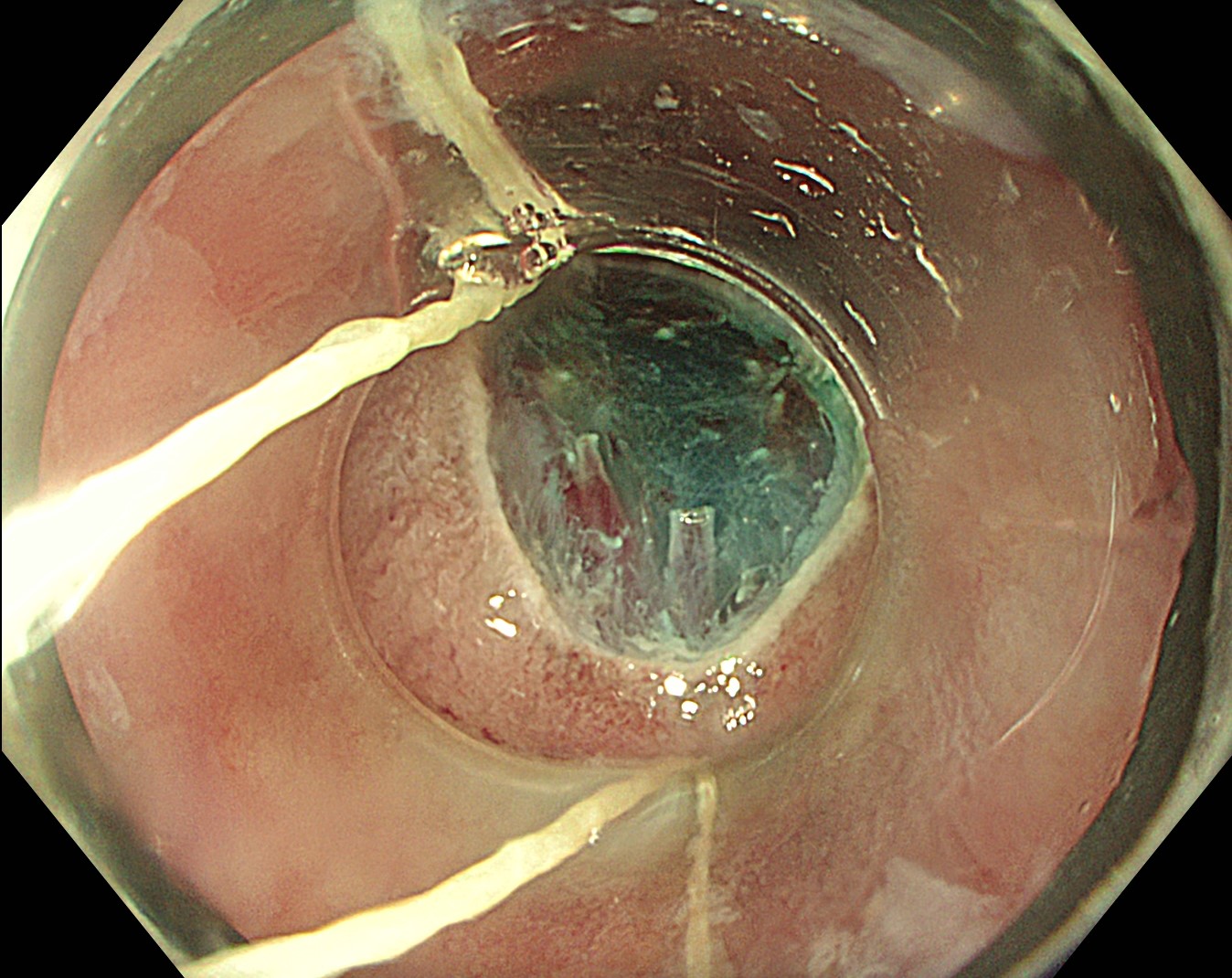
7. EMR
An EMR is performed using a multi-band mucosectomy device
8. Base
A clean base was seen after resection
9. Specimen
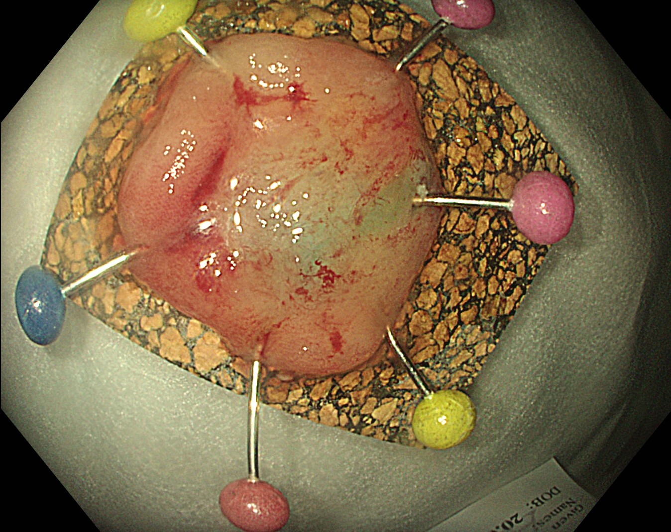
The specimen is seen pinned to a cork
Case video
On initial views with WLI, a subtle depressed area is seen at the site of recent biopsy. Upon closer examination with TXI and magnification, the depressed lesion is more clearly seen. The lesion is then examined with NBI and high-magnification using the underwater technique, which reveals tortuous vessels at the base signifying the presence of pT1a adenocarcinoma. A single-piece EMR is then successfully performed.
Overall Comment
This case involved an 81-year-old man with Barrett’s esophagus and a history of previous endoscopic submucosal dissection for invasive adenocarcinoma. On surveillance endoscopy a few small subtle areas of nodularity were biopsied, with one area demonstrating the presence of adenocarcinoma. At repeat endoscopy 10 days later there were no nodules, however there were a few small depressed areas at the site of recent biopsies. These areas were more conspicuous using TXI. Once localized, the area of concern was carefully examined using WLI, TXI, NBI and high-magnification. On inspection using NBI with high-magnification and the underwater technique, tortuous vessels were noted at the base of the depressed area, representing residual adenocarcinoma. A single-piece EMR was performed, with histology confirming the presence of pT1a adenocarcinoma with clear deep and lateral margins.
* Specifications, design and accessories are subject to change without any notice or obligation on the part of the manufacturer.
Prof. Stefan Seewald Case 6: Peroral endoscopic myotomy (POEM)
Prof. Philip Chiu
- Keyword
- Content Type