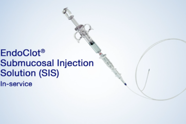
1. Injection
(1) 23G Injection needle
(2) Injection Solution: Normal saline + blue dye
*Do not add epinephrine
(3) Injection Point: 2cm above from the starting point of myotomy. Usually on anterior wall just below compression by left main bronchus approximately 30cm endoscopy insertion from patient’s teeth.
2. Incision
(1) TriangleTipKnife J (OLYMPUS: KD-645L) is used for POEM.
(2) Setting for incision
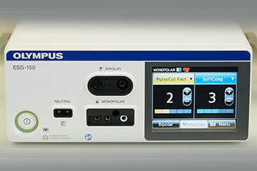
Electrosurgical unit (OLYMPUS ESG-300): PulseCut, 30W, Effect 2.
*Interrupted output is useful
(3) Always the knife should be moved very slowly and carefully.
(4) 2cm longitudinal incision is required to introduce endoscope into submucosal space smoothly.
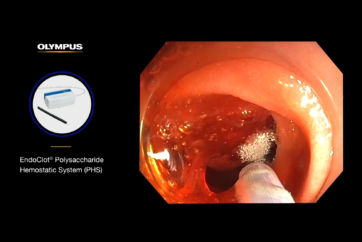
3. Tunneling
(1) Setting for tunneling
Electrosurgical unit (OLYMPUS ESG-300): SprayCoag, 30W, Effect 3.
*Very short step is useful
(2) Dissect close to the muscle to avoid mucosal injury keeping away the knife from the mucosa.
(3) Inject 5 to 10 ml additional solution using jet function of TriangleTipKnifeJ into submucosal layer accordingly.
(4) Use Coagrasper for hemostasis, after identification of bleeding spot and grasping it, carefully activate energy source avoiding unintentional injury to the mucosal layer.
(5) Keep the muscle on the top of the display and the mucosa on the bottom. This is the very important critical view.
(6) One third of circumferential dissection is recommended.
(7) Submucosal tunnel should be created down to gastric cardia at least 3cm over EGJ.
(8) “Spindle vein” is characteristic to submucosal layer at gastric cardia
4. Myotomy
(1) Setting for myotomy
Electrosurgical unit (OLYMPUS ESG-300): SprayCoag, 30W, Effect 3.
*Very short step is useful
(2) Cut only circular muscle and leave the longitudinal muscle which can act as a safety margin to mediastinal structure.
(3) Begin to incise carefully as the thickness of the circular muscle cannot be identified at beginning of myotomy.
(4) After cutting the circular muscle, sometime the longitudinal muscle is torn, but no clinical issue can happen.
(5) Keep the tip of the TriangleTipKnifeJ clean for smooth incision. It is efficient to prepare 2 knives and use them interchangeably because one of them can be cleaned when it is not used.
(6) The circular muscle should be fully cut.
(7) Even in achalasia patients circular muscle at LES is not thick. This thin circular muscle is incompetent to relax during swallowing and should be cut completely.
(8) In gastric side longitudinal muscle layer becomes obscure and eventually it becomes full layer resection but nothing adverse happens.
(9) At least 3 cm myotomy for gastric side is recommended. It enables complete release of LES muscle.
(10) When anatomy becomes unclear in gastric side, inject blue dye added saline into submucosal layer. It creates good contrast between submucosa and muscle layer.
5. Confirmation
(1) With the retroflex view, the relaxed GEJ can be confirmed.
(2) Blue dye injected for tunneling is also observed and you can confirm that the length of the myotomy is enough to be toward gastric side.
6. Confirmation (Double scope method)
(1) After creating a submucosal tunnel, the extent of the tunnel can be confirmed by applying double scope method.
(2) Additional thin scope is needed to perform this method. The thin scope is intubated into the esophagus and advanced down to the stomach.
(3) The standard scope is advanced to the distal end of the submucosal tunnel.
(4) In the retroflexion view of the thin scope in the stomach, the tip of the standard scope in the submucosal tunnel can be visualized. Transluminal light function can be used to clearly visualize the position of the standard scope.
(5) By applying double scope method, incomplete myotomy (short submucosal tunnel) and excessive myotomy into the gastric side (long submucosal tunnel) can be avoided.
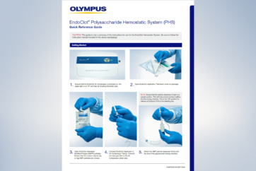
7. Closure
(1) Put the first clip on the distal end of mucosal incision. First clip creates a fold of mucosa which makes subsequent placement of clips easier. Apply the second clip and others to proximal side to close the opening tightly. Typically 7 clips are needed.
(2) For the first clip, Olympus QuickClipPro is good with reopening function, and for other clips, Olympus EZ Clip is better with smaller clip tail.
(3) The oblique cap on the tip of the endoscope helps to apply the proper tension onto tissues for clipping.
(4) Tight closure is required to prevent the leakage of esophageal content into mediastinum.
(5) After closing the defect, the tight closure should be confirmed from both side of closed mucosa.
- Content Type