Oropharynx Cancer (Posterior Wall) Aged 54, male
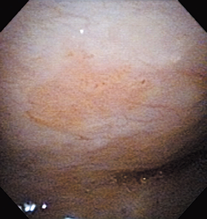
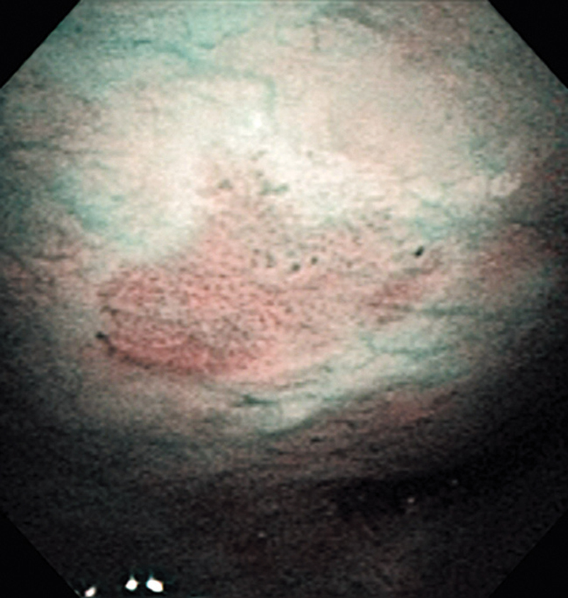
Comment:
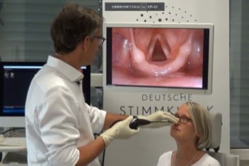
The lesion was detected in the oropharyngeal posterior wall in a laryngopharyngoscopic NBI examination during follow-up after treatment of a carcinoma in the floor of the mouth.
It was recognized under NBI as a lesion with a brownish area, and the close-up view additionally showed scattered brown dots. In the conventional white light image, the same area was seen as a reddening lesion.
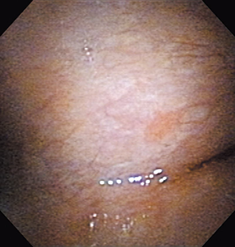
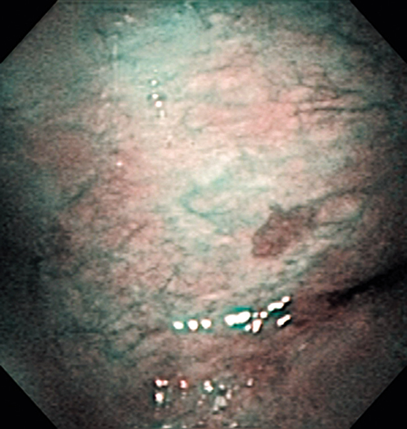
Oropharynx Cancer (Posterior Wall) Aged 72, male
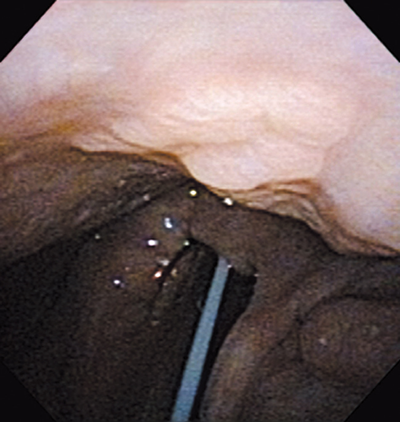
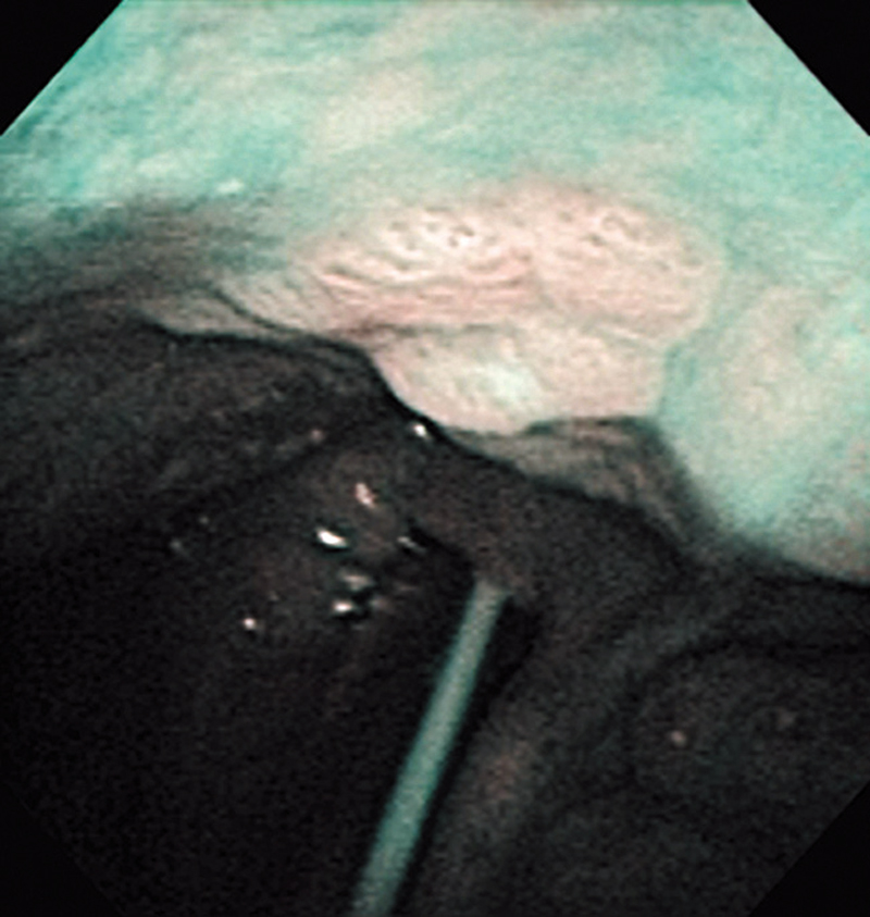
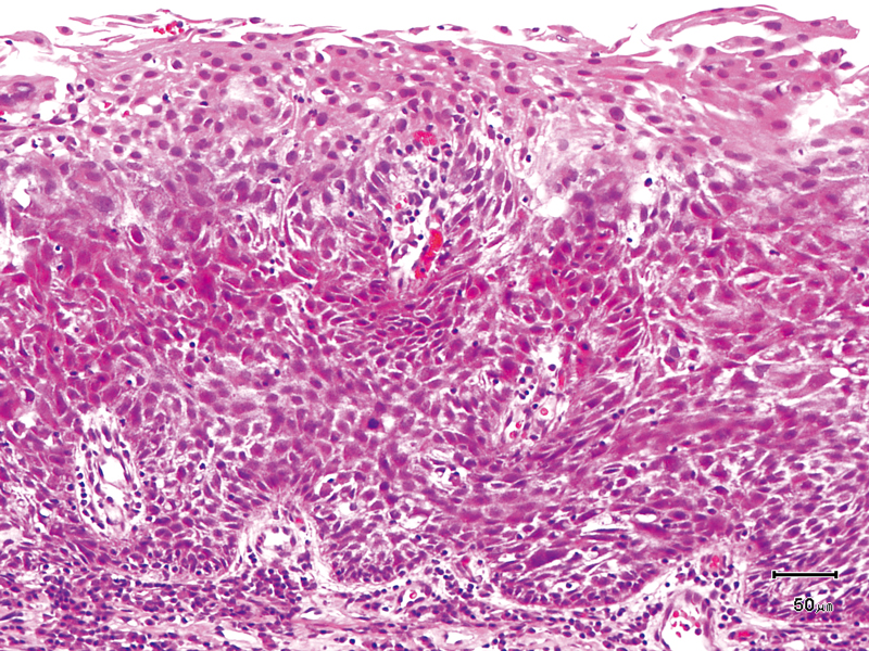
Comment:
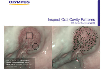
The lesion was detected on the oropharyngeal posterior wall in a periodic laryngopharyngoscopic NBI examination after an esophageal carcinoma surgery.
The NBI image showed a brownish, slightly-elevated lesion. In the conventional white light image, the same area was recognized as a slightly-whitish elevated lesion.
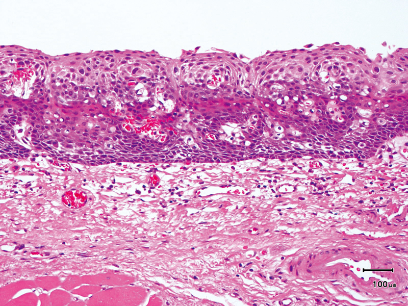
The lesion was treated with endoscopic mucosal resection and diagnosed as a squamous cell carcinoma in situ.
- Keyword
- Content Type