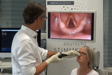
How NBI Works
The Underlying Principle
Normal white light (WL) contains all colors. When WL hits the surface of a tissue, all colors are absorbed. Thus the image remains with lack of contrast. With NBI this is different.
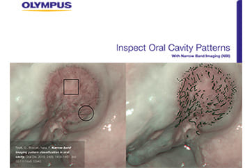
NBI uses only blue and green light. When blue and green light hit the surface of the tissue, it is highly absorbed by hemoglobin in the blood vessels. While the blue light is absorbed by the capillaries in the mucosa, the green light reaches deeper to the submucosal area, where it is reflected by the blood vessels. This is why NBI creates a significantly higher contrast between blood vessels and the surrounding tissue than WL. The NBI images therefore show more contrast than WL images. Since small tumors are often surrounded by many blood vessels, NBI helps to detect these tumors at an early stage and to analyze these areas accordingly. Thus, NBI supports the early and optical diagnosis of oral, pharyngeal and laryngeal cancer lesions, which as a result allows better treatment and more accurate follow-ups.
Numerous studies highlight the clinical value of NBI, especially with regard to the characterization of suspicious mucosal areas and the detection of cancerous lesions.

- Content Type