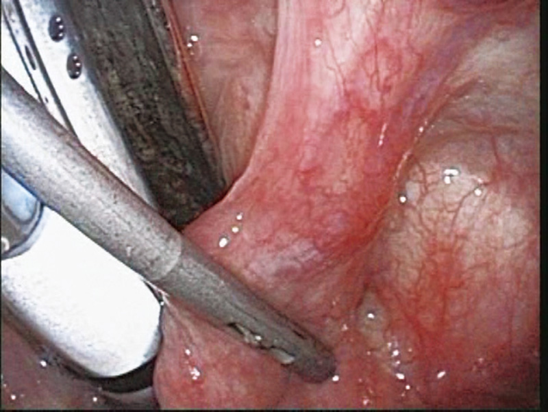
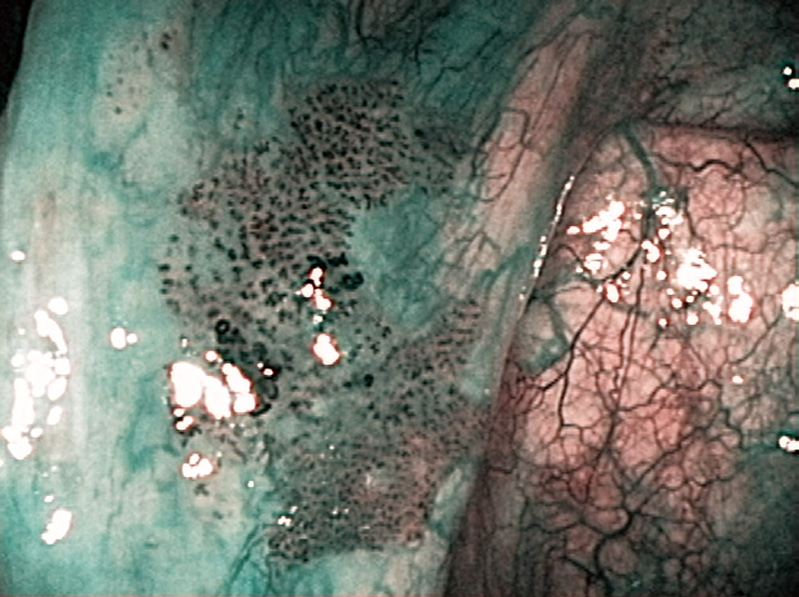
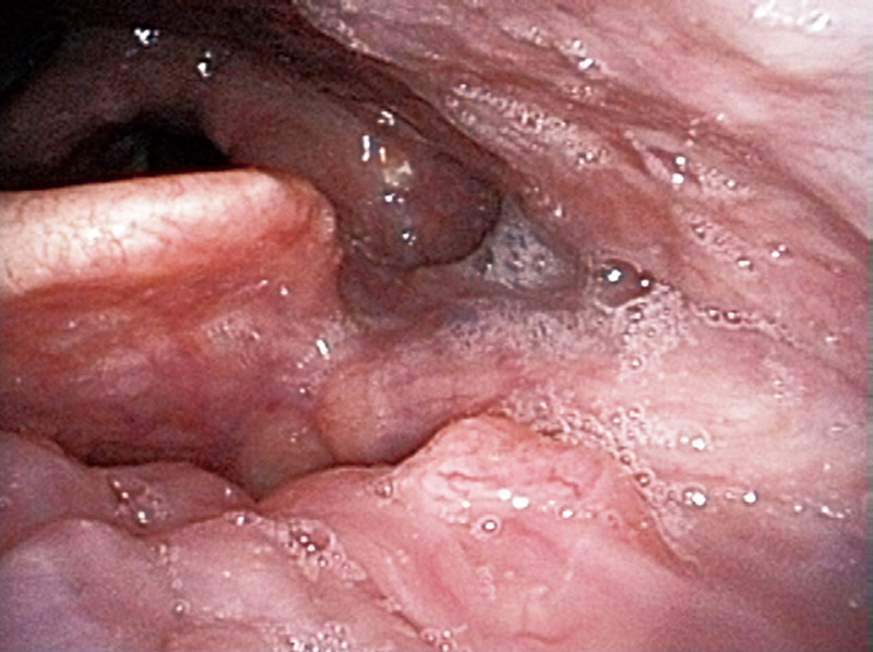
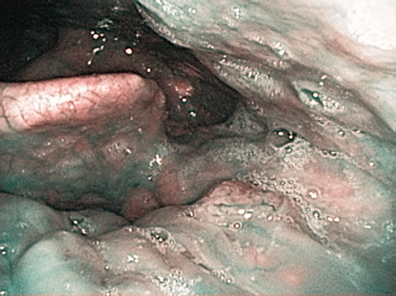
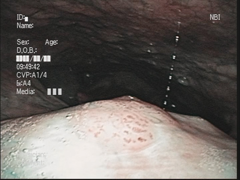
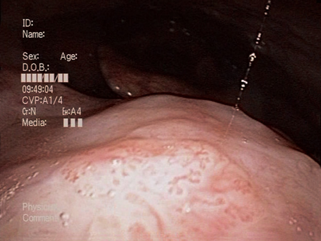
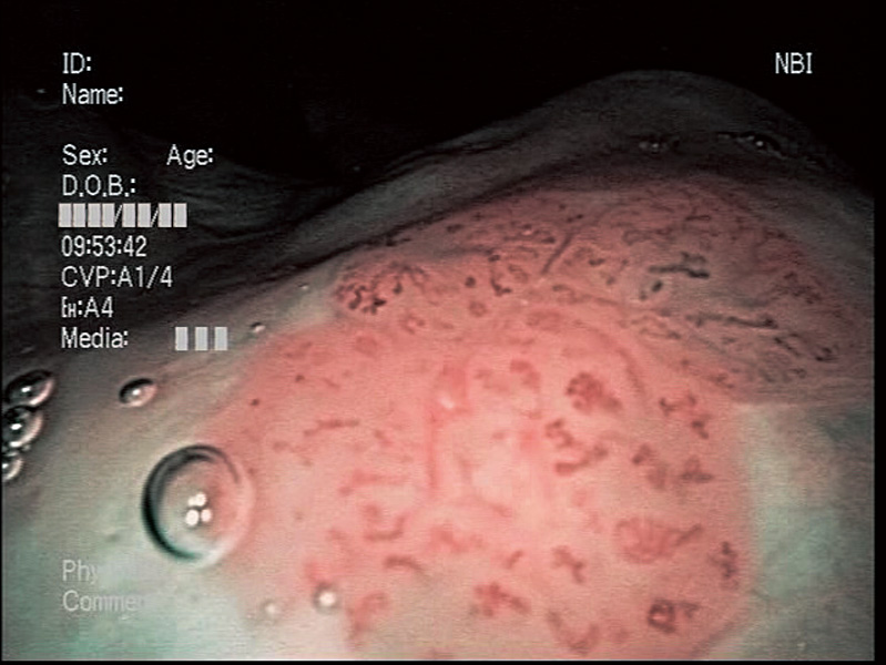
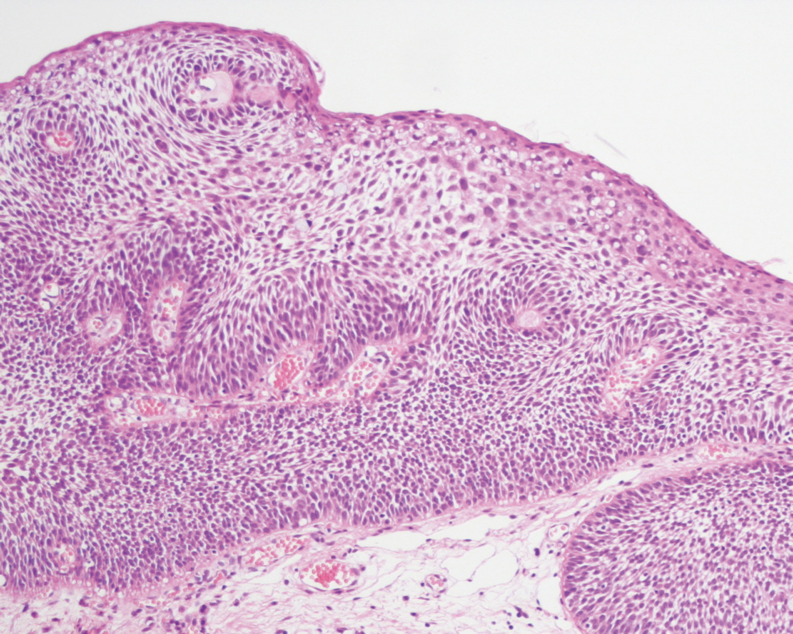
Hypopharyngeal Intraepithelial Carcinoma (Right Pyriform Sinus) Aged 77, male
general anesthesia
close-focus view
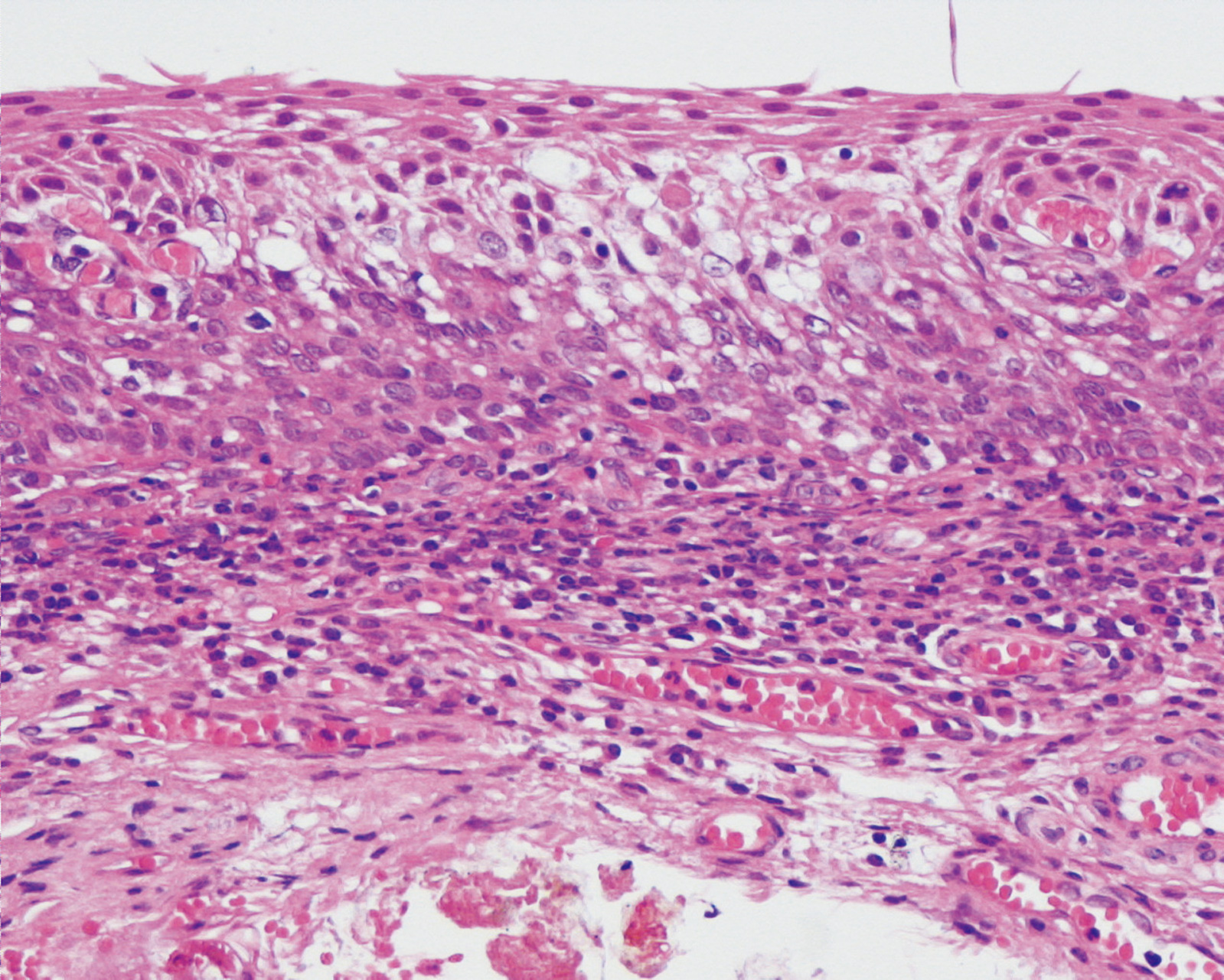
pathology
Comment:
These are the peroral endoscopic findings observed under general anesthetic. In the distant view under white light, a flat reddish lesion was observed.
In NBI, a brownish area presented in the same region. In the NBI close-focus view, intraepithelial abnormal vessels were visualized as scattered brownish dots, and their extension, dilatation, weaving and different shapes were recognized. The branching vessels in the subepithelial layer of the normal mucosa were visualized in green, presenting clear contrast to the lesion area.
The lesion was treated with ELPS(Endoscopic Laryngo Pharyngeal Surgery) resection, and pathologically diagnosed as an intraepithelial carcinoma.
Images and comments by Dr. Y. Satou <ENF-VQ>
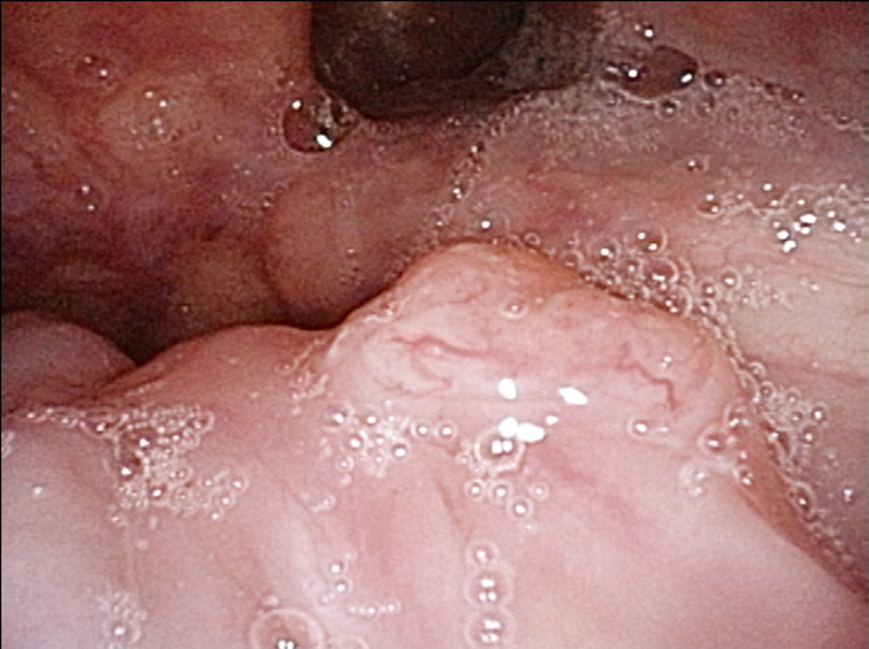
Oropharyngeal Cancer (Left Tongue Base) Aged 60, male
close-focus view
pathology
Comment:
Diagnosed as a squamous cell carcinoma after a biopsy of the left cervical lymph node in September 2005. As primary unknown cancer of the cervical lymph node had previously been detected, nasal endoscopic examinations had been performed every one to three months, but no noticeable finding had been recognized in the laryngopharynx. Two years and six months after that diagnosis, a 5 mm small tumor mass was recognized in nasal NBI endoscopy in the left tongue base and the NBI close-focus view suggested enlarged abnormal vessels. A biopsy was performed and the lesion was diagnosed as a moderately- to poorly-differentiated squamous cell carcinoma.
Images and comments by Dr. Y. Satou <ENF-VQ>
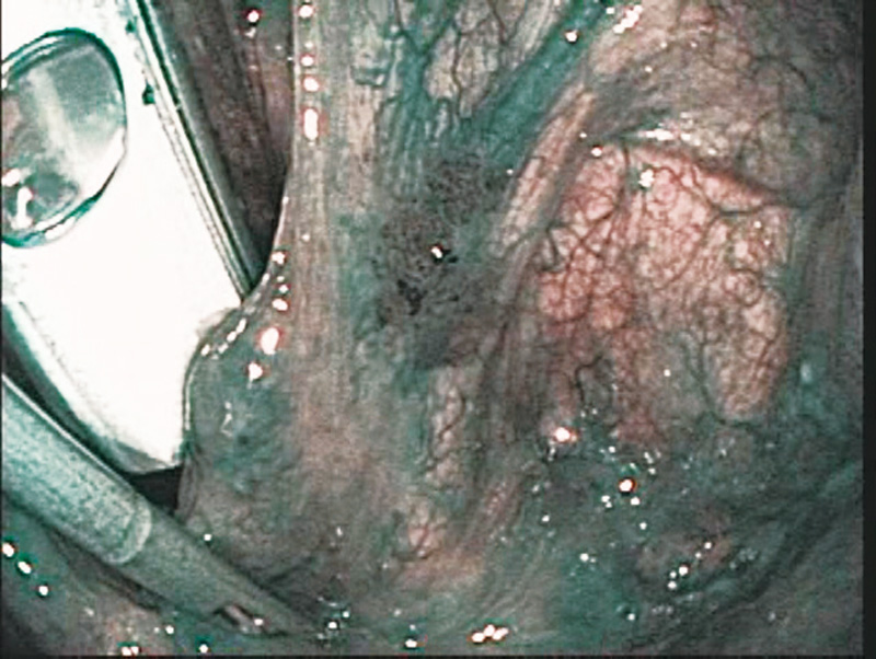
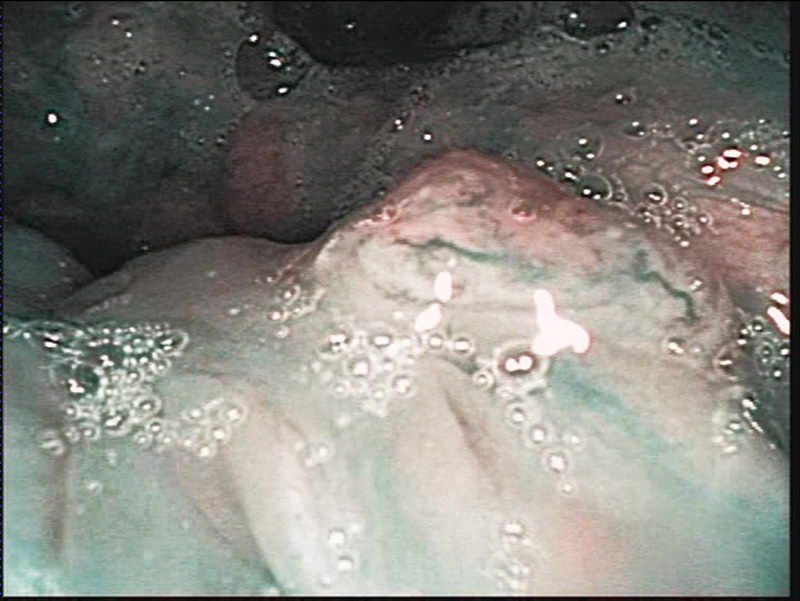
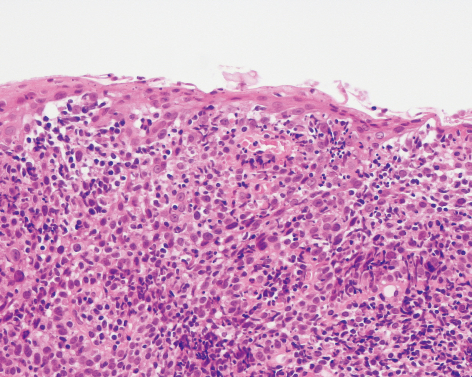
Epipharyngeal Intraepithelial Carcinoma (Soft Palate Upper Surface) Aged 60, male
close-focus view
pathology
Comment:
The patient visited us complaining abnormal sensation in the laryngopharynx. Nasal NBI observation showed a brownish area with clear boundaries in the midline region of the soft palate upper surface. In the NBI close-focus view, the intraepithelial abnormal vessels showed signs of extension, dilatation, weaving and different shape.
The lesion was pathologically diagnosed as an intraepithelial carcinoma.
Images and comments by Dr. Y. Satou <ENF-VQ>
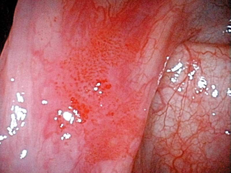
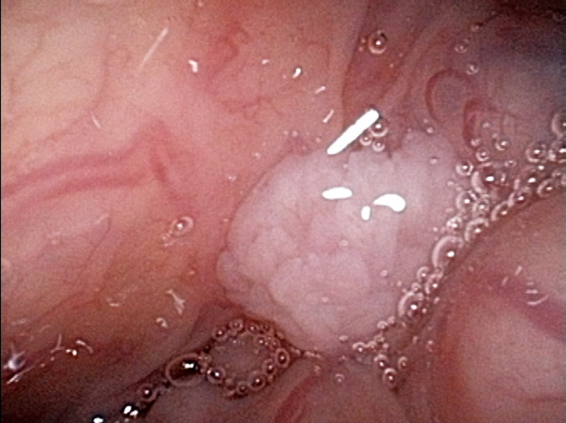
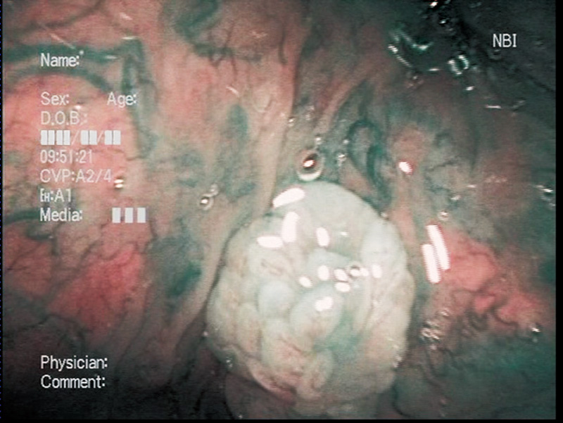
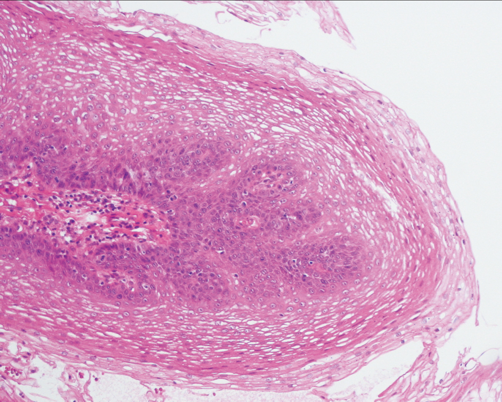
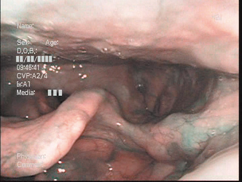
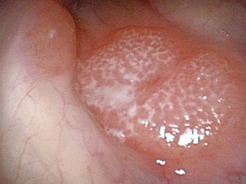
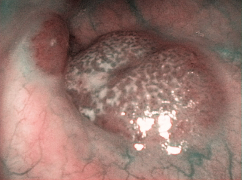
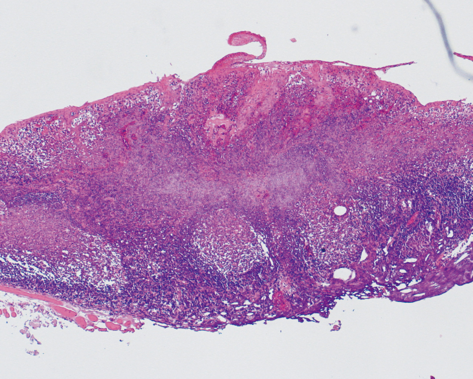
Oropharyngeal Papilloma (Left Epiglottic Vallecula) Aged 61, male
pathology
Comment:
In white light observation, a white lesion was recognized in the left epiglottic vallecula. The NBI close-focus view showed lobular white tumor mass within which thin and constant capillaries were recognized and therefore papilloma was suspected. A biopsy was performed and it was pathologically diagnosed as a papilloma.
Images and comments by Dr. Y. Satou <ENF-VQ>
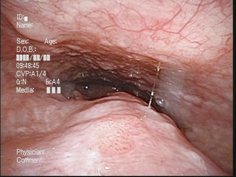
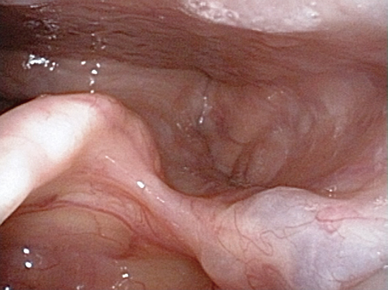
Lymphoid Follicles in the Hypopharynx (Left Pyriform Sinus) Aged 74, female
close-focus view
pathology
Comment:
The patient visited a local hospital complaining abnormal sensation in the laryngopharynx, and was referred to us on suspicion of hypopharyngeal cancer of the left pyriform sinus. Because no abnomal vascular proliferation was observed in either while light or NBI close-focus observation, lymphoid follicles were suspected.
The lesion was treated with endoscopic resection upon the patient’s request, and pathologically diagnosed as lymphoid follicles.
Images and comments by Dr. Y. Satou <ENF-VQ>
- Keyword
- Content Type