Takashi Kawai, MD, PhD
Department of Gastroenterological Endoscopy
Tokyo Medical University Hospital
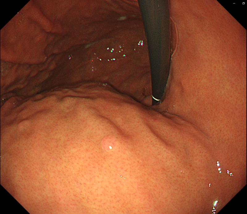
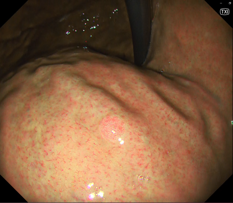
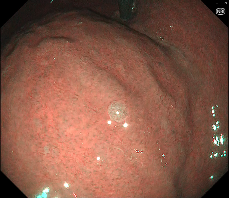
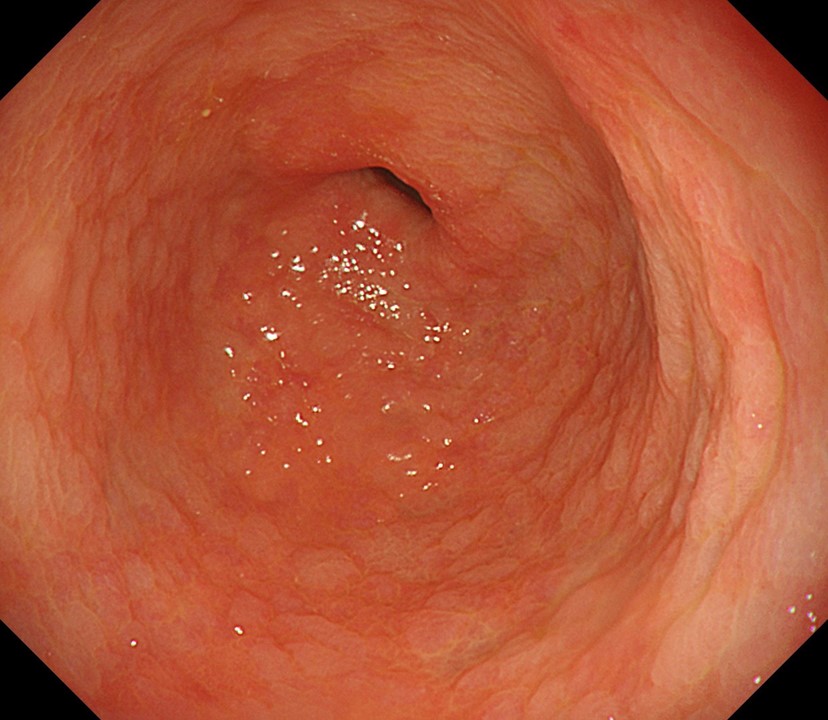
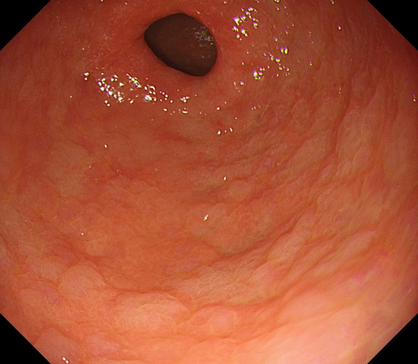
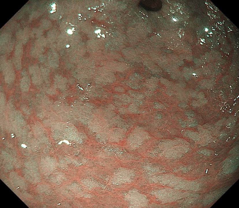
Scope : GIF-1200N / EVIS X1
Case : Regular arrangement of collecting venules (RAC)
Organ : From the middle of the gastric body to the lesser curvature of the upper stomach
Patient Information : M, 60s
Medical History : H. pylori eliminated
WLI
TXI
NBI
Overall comment
Under white light, a regular arrangement of collecting venules (RAC) was observed. When the mode was switched to Texture and Color Enhancement Imaging (TXI), the RAC was depicted even more clearly. However, when the mode was switched to Narrow Band Imaging (NBI), the RAC became virtually invisible. Switching back to white light observation revealed a fundic gland polyp in the same location. When observed in the TXI mode, the surface structure of the polyp was more clearly recognizable. In the NBI mode, the mucosal microstructure of the polyp could be observed. We found TXI also useful for observation of RAC.
Scope : GIF-1200N / EVIS X1
Case : Intestinal metaplasia
Organ : Pyloric antrum of the stomach
Patient Information : M, 60s
Medical History : Endoscopic treatment of gastric cancer
WLI
WLI
NBI
Overall comment
Multiple grayish-white flat elevations of different sizes were plainly visible against a background of atrophied gastric mucosa throughout the pyloric antrum of the stomach. In Narrow Band Imaging (NBI) observation, the intestinal metaplasia was recognized as an area exhibiting brownish to pale colors.
* Specifications, design and accessories are subject to change without any notice or obligation on the part of the manufacturer
Takashi Kawai, MD, PhD Case 8: Low grade dysplasia (Duodenum)
Dr. Jae Young Jang
- Content Type