Gastric Case 7

Dr. Khanh Do-Cong Pham, FASGE, FESGE
Department of Medicine
Haukeland University Hospital
Bergen, Norway
Scope: GIF-EZ1500
Case: Two gastric neuroendocrine tumors (NET)
Organ: Stomach
Patient Information: F, 80s, Two gastric NETs
Medical History: Hypertention, cardial insuficiency, asthma
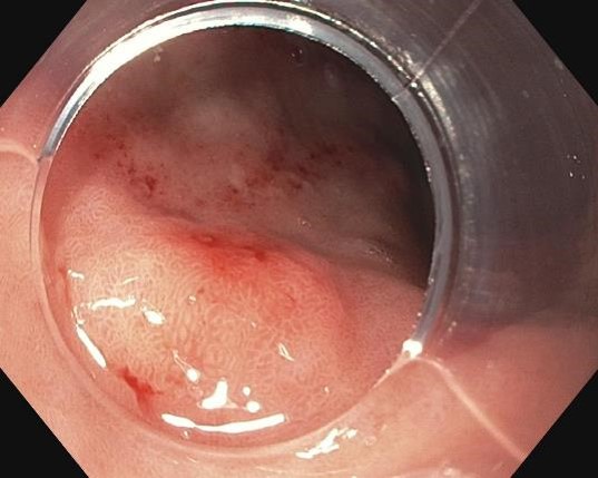
1. 1st Gastric NET (WLI)
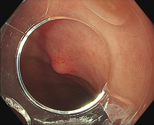
The tumor appears light yellowish and slightly elevated.
2. 1st Gastric NET (WLI)
Using the cap for tactile impression, the tumor is firmer than the surroundings.
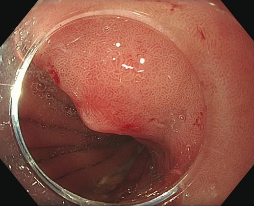
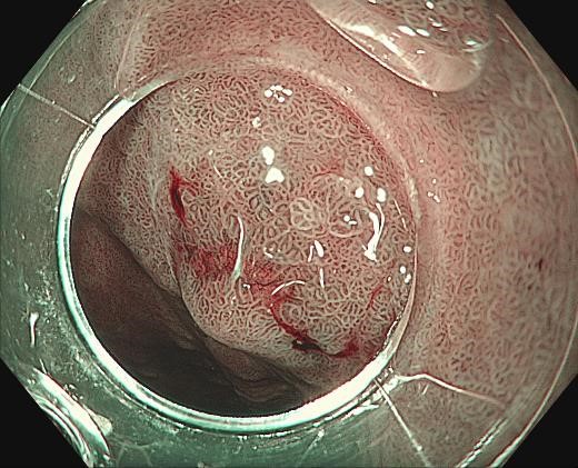
3. 1st Gastric NET (WLI, Near Focus, Under water)
The Near Focus allow close up evaluation. The tumor can be seen beneath of the thinner part of the mucosa.
4. 1st Gastric NET (NBI)
The 1st Gastric NET in NBI.
The 1st Gastric NET in NBI close-up. The tumor is more apparent where the mucosa is thin.
6. 1st Gastric NET (TXI mode 1)
The TXI mode enhance the color difference.
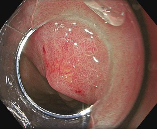
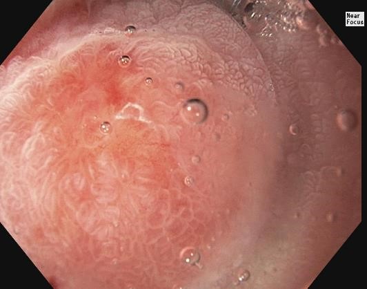
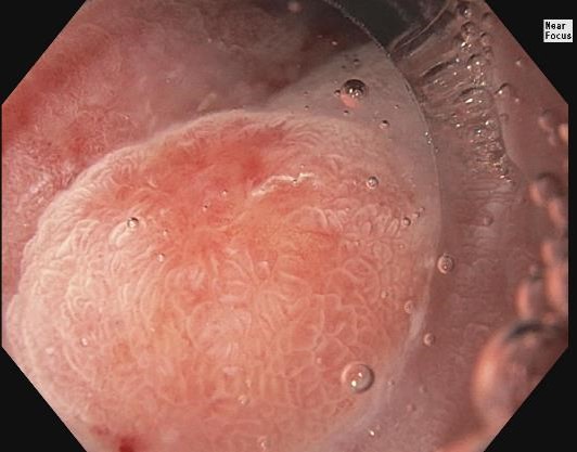
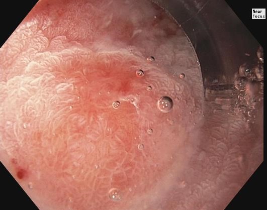
7. 2nd Gastric NET (WLI)
Slight elevation, with central thinning of the mucosal surface
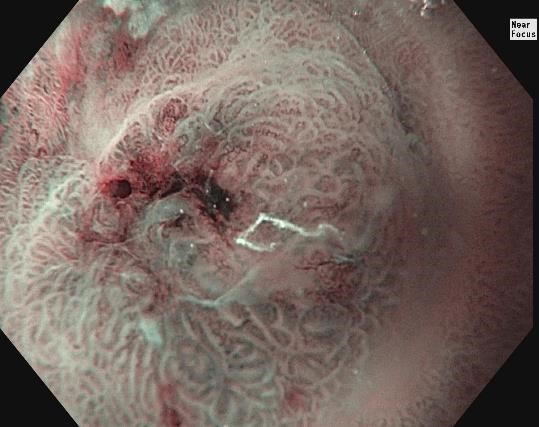
The 2nd Gastric NET in near focus and NBI
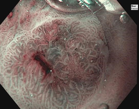
The 2nd Gastric NET in near focus and NBI
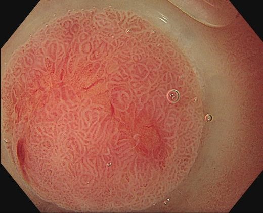
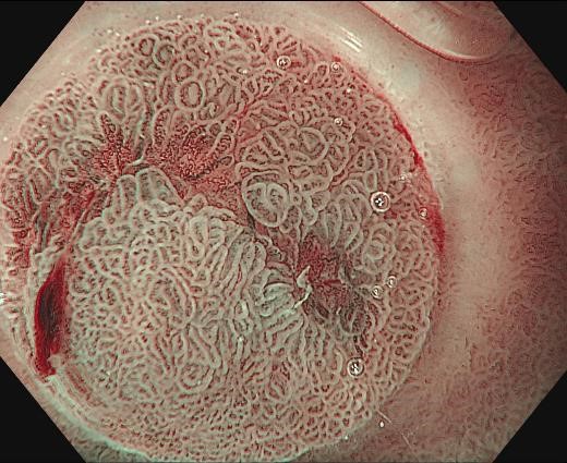
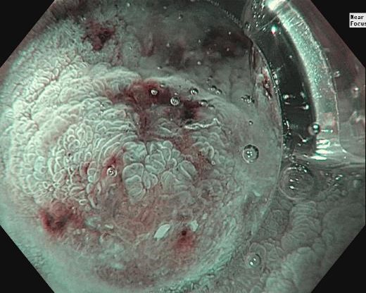
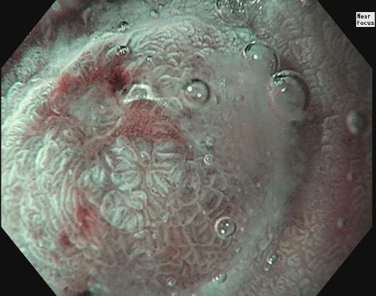
Acetic acid 2-3% was applied to enhance the mucosal surface. There is intestinal metaplasia surrounding the tumor.
Acetic acid 2-3% was applied to enhance the mucosal surface. There is intestinal metaplasia surrounding the tumor.
Acetic acid 2-3% was applied to enhance the mucosal surface. There is intestinal metaplasia surrounding the tumor.
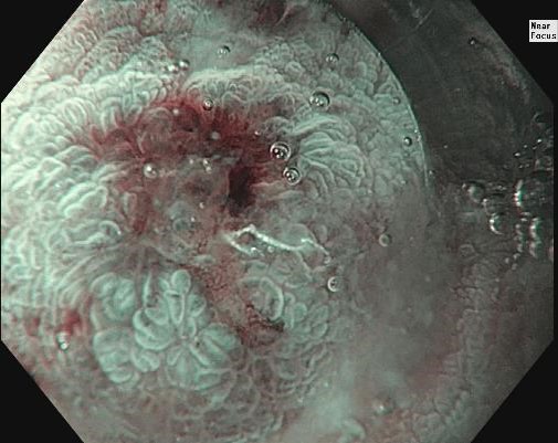
Combining NBI, Near Focus, and acetic acid enhances the surface structures in detail.
Combining NBI, Near Focus, and acetic acid enhances the surface structures in detail.
Combining NBI, Near Focus, and acetic acid enhances the surface structures in detail.
Case video
We demonstrate in the video how gastric neuroendocrine tumors may look like with the different properties of the EVIS X1 and the GIF-EZ1500 gastroscope.
Overall Comment
Most gastric neuroendocrine tumors (NETs) are associated with chronic atrophic gastritis and can be identified through high-definition white light endoscopy. NETs typically appear as small round “hills” in a flat landscape (Paris Is or Is+IIc). The mucosa on the surface tends to be thin for larger lesions, and the tumor presents as a subepithelial mass with a yellowish hue. Narrow Band Imaging (NBI) and TXI can be used for easier recognition of gastric NETs.
* Specifications, design and accessories are subject to change without any notice or obligation on the part of the manufacturer
Dr. Hisashi Doyama Case 8: Early stomach cancer
Prof. Dr. Liu Zhiguo
- Keyword
- Content Type