Esophageal Case 3
Prof. Stefan Seewald
GastroZentrum Hirslanden, Zurich, Switzerland
Disclaimer:
- NBI™ and TXI™ Technologies are not intended to replace histopathological sampling as a means of diagnosis
- The positions and statements made herein by Prof. Seewald are based on Prof. Seewald’s experiences, thoughts and opinions. As with any product, results may vary, and the techniques, instruments, and settings can vary from facility to facility. The content hereof should not be considered as a substitute for carefully reading all applicable labeling, including the Instructions for Use. Please thoroughly review the relevant user manual(s) for instructions, risks, warnings, and cautions. Techniques, instruments, and setting can vary from facility to facility. It is the clinician’s decision and responsibility in each clinical situation to decide which products, modes, medications, applications, and settings to use.
- The EVIS X1™ endoscopy system is not designed for cardiac applications. Other combinations of equipment may cause ventricular fibrillation or seriously affect the cardiac function of the patient. Improper use of endoscopes may result in patient injury, infection, bleeding, and/or perforation. Complete indications, contraindications, warnings, and cautions are available in the Instructions for Use (IFU)
Scope: GIF-EZ1500
Case: Squamous Cell Carcinoma
Organ: Esophagus
Patient information: M, 70s
Medical history: Smoker
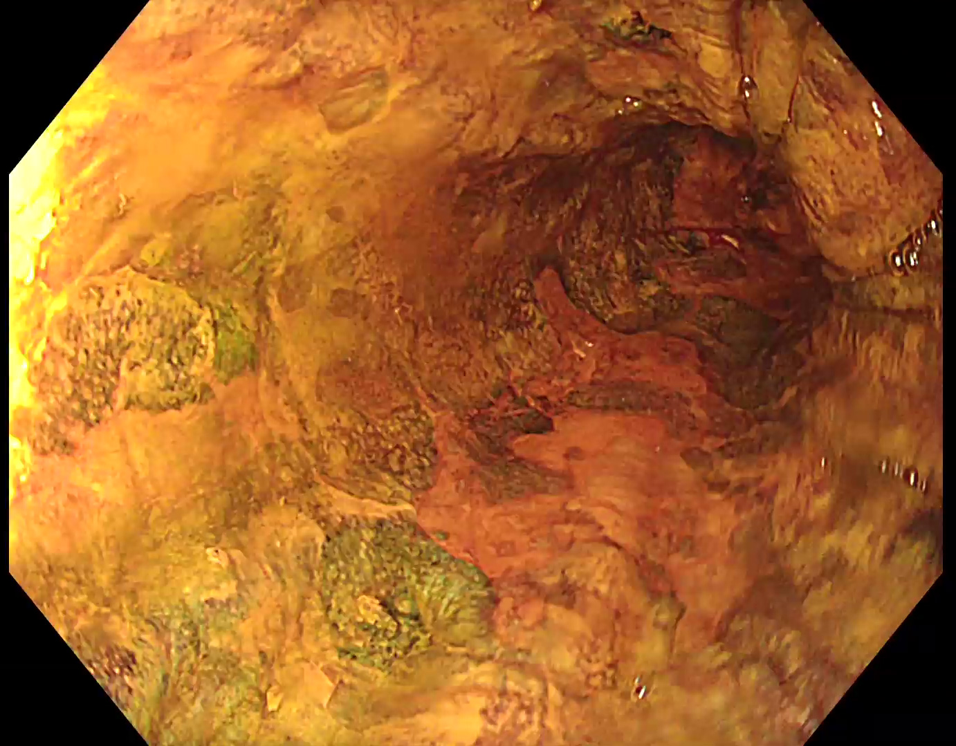
1. WLI
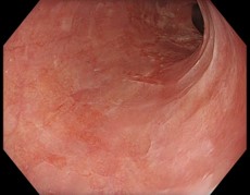
A suspicious reddish lesion can be identified at 6 to 9 o’clock.
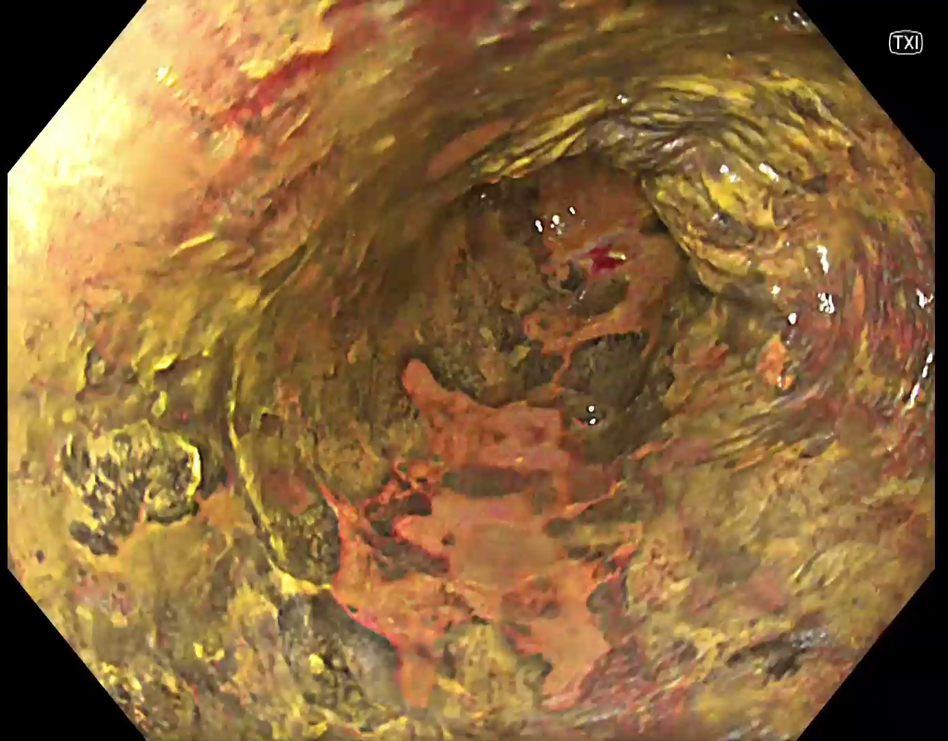
2. TXI™ technology
TXI™ technology suggests a larger area that may be interpreted as reflux esophagitis at the first glance.
3. NBI™ technology
However, with NBI technology abnormal intrapapillary capillary loops (IPCLs) can be identified which require closer examination.
4. Near Focus with NBI™ technology
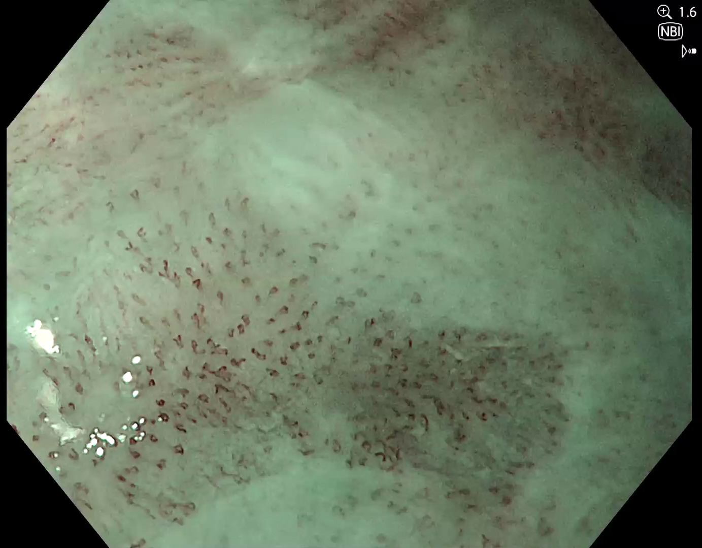
Abnormal IPCLs can be confirmed under near focus, suggesting a squamous cell carcinoma.
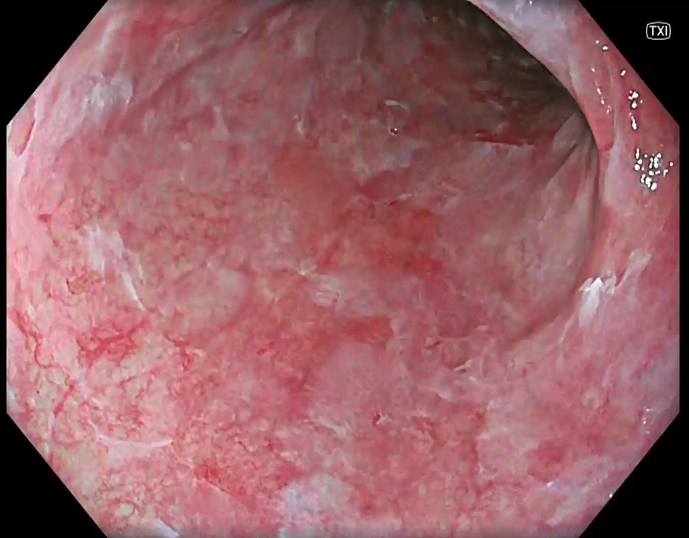
5. Lugol staining (WLI)
To confirm the extend of the carcinoma, Lugol staining was applied. Various Lugol-voiding lesions can be identified.
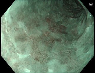
6. Lugol Staining (TXI™ technology)
Under TXI™ technology mode 1, the delineation is supported by stronger color contrast of the carcinoma. The pink color sign is more prominent compared to white light.
Case video
Overall Comment
This case presents an incidentally detected squamous cell carcinoma, NBI technology was helpful for detection and characterization in this case. TXI technology in combination with Lugol was helpful to delineate the lesion.
* Specifications, design and accessories are subject to change without any notice or obligation on the part of the manufacturer.
Prof. Stefan Seewald Case 4: Esophageal papilloma
Prof. Stefan Seewald
- Keyword
- Content Type


