Esophageal Case 4
Prof. Stefan Seewald
GastroZentrum Hirslanden, Zurich, Switzerland
Disclaimer:
- NBI™ and TXI™ Technologies are not intended to replace histopathological sampling as a means of diagnosis
- The positions and statements made herein by Prof. Seewald are based on Prof. Seewald’s experiences, thoughts and opinions. As with any product, results may vary, and the techniques, instruments, and settings can vary from facility to facility. The content hereof should not be considered as a substitute for carefully reading all applicable labeling, including the Instructions for Use. Please thoroughly review the relevant user manual(s) for instructions, risks, warnings, and cautions. Techniques, instruments, and setting can vary from facility to facility. It is the clinician’s decision and responsibility in each clinical situation to decide which products, modes, medications, applications, and settings to use.
- The EVIS X1™ endoscopy system is not designed for cardiac applications. Other combinations of equipment may cause ventricular fibrillation or seriously affect the cardiac function of the patient. Improper use of endoscopes may result in patient injury, infection, bleeding, and/or perforation. Complete indications, contraindications, warnings, and cautions are available in the Instructions for Use (IFU)
1) Data on file Data on file with Olympus (DC00489968)
Scope: GIF-EZ1500
Case: Esophageal papilloma
Organ: Esophagus
Patient information: F, 60s
Medical history: Heartburn
1. WLI
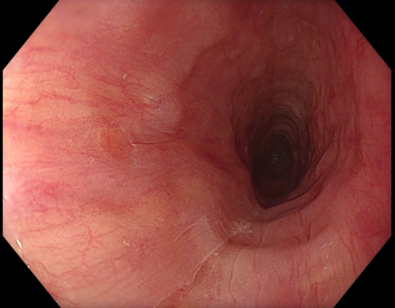
A very small reddish area at 9 o’clock appears suspicious.
2. TXI™ technology
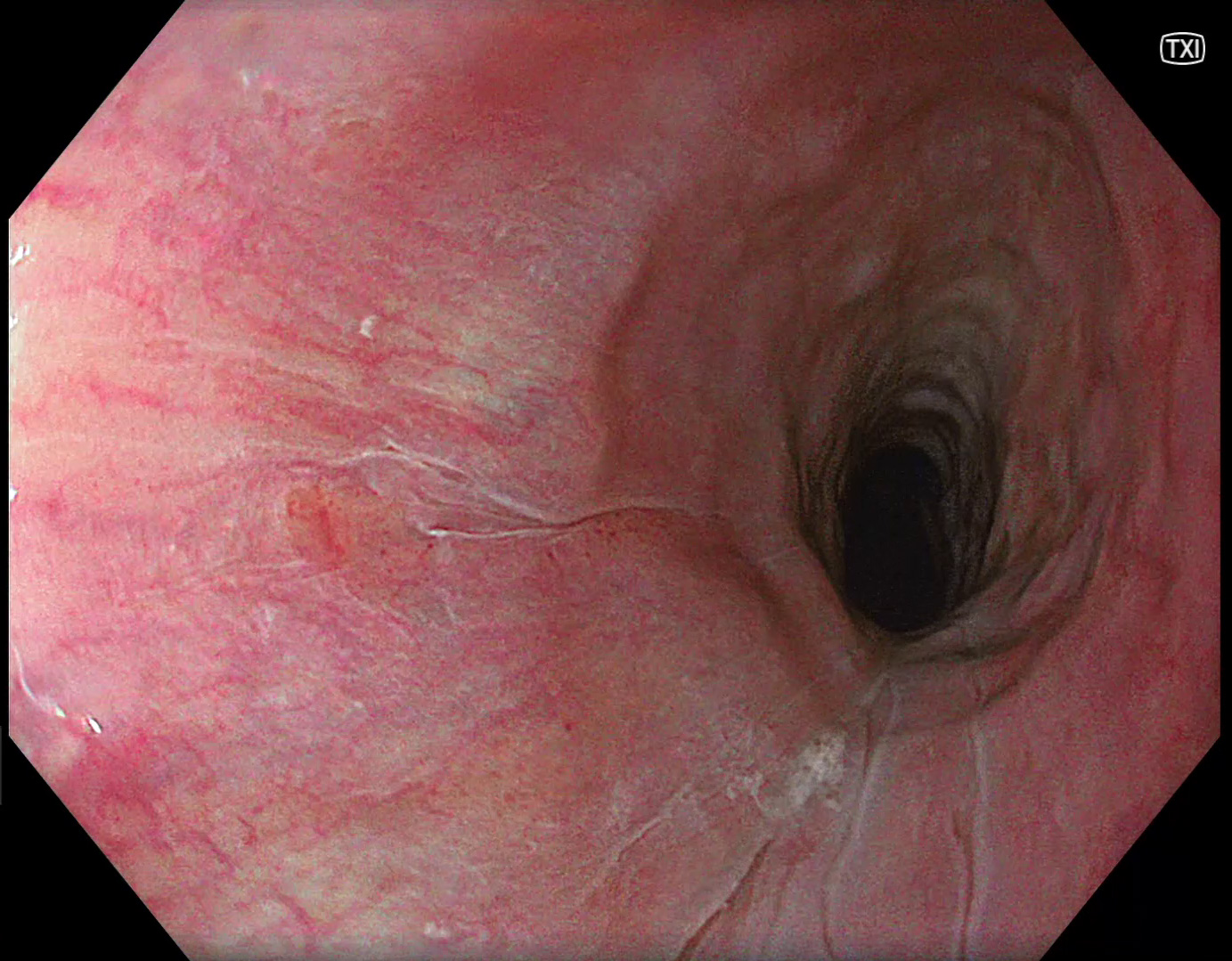
Using TXI™ technology increases the color contrast, meaning the reddish area is becoming more prominent1.
3. NBI™ technology
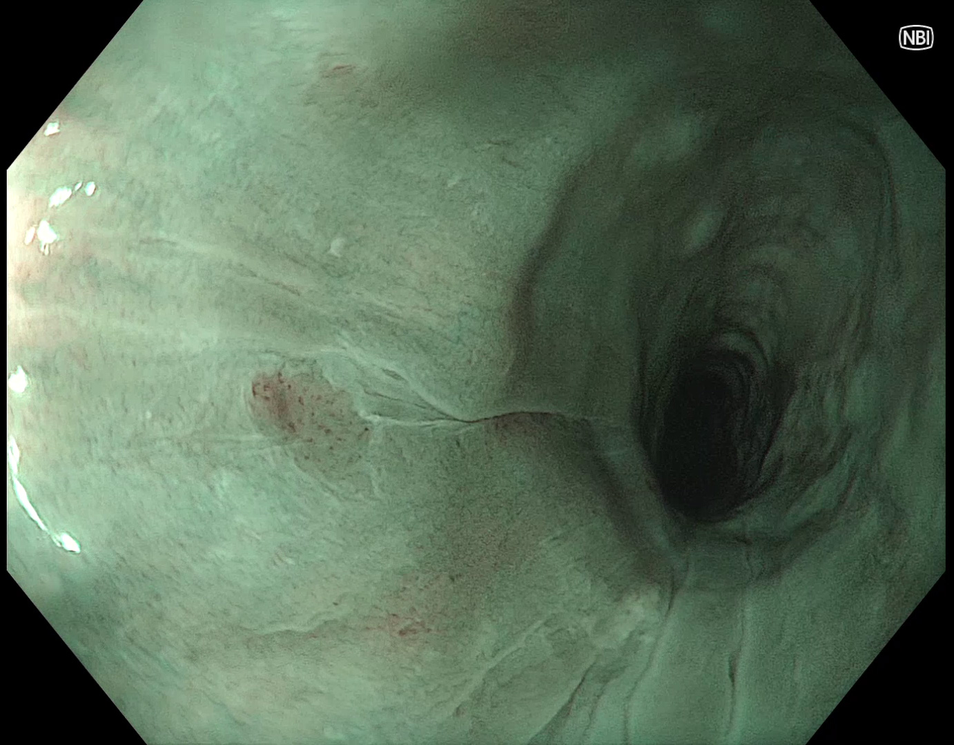
NBI™ technology reveals a small brownish area.
4. Near focus with NBI™ technology
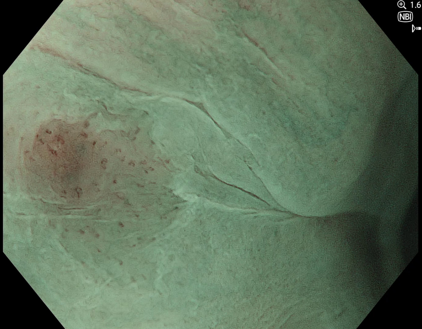
Enlarged IPCLs with brownish background mucosa can be seen.
5. WLI
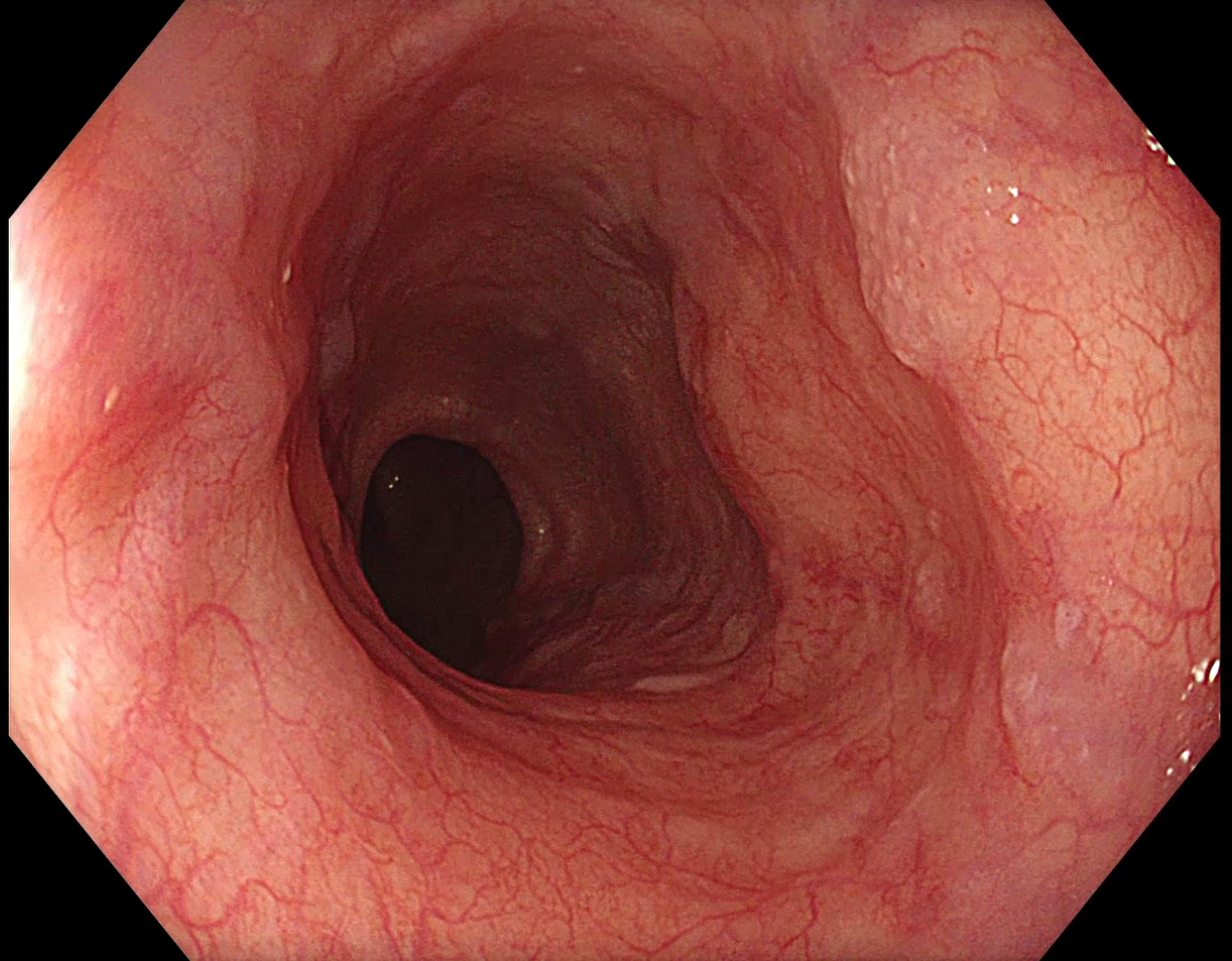
In the middle esophagus, further papillomas can be easily identified by WLI.
6. TXI™ technology
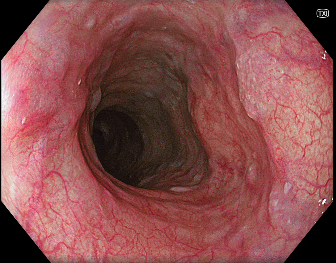
TXI™ technology is enhancing the distinct texture component of these lesions1.
7. NBI™ technology
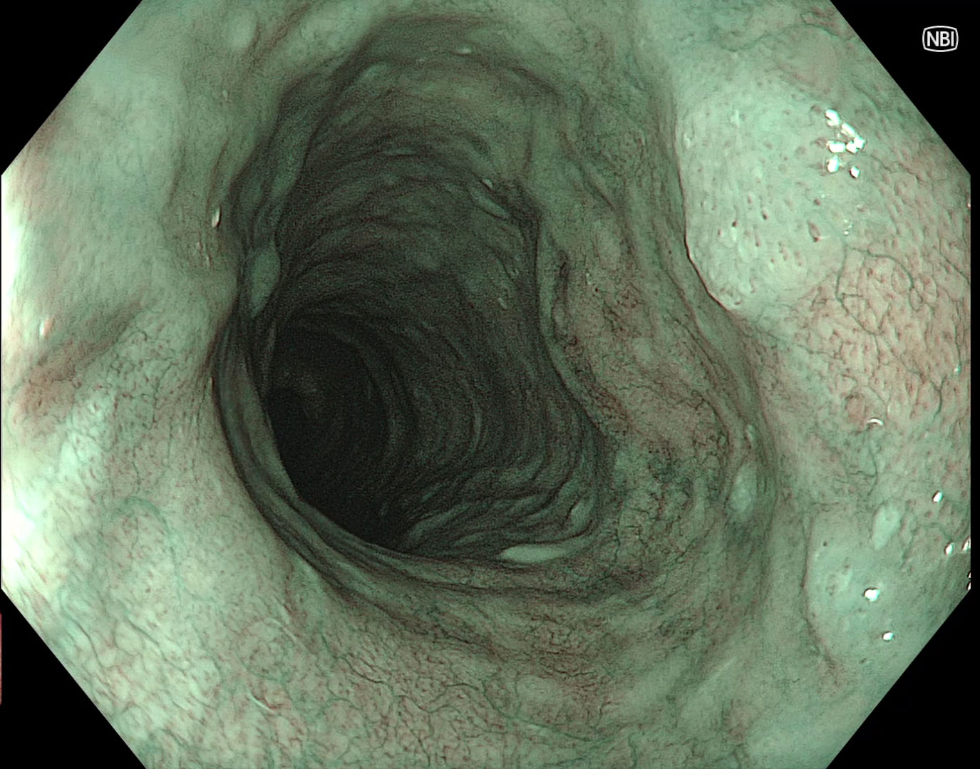
NBI™ technology demonstrates no pathological IPCLs.
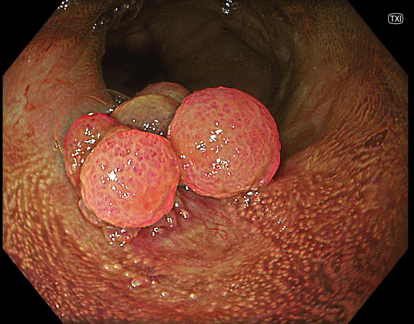
8. WLI
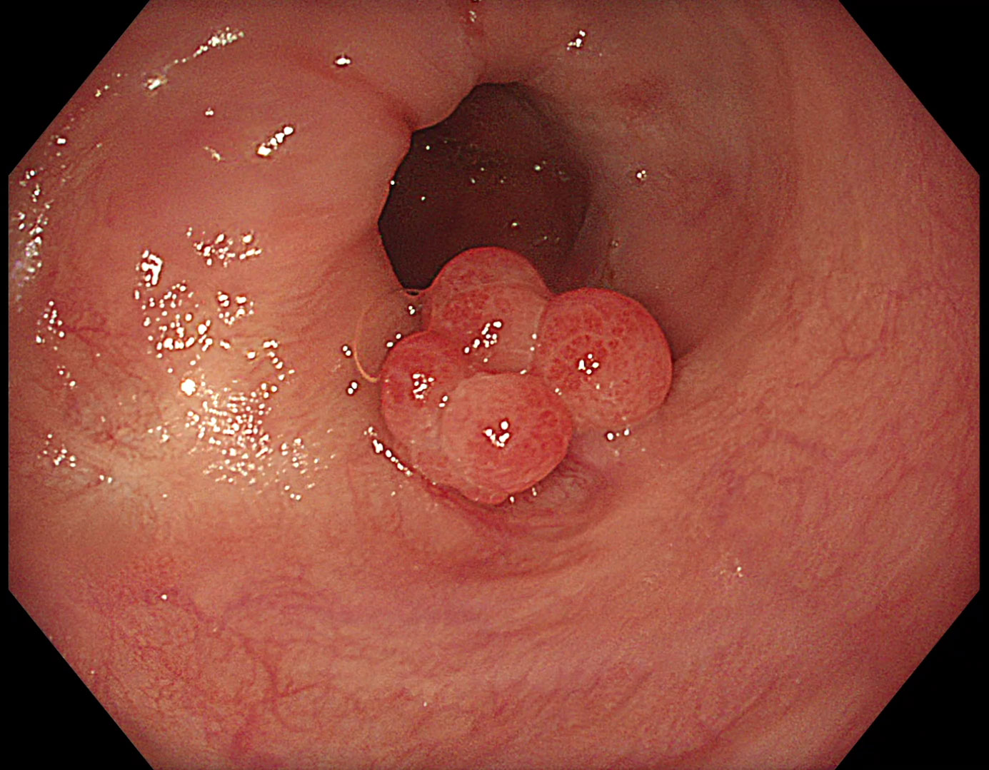
In the lower esophagus, multiple papillomas can be identified.
9. TXI™ technology
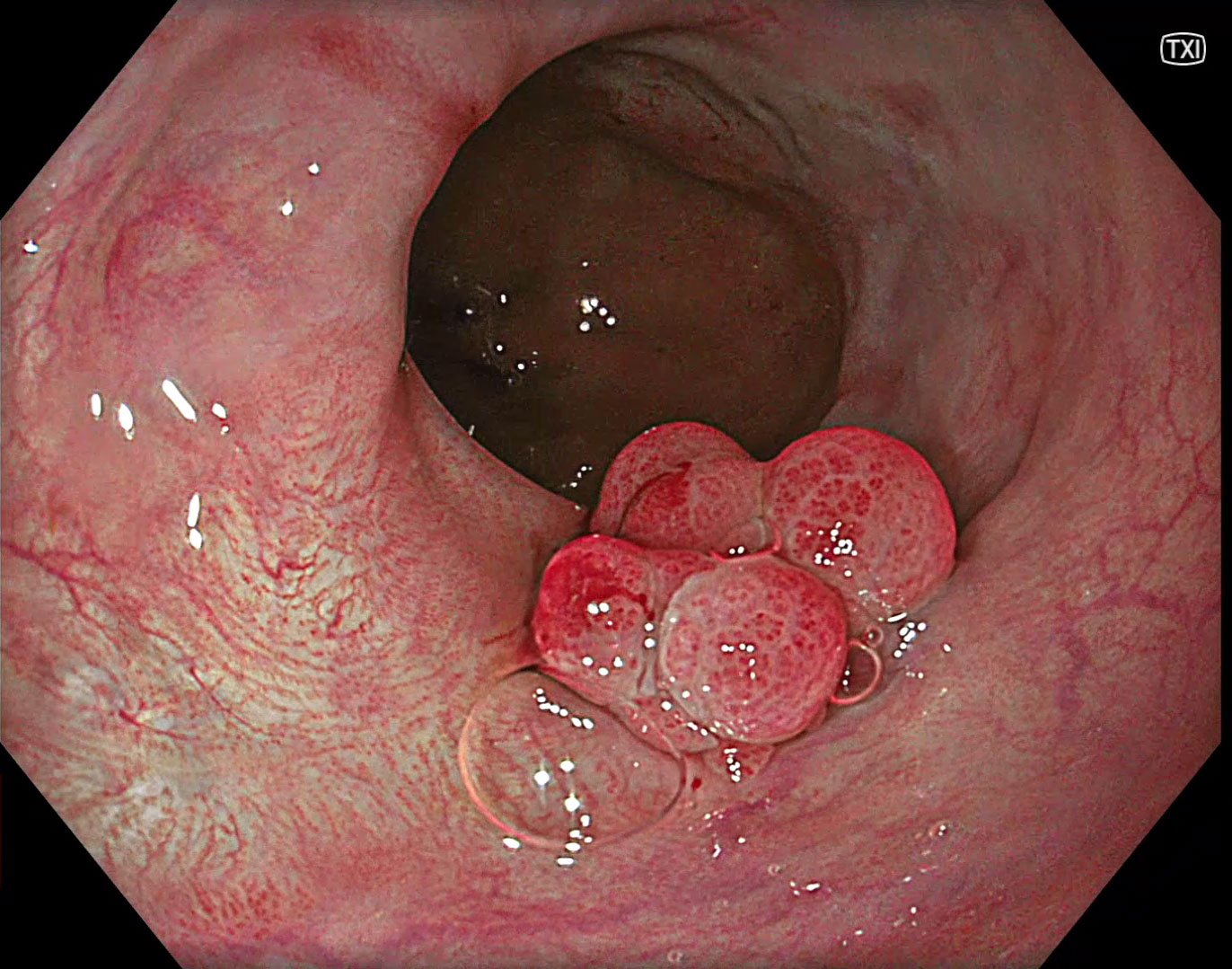
Under TXI™ technology the inflammatory aspect is enhanced by increased color contrast1.
10. NBI™ technology
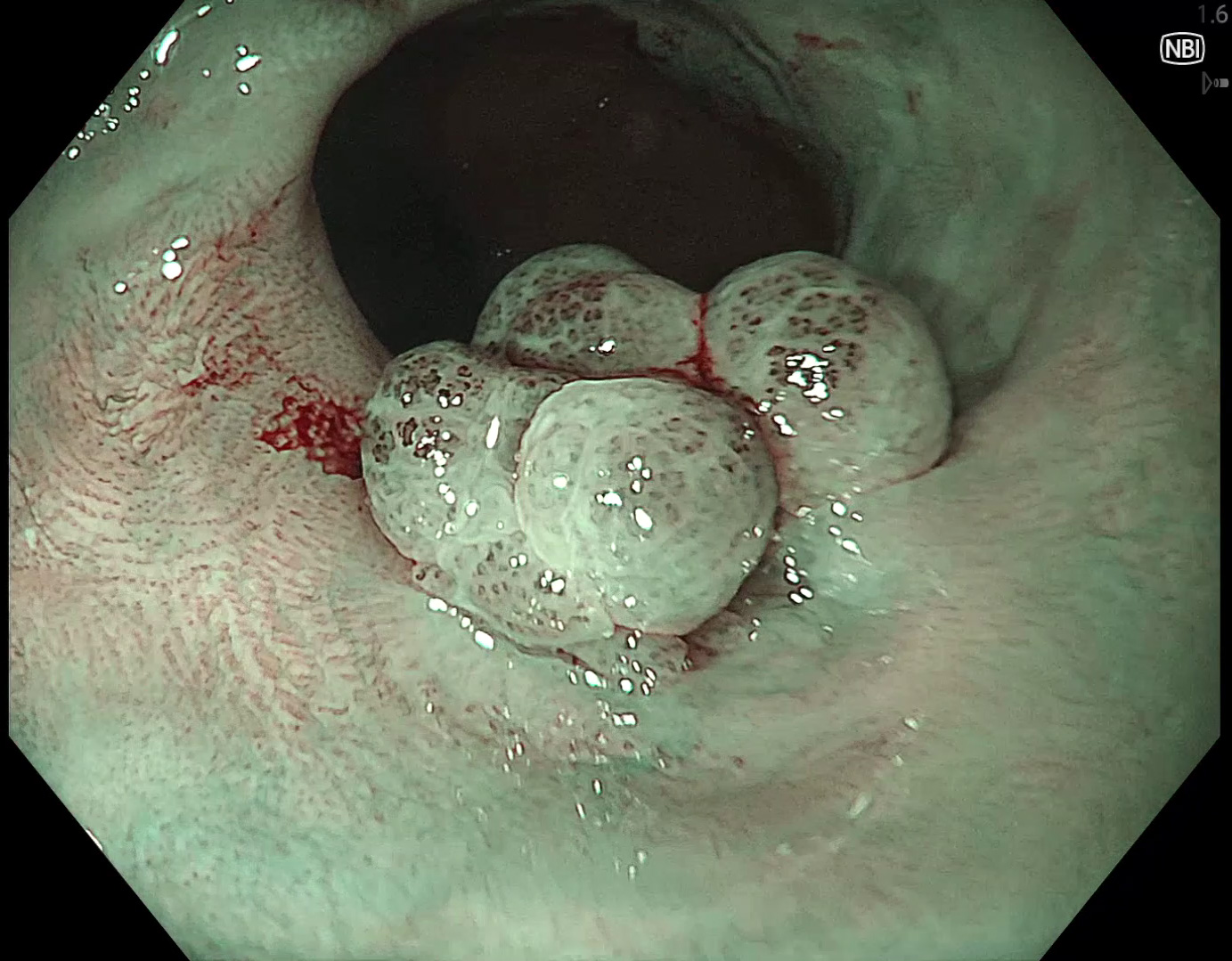
Inflammatory changes around the papilloma can be seen by dilatated IPCLs.
11. Near focus with NBI™ technology
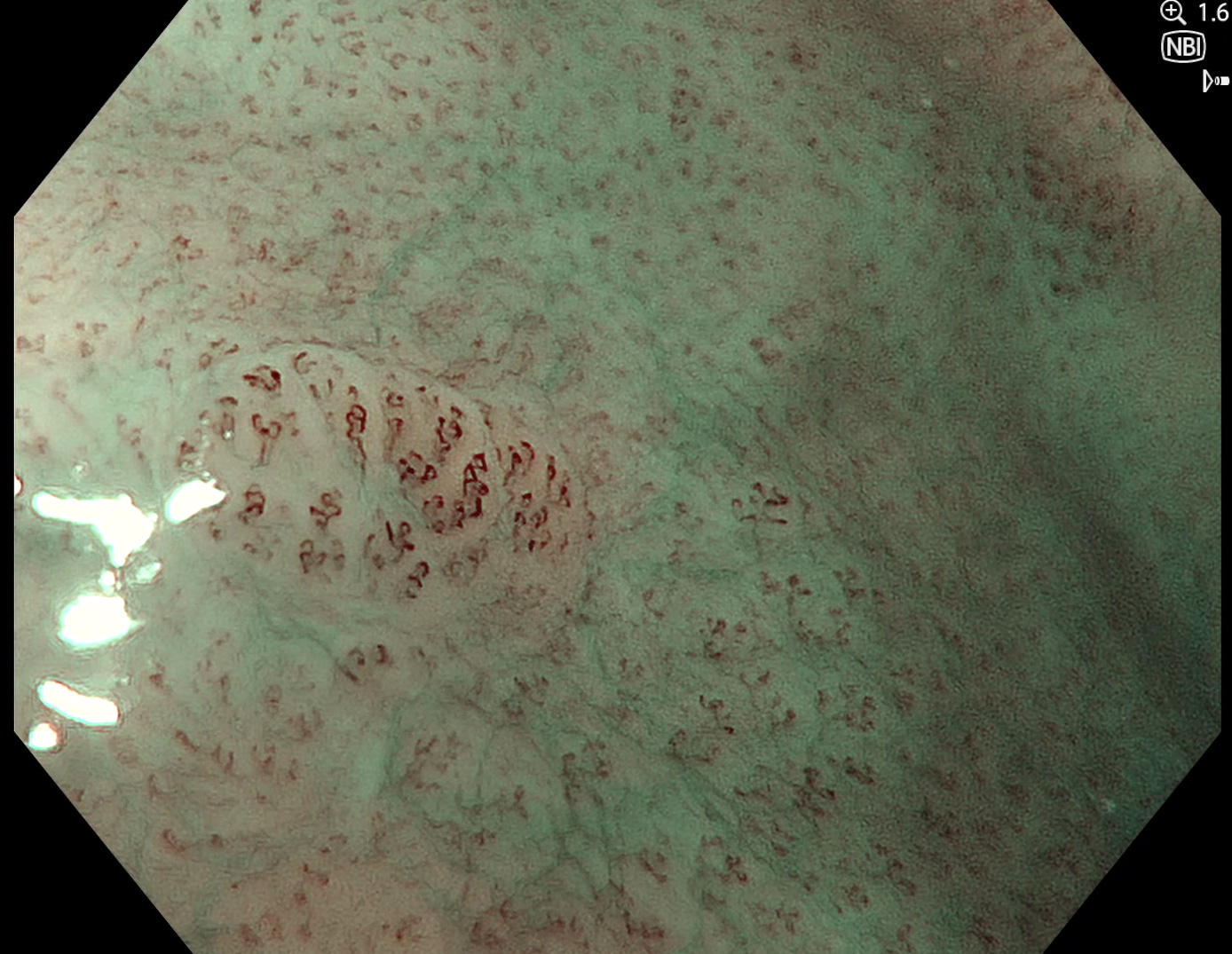
There are additional subtle lesions with IPCL dilatations.
12. Lugol staining and TXI™ technology
Lugol staining and TXI ™ technology shows no typical Lugol voiding behavior.
Case video
* Specifications, design and accessories are subject to change without any notice or obligation on the part of the manufacturer.
Prof. Stefan Seewald Case 5: Early adenocarcinoma in Barrett’s esophagus
Prof. Rajvinder Singh
- Keyword
- Content Type


