Gastric Case 2

Dr. D Nageshwar Reddy
AIG Hospitals, Hyderabad, India

Dr. Hardik Rughwani
AIG Hospitals, Hyderabad, India
Disclaimer:
- TXI™ Technology is not intended to replace histopathological sampling as a means of diagnosis
- The positions and statements made herein by Dr. Reddy and Dr. Rughwani are based on their experiences, thoughts and opinions. As with any product, results may vary, and the techniques, instruments, and settings can vary from facility to facility. The content hereof should not be considered as a substitute for carefully reading all applicable labeling, including the Instructions for Use. Please thoroughly review the relevant user manual(s) for instructions, risks, warnings, and cautions. Techniques, instruments, and setting can vary from facility to facility. It is the clinician’s decision and responsibility in each clinical situation to decide which products, modes, medications, applications, and settings to use.
- The EVIS X1™ endoscopy system is not designed for cardiac applications. Other combinations of equipment may cause ventricular fibrillation or seriously affect the cardiac function of the patient. Improper use of endoscopes may result in patient injury, infection, bleeding, and/or perforation. Complete indications, contraindications, warnings, and cautions are available in the Instructions for Use (IFU)
1) Data on file with Olympus (DC00489968)
Scope: GIF-EZ1500
Case: Multiple gastric polyps (Benign)
Organ: Stomach
Patient information: F, 50s
Medical history: History of chronic iron deficiency anemia and dyspepsia
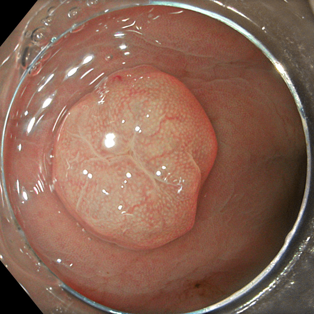
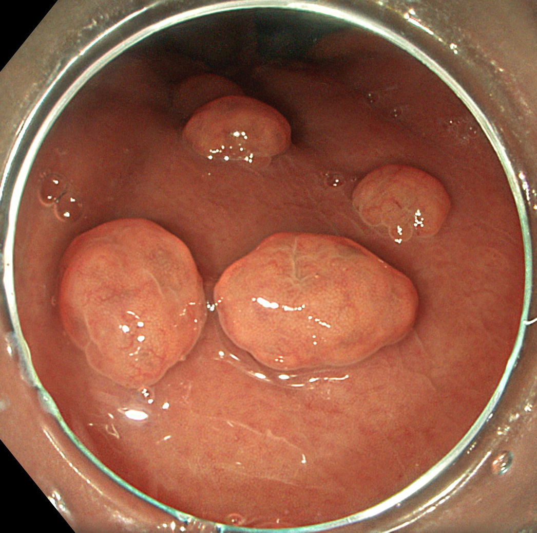
1. WLI
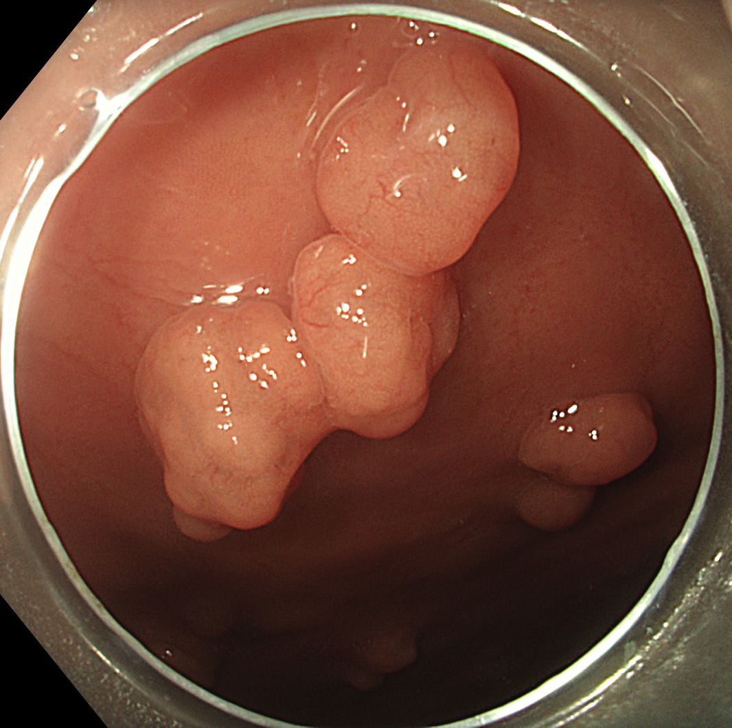
Multiple sessile polyps of <1cm sizes noted in the body of the stomach
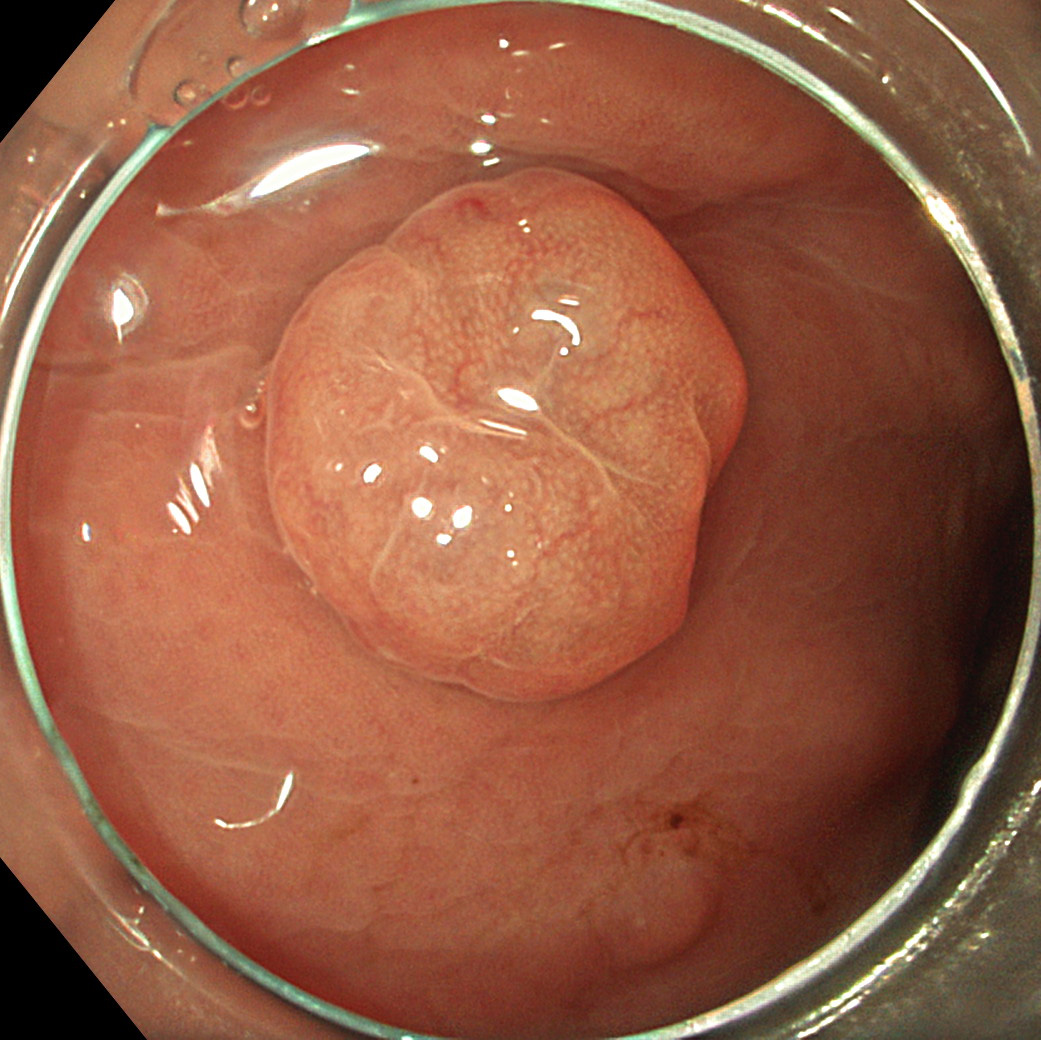
2. TXI™ technology mode 1
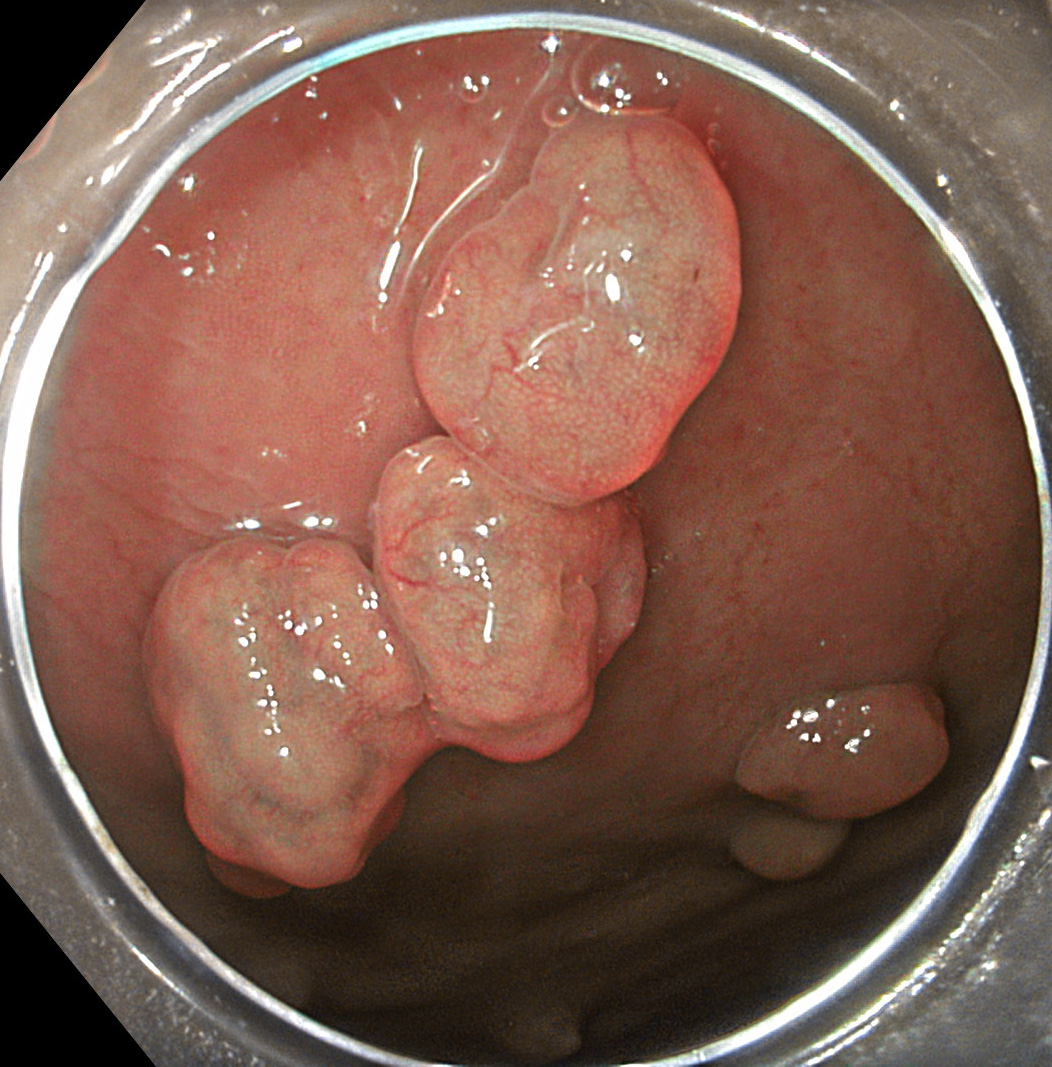
TXI™ technology mode 1 image shows color and structural image enhancement compared to WLI. There is increased brightness in the darker areas.
3. TXI mode 2
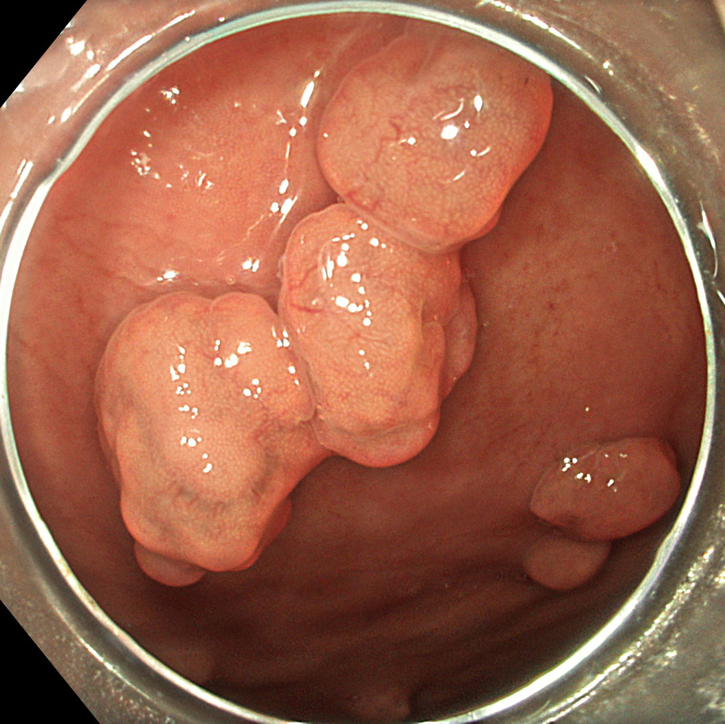
TXI™ technology mode 2 shows enhancement of brightness and texture compared to WLI. The color tone of mode 2 is more similar to WLI than mode 1.
4. WLI
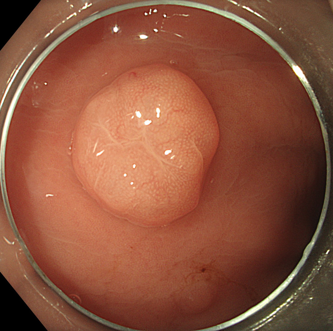
Single sessile polyp of >1cm size noted in the body of the stomach
5. TXI™ technology mode 1
TXI™ technology mode 1 shows color and structural image enhancement compared to WLI. There is increased brightness in the darker areas.
6. TXI™ technology mode 2
TXI™ technology mode 2 shows enhancement of brightness and texture compared to WLI. The color tone of mode 2 is more similar to WLI than mode 1.1
7. WLI
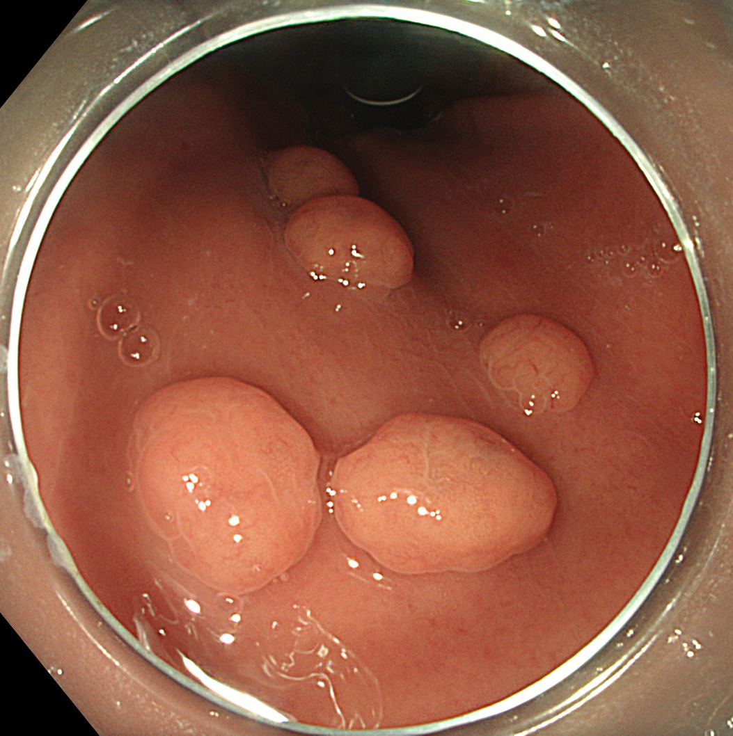
Multiple sessile polyps of <1cm sizes noted in the body of the stomach
8. TXI™ technology mode 1
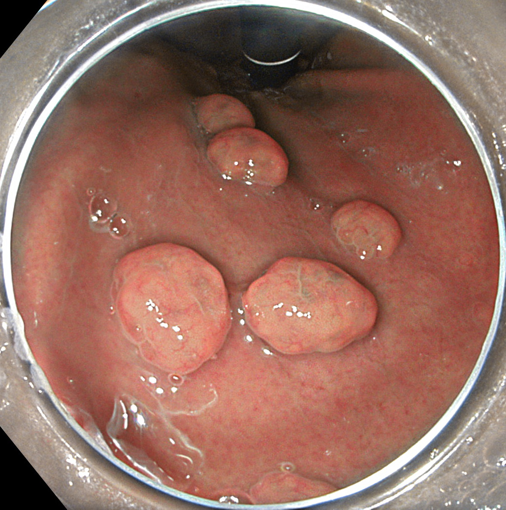
TXI™ technology mode 1 shows color and structural image enhancement compared to WLI. There is increased brightness in the darker areas.
9. TXI™ technology mode 2
TXI™ technology mode 2 shows enhancement of brightness and texture compared to WLI. The color tone of mode 2 is more similar to WLI than mode 1.1
Overall Comment
TXI™ technology can enhance brightness selectively in dark areas of endoscopic images and can enhance subtle tissue differences1 such as slight morphological and/or color changes over conventional enhancement methods including structure enhancement and color enhancement. Comparing TXI™ technology with conventional white light imaging (WLI) for real-world image processing, TXI™ technology can achieve image enhancement with characteristics of WLI appearance while preventing over-enhancement observed in other methods. TXI™ technology has two modes, mode1 and mode 2. Enhancement of brightness and texture is similar in both modes. The color contrast is enhanced with TXI mode 1 whereas TXI mode 2 has a more natural color tone as no color enhancement is performed. Overall, TXI™ technology provides a balance of improving image features important for the physician searching for abnormalities (selective increase in brightness, color separation, and texture enhancement) while minimizing gross changes that may negatively impact familiarity (naturalness).
* Specifications, design and accessories are subject to change without any notice or obligation on the part of the manufacturer.
Prof. Stefan Seewald Case 3: Early gastric cancer
Dr. D Nageshwar Reddy
Dr. Hardik Rughwani
- Keyword
- Content Type


