Colorectal Case 2

Prof. Stefan Seewald
GastroZentrum Hirslanden, Zurich
Scope:CF-EZ1500DI
Case:LST-NG
Organ: Colon
Patient information: M, 60
Medical history: Preventive colonoscopy
1. LST-NG in WLI
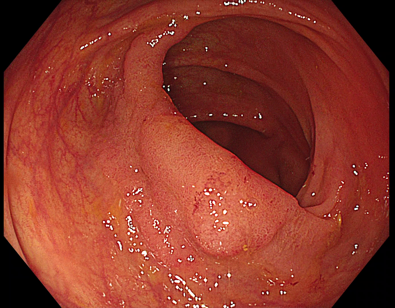
LST-NG spreading across a fold
2. LST-NG with TXI
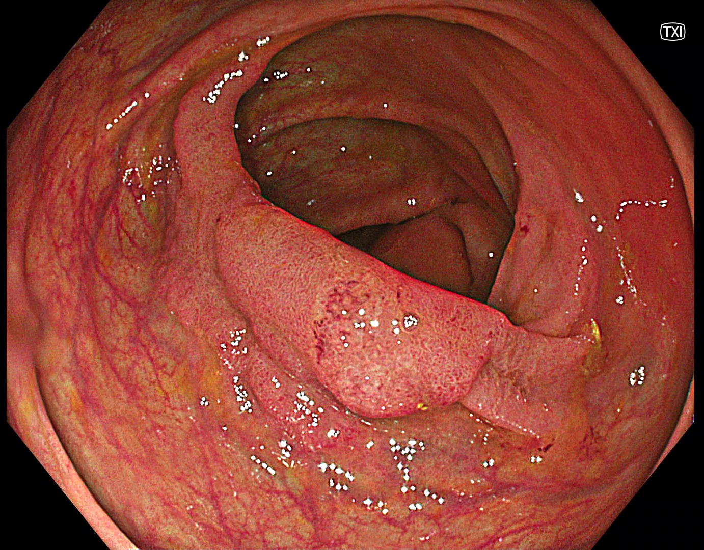
In TXI, the margins of the lesions become more evident. Stool is not interfering with TXI imaging.
3. LST-NG in NBI
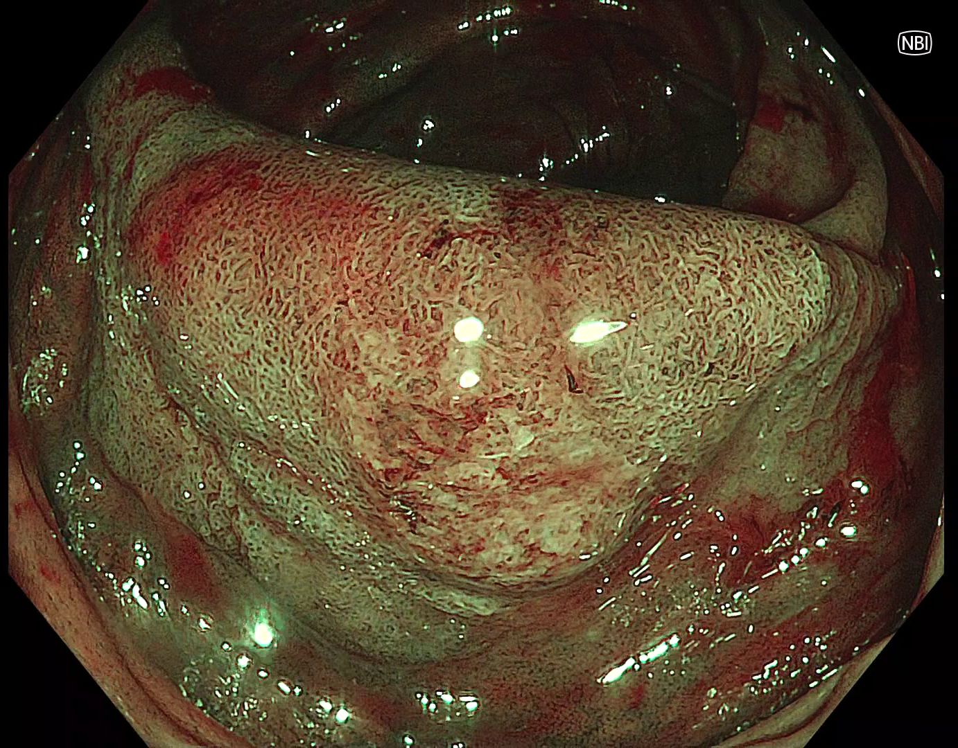
NBI allows for analysis of superficial vessel pattern but is highly affected by remaining stool. NBI requires excellent bowel preparation and thorough flushing.
4. TXI after injection
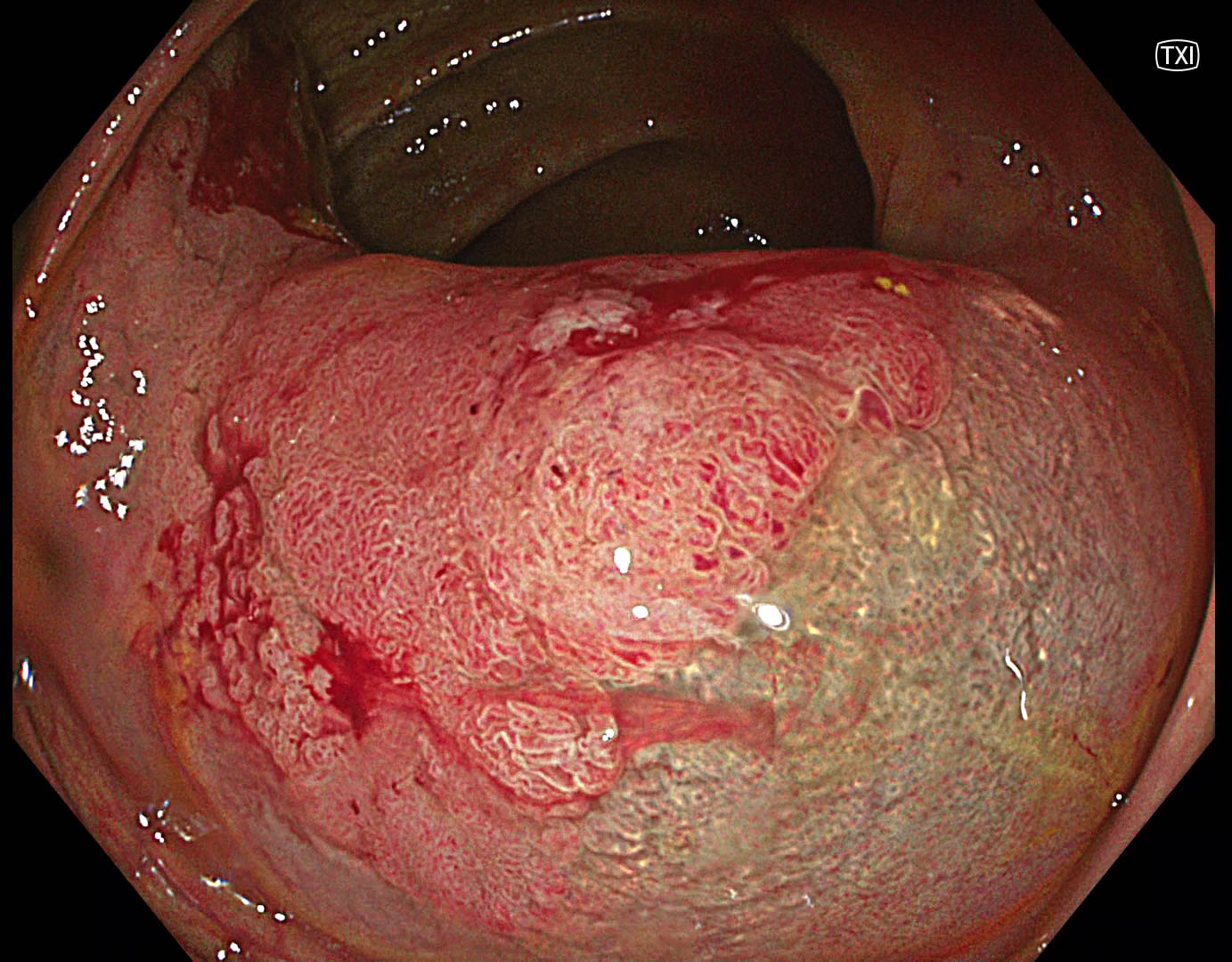
After injection, TXI is delivering a better visibility of the demarcation line.
5. TXI during endoscopic resection
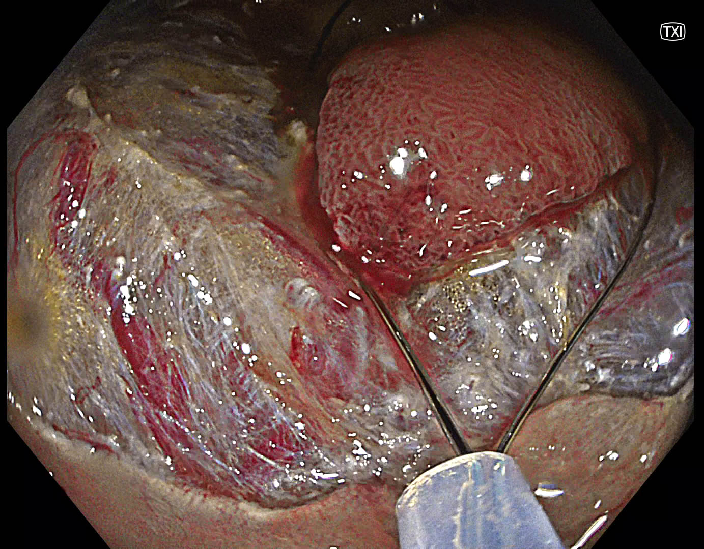
TXI is enhancing visibility of submucosal fibers and blood vessels in the submucosal space.
6. Bleeding spot with RDI
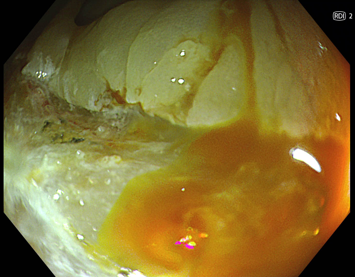
RDI is helping to identify bleeding spots and allows for precise coagulation.
Case video
Overall Comment
In the presented case, TXI was beneficial for delineation and resection. RDI was helpful for efficient coagulation of bleeding.
* Specifications, design and accessories are subject to change without any notice or obligation on the part of the manufacturer.


