
Department of Respiratory and Critical Care Medicine Director,

Department of Respiratory and Critical Care Medicine
Disclaimer:
- RDI™ and TXI™ Technologies are not intended to replace histopathological sampling as a means of diagnosis
- The positions and statements made herein by Dr. Valipour and Dr. Sperk are based on their respective experiences, thoughts and opinions. As with any product, results may vary, and the techniques, instruments, and settings can vary from facility to facility. The content hereof should not be considered as a substitute for carefully reading all applicable labeling, including the Instructions for Use. Please thoroughly review the relevant user manual(s) for instructions, risks, warnings, and cautions. Techniques, instruments, and setting can vary from facility to facility. It is the clinician’s decision and responsibility in each clinical situation to decide which products, modes, medications, applications, and settings to use.
- The EVIS X1™ endoscopy system is not designed for cardiac applications. Other combinations of equipment may cause ventricular fibrillation or seriously affect the cardiac function of the patient. Improper use of endoscopes may result in patient injury, infection, bleeding, and/or perforation. Complete indications, contraindications, warnings, and cautions are available in the Instructions for Use (IFU)
- Dr. Valipour and Dr. Sperk, the authoring physician(s) of this presentation, are/ is a paid consultant(s) to the Olympus Corporation.
Scope:BF-1TH1100
Patient information: Female, 61 years old
Medical history: CT scan revealed atelectasis of the left upper lobe, caused by a central tumor mass coiling around the left pulmonary artery.
1. Superficial bleeding after forceps biopsy
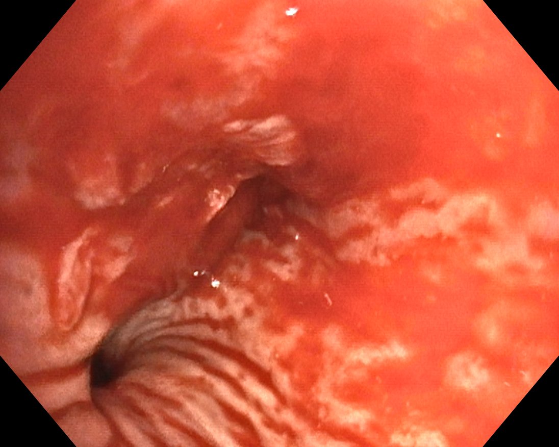
Superficial bleeding after forceps biopsy of a mucosal tumor mass in the transition area between the left main bronchus and the left upper lobe. The area of damaged mucosal tumor mass after forceps biopsy is better visible in this mode (TXI™ technology), whereas the origin of active bleeding cannot be exactly detected.
2. Superficial bleeding after forceps biopsy.
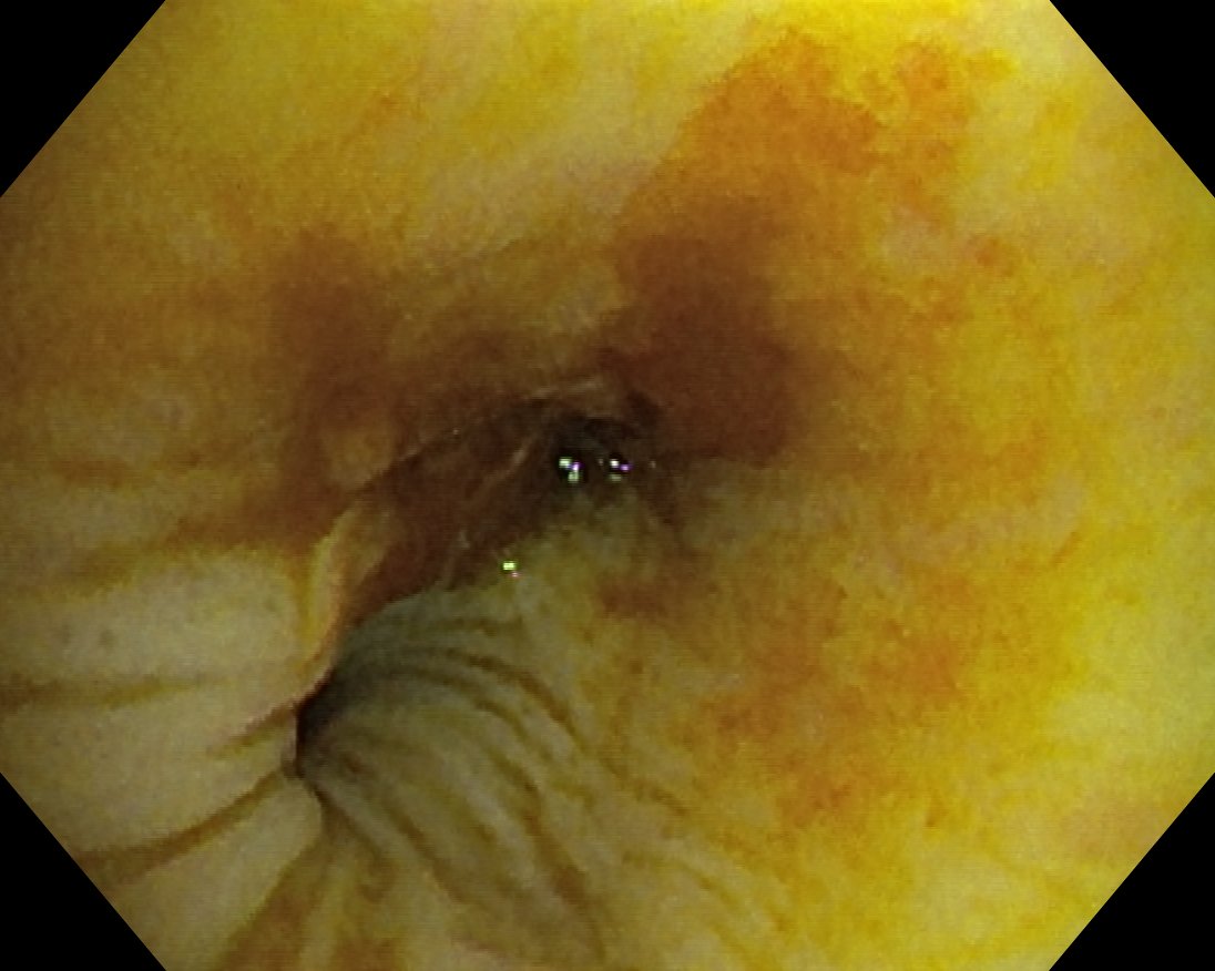
RDI™ technology mode facilitates detecting the source of active bleeding after forceps biopsy of a mucosal tumor mass.
Case video
Overall Comment
TXI™ technology mode provides a good visibility of the area of damaged mucosal tumor mass after forceps biopsy, whereas the source of active bleeding cannot be exactly detected.
RDI™ technology mode facilitates detecting the source of active bleeding after forceps biopsy of a mucosal tumor mass leading to a faster interventional response to bleeding complications by the health care professional.
- Keyword
- Content Type