Preparation
■ Where to perform the procedure
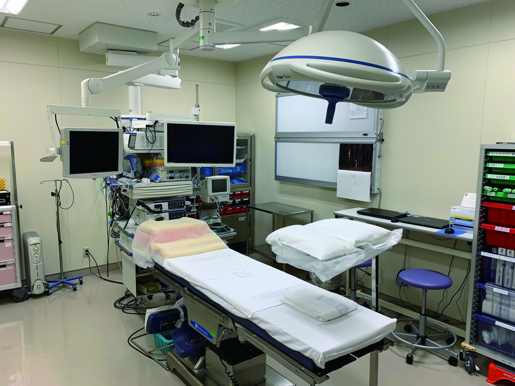
When sufficient consideration is taken to ensure sterility, thoracoscopy under local anesthesia can be performed in a standard endoscopy suite or in a treatment room (Photo 1).
■ Thoracoscope
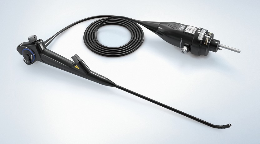
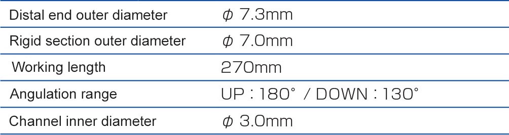
Because lung volume cannot be controlled under local anesthesia, the observation range is restricted. This means a semi-flexible thoracoscope with a flexible tip (Photo 2) is easier to use than a rigid thoracoscope. Additionally, a video thoracoscope offers a brighter field of view, making observation easier.
■ Required equipment
The primary equipment required for the procedure is listed below (items necessary for disinfection and anesthesia are omitted).
・Pulse oximeter, sphygmomanometer, electrocardiogram monitor
・Endoscopy system (thoracoscope, light source, video monitor, image recorder)
・Extracorporeal ultrasound system, oxygen, suction system
・Electrosurgical generator and knife (as required)
Sterile items
Sterilization method
- V-PRO® maX (Flexible cycle)*1
- STERRAD® 100S (Long cycle)*2
- STERRAD® NX® (Advanced cycle)*2, *3
- STERRAD®100NX® (Duo cycle)*2, *3
*1 Olympus products have been validated by STERIS Corporation for the microbiological efficacy. As for the sterilizer, contact STERIS Corporation. Also, refer to the “V-PRO® maX” instruction manual.
*2 Olympus products have been validated by ASP (Advanced Sterilization Products) for the microbiological efficacy. As for the sterilizer, contact ASP. Also, refer to the “STERRAD® 100S/NX®/100NX®” instruction manual.
*3 The endoscope and accessories listed as compatible with STERRAD® NX®/100NX® can be processed with or without ALLClearTM Technology. For further details, contact ASP.
The two-way angulation capability of this thoracoscope allows you to obtain a wider field of view. Thanks to its compatibility with bronchoscopy systems, it can also be used in an endoscopy suite.
■ Staff preparation
Thoracoscopy under local anesthesia is usually performed by a total of four individuals – the endoscopist, an assistant, and two ancillary staff members. The endoscopist and assistant must wash their hands (i.e. surgical hand scrub) in the same manner as for an ordinary surgical operation, and they must wear sterile surgical gowns, sterile gloves, masks, and caps.
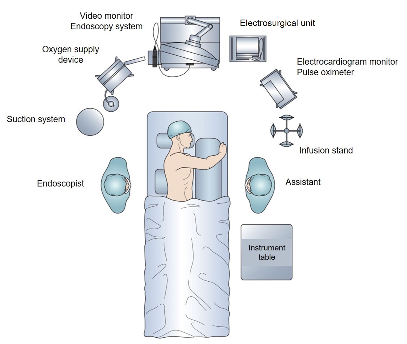
■ System setup (layout)
The video monitor should be placed at the patient’s head side so that the endoscopist and assistant can view it easily while the instrument table should be placed near the endoscopist or assistant (Fig. 1, Photo 4).
■ Patient position
Thoracoscopy under local anesthesia is usually performed with the patient in a lateral position with the unaffected side down. This allows extensive observation both in the anterior and posterior regions. If the patient cannot be placed in the lateral position, use a mat or similar support to incline their body so that they are in a position as close to the lateral position as possible.
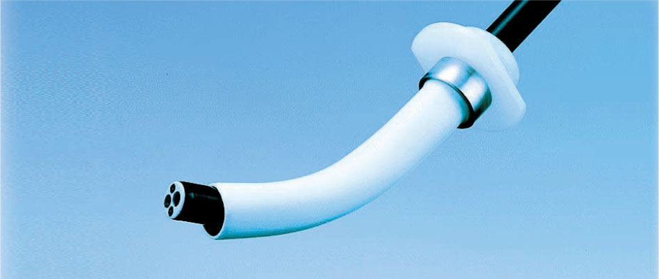
The flexible design of this trocar protects the thoracoscope’s distal-end bending section, while reducing intercostal stimulation.
- Content Type