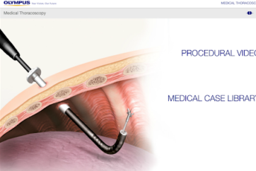
Treatments in thoracoscopy under local anesthesia
■ Separating the adhesion
With acute empyema, the pleural cavity tends to form compartments rapidly due to fibrin deposition, which often makes it difficult to perform drainage. Recovery and improvement can be facilitated by destroying the fibrin membrane to drain the pleural effusion 6,7. This should be performed as early as possible from the onset. Based on our experience, favorable treatment results are shown in cases where the treatment was performed within about two weeks from the onset.
The specific details of this treatment are as follows. Insert forceps with larger cups in the scope channel. Repeat forceps manipulation by pressing the opened cups against the fibrin membrane to destroy the membrane. Fibrinous adhesion can be destroyed and removed easily. However, chronic adhesion with hypervascularization causes bleeding when it is cut so do not touch it in the procedure under local anesthesia.
■ Pleurodesis (talc poudrage)
To control pleural effusion caused by carcinomatous pleuritis, chemical pleurodesis using various anticancer agents such as tetracycline antibiotics and doxorubicin is performed while pleurodesis with OK-432 is also performed. The results, however, are not necessarily satisfactory. Recurrent pooling of pleural effusion often occurs.
Talc, on the other hand, can be quite effective. In particular, when sprayed uniformly over the pleural surfaces using a thoracoscope, adhesion of the visceral and parietal pleurae can be achieved (not indicated in Japan). Sterile talc powder is available in Europe and North America. We hope that it will be introduced to Japan as soon as possible.
Prevention and management of complications
To date, we have performed this procedure on more than 1,000 patients. In only one case did we experience a problematic complication in which RPE occurred. The mortality rate for this procedure is reported to be 0.34%. When restricted to diagnostic purposes only, the rate is 0%7,8.
Major complications such as empyema, bleeding, air leak, postprocedural pneumothorax, and pneumonia are reported to occur in 1.8% of cases, while minor complications such as subcutaneous emphysema, minor bleeding, and skin infection affect 7.3%8. In the following sections, we will look at prevention and management of major complications that may occur.
■ Bleeding
Although we have not experienced bleeding serious enough to require hemostasis, as a precaution we recommend pressing the bleeding point with an epinephrine-soaked cotton ball. Coagulation with an electrosurgical knife or bipolar device is another option, but this can be difficult to do under local anesthesia because pain is likely to occur in the pleura. Keep in mind that hemostasis of arterial bleeding is not easy. Hence, any treatment that may cause bleeding and damage the vascular system must always be avoided.
Usually, the intercostal arteries and veins can be seen through the parietal pleura, so you can avoid them when performing biopsy. While there is little chance of damaging vessels in patients with thickened pleura, it is best to avoid biopsying regions where vessels are present. You can do so by checking the locations of the ribs with forceps. Do not touch anything in patients with complete pleural adhesion as there is hypervascularization inside the adhesion, making it likely that dissection will cause bleeding.
■ Pain
Patients rarely feel pain during a biopsy when the lesion is located on the pleura and the biopsy site is thickened. However, pain often accompanies a biopsy if the biopsy site is close to normal tissue. Before performing biopsy, we touch the tissue with the tip of forceps to confirm whether the patient feels pain. If they feel pain, we replace the forceps with an anesthetic spray tube and spray 1% lidocaine directly on the biopsy site.
■ Pneumothorax
In most cases, you should not perform a biopsy from the visceral pleura in thoracoscopy under local anesthesia. Since cases with severe adhesion are not indicated, the risk of pneumothorax is low. In patients with limited pleural effusion and adhesion, be careful when accessing the thoracic cavity as it is possible to damage the lung parenchyma while puncturing the chest wall. It is a good idea to perform extracorporeal ultrasonography right before the examination to confirm the conditions beneath the puncture site.
■ Re-expansion pulmonary edema (RPE)
As mentioned above, a massive pleural effusion should – if possible – be drained little by little until the day before the procedure. When a large amount of the liquid is drained during the procedure, try to release the clamp gradually so that the lung can re-expand incrementally. Watch for a pulmonary edema, which may emerge from within 30 minutes to a few hours after the procedure. If possible, perform the procedure in the morning so that you can better monitor the patient afterwards. If an edema does appear, it can usually be controlled with steroids.
■ Infection
To prevent intrathoracic infection in cases where a pleural drainage tube has already been inserted, do not use the same entry port. Instead, make a new puncture at another site. We do not generally administer antibiotics preventively after the procedure.
■ Other
In cases where the chest wall subcutaneous tissue or muscle layer is thin, subcutaneous emphysema may sometimes be seen. In cases where a malignant mesothelioma or carcinomatous pleuritis is present, a tumor may infiltrate from the periphery of the punctured hole. In either case, insert a larger diameter tube after the procedure. Suture and secure the surrounding area carefully. Then immobilize the tube by applying pressure with gauze.
Future prospects
In Europe and North America, the concept of medical thoracoscopy has been popular for quite some time and even internists have performed pleural effusion diagnosis. In Japan, on the other hand, there seems to be a deep-seated consensus that thoracoscopy should be performed by a surgeon, making it more difficult to popularize.
The fact that thoracoscopy can be performed under local anesthesia makes it a tremendously flexible procedure which can be applied to an ever wider range of clinical applications, expanding the potential for diagnosis and treatment of respiratory diseases.
<References>
1. Ishii H. Diagnosis of respiratory diseases using thoracoscopy. 17th Respiratory Disease Seminar (in Japanese [ed. by the postgraduate education committee of the Japanese Respiratory Society]). 1996; 38–49.
2. Ishii H, Kitamura S. Usefulness of thoracoscopy from the viewpoint of internal medicine: diagnosis of pleural lesions, lung tumor-like lesions and diffusive lesions (in Japanese). Jpn J Thorac Dis. 1996; 34: 159.
3. Ishii H. Usefulness of thoracoscopy under local anesthesia (in Japanese). Kekkaku. 2000; 75: 51–56.
4. Kennedy L, Sahn SA. Talc pleurodesis for the treatment of pneumothorax and pleural effusion. Chest. 1994; 106: 1215–22.
5. Aelony Y, et al. Thoracoscopic talc poudrage pleurodesis for chronic recurrent pleural effusions. Ann Int Med. 1991; 115: 778–82. Lung Dis. 1993; 74: 225–239.
6. Bhatnagar R, Corcoran JP, Maldonado F, Feller-Kopman D, Janssen J, Astoul P, Rahman NM. Advanced medical interventions in pleural disease. Eur Respir Rev. 2016; 25: 199–213.
7. Sumalani KK, Rizvi NA, Asghar A. Role of medical thoracoscopy in the management of multiloculated empyema. BMC Pul Med. 2018; 18: 179.
8. Rahman NM, Ali NJ, Brown G, Chapman SJ, Davies RJ, Downer NJ, Gleeson FV, Howes TQ, Treasure T, Singh S, Phillips GD. Local anaesthetic thoracoscopy: British Thoracic Society pleural disease guideline 2010. Thorax. 2010; 65: Suppl 2: 54–60.
9. Boutin C, et al. Thoracoscopy in malignant effusions. Am Rev Respir Dis. 1981; 124: 588–592.
10. Boutin C, et al. Diagnostic and therapeutic thoracoscopy: techniques and indications in pulmonary medicine. Tuberc Lung Dis. 1993; 74: 225–239.
- Content Type


