Dr. Takahiro Nakajima
Department of General Thoracic Surgery, Associate Professor,
Dokkyo Medical University
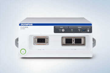
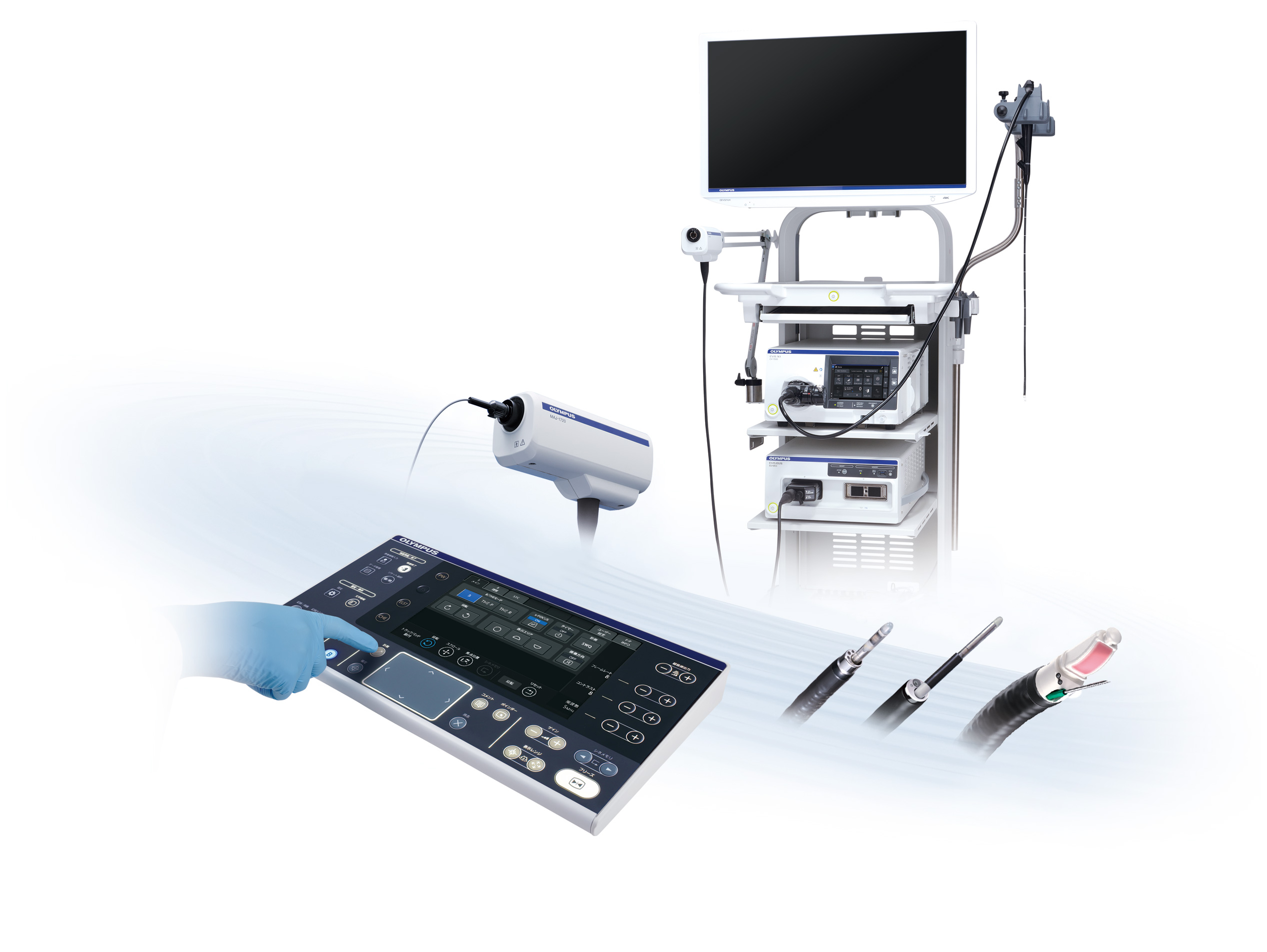
1. New Endoscopic Ultrasound Center EU-ME3
As a pioneer in endoscopic ultrasound technology, Olympus aims to leverage its expertise to enhance the care pathway in respiratory disease management. The EU-ME3 ultrasound processor is unique in its ability to support a wide range of Endobronchial Ultrasound Bronchoscopy (EBUS) procedures, such as EBUS-guided transbronchial needle aspiration (EBUS-TBNA) and radial EBUS for peripheral bronchoscopy procedures.
The EU-ME3 provides outstanding image quality and functionality – the image quality has been substantially enhanced compared to the prior generation model (EU-ME2), providing enhanced visualization, and therefore supporting more reliable diagnosis and treatment.
The Endoscopic ultrasound procedure typically utilizes a variety of observation modes depending on procedure type. In addition to the basic observation mode, the EU-ME3 features an elastography function which visualizes the amount of strain in the tissue (tissue stiffness) during compression and retraction, making it possible to obtain more information about tissue properties.
i-ELST* is a new technology incorporated into the EU-ME3 that makes it easier to display elastic images, even when displacement due to pulsation is modest.
Smart and customizable user settings make it easy to fulfil needs for multiple specialties and personal requirements. The backlit keyboard includes a simple, easy-to-use large touch panel and trackpad, designed to support better operability and easier cleaning.
Recently, Dr. Takahiro Nakajima at Dokkyo Medical University, Japan used the EU-ME3 system on a trial basis for a month. Today, Dr. Nakajima talks about how the current challenges of ultrasound bronchoscopy affect his procedures and the improvements achieved with the use of the EU-ME3 system.
2. EU-ME3 Clinical Usefulness of Each Function
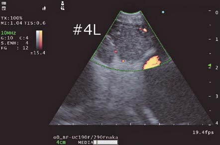
B-mode
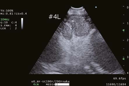
Obtaining improved B-mode images, which is the basis of all observation modes, will contribute the overall improvement of Endobronchial ultrasound-transbronchial needle aspiration (EBUS-TBNA) examinations. The EU-ME3 demonstrates improved ultrasound image resolution and depth compared to its predecessor, the EU-ME2. As a result, not only are the margins of the structure of interest more clearly delineated, but the texture of the lymph node cross-section can also be distinctly identified.
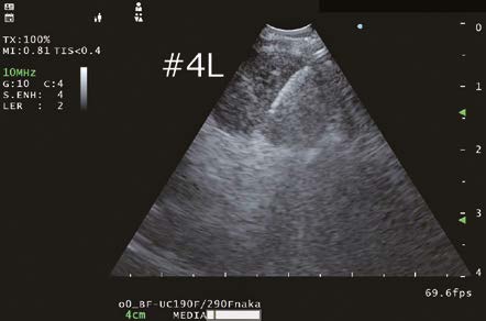
During an EBUS-TBNA case, the position of the aspiration needle, especially the tip of the needle was well visualized and artifacts due to the needle itself were reduced. The enhanced image quality of the B-mode may contribute to making EBUS-TBNA procedures safer and more effective.
Doppler Modes
Doppler modes are also available with EU-ME3. In my observations, it appears that the Doppler image generated with EU-ME3 exhibited improved resolution compared to EU-ME2. The color blurring was reduced, and the image appeared crisper and clearer. When performing EBUS-TBNA in my facility, we typically use the H-Flow mode which displays blood flow with minimal protrusion from blood vessels to confirm vascular intervention with H-Flow mode before puncturing a lymph node.
Color Flow: Displays direction and velocity of blood flow in color
Power Flow: Displays blood flow intensity in color
H-Flow: Displays direction and intensity of blood flow in color
Pulsed Wave Doppler (PWD): Displays blood flow velocity at a specified location in graph
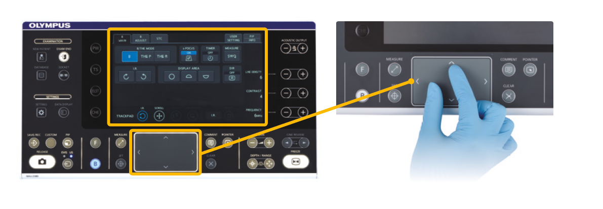
Keyboard
The EU-ME3 features an upgraded keyboard with a trackpad and a larger built-in LCD touch panel, whereas the EU-ME2 had an integrated trackball as its keyboard interface. The implementation of the trackpad in the EU-ME3 has notably enhanced operability during procedures, especially when performed with gloved hands. I have personally experienced that operability remains unaffected while wearing gloves.
Furthermore, the main panel of the keyboard is flat-shaped, making it easier to clean and maintain hygiene standards.
Additionally, the larger LCD touch panel, along with the display of necessary functions in a larger size, enables more intuitive operation in various situations encountered during a procedure.
3. Clinical Experience with EU-ME3
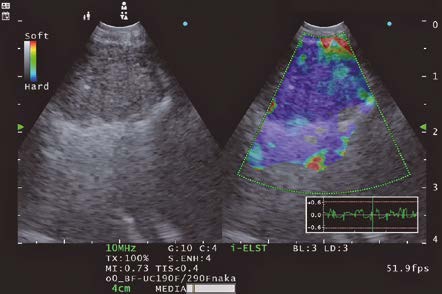
ELST Mode (i-ELST)
Case) 70 years old male patient presented himself 6 years and 10 months after thoracoscopic resection surgery for lung cancer in the right lower lobe.
A nodule in the left lung and an enlarged mediastinal lymph node was identified at a different hospital. The patient was subsequently referred to our hospital for further evaluation and management.
In this case, blood flow was observed in the upper right of the lymph node where the needle would pass through, so it was necessary to avoid this area when puncturing the lymph node.
The selection of the puncture site was determined by using the ELST mode. The EU-ME3 with its newly equipped “i-ELST function”, enables to display stable ELST images even with minute vibrations. The ELST images were green at the center of the lymph node and predominantly blue at the margins of the lymph node.
It has been reported that a combination of ELST and B-mode images is useful for identifying the most suspected site of the lymph node when performing EBUS-TBNA.*
As described above, we were able to collect viable tumor cells and the core by adjusting the puncture site and puncturing the node referring to the ELST mode images.
Small cell carcinoma was suspected by rapid on-site cytologic evaluation during EBUS-TBNA and immediately confirmed by a pathologist. We determined pleural dissemination and diagnosed stage IV small cell carcinoma. Chemotherapy with CBDCA + VP-16 + Durvalumab was started.
References:
1) Fujiwara T, Nakajima T, et al. The combination of endobronchial elastography and sonographic findings during endobronchial ultrasound-guided transbronchial needle aspiration for predicting nodal metastasis. Thorac Cancer. 2019;10(10):2000-5.
4. Future of EU-ME3
The EU-ME3 has improved the image quality of ultrasound images in various modes including B-mode. Through my experiences, I found that EU-ME3 is expected to (1) make ultrasound images clearer in B-mode, making it easier to classify image findings; (2) in Doppler mode, the enhanced imaging with better resolution and less color blurring will enable physicians to control a puncture site more accurately; and (3) in Elastography mode, the stable elastic image displayed by i-ELST function allows physicians to determine a puncture site easily.
Moreover, the utilization of Elastography mode during procedures or playback measurements with recorded Elastography mode images from the EU-ME3’s internal memory allows for the collection of Strain Ratio* and Strain Histogram**. As previously reported2),3), Elastography offers essential parameter data to assess tissue stiffness (elasticity) in a specific region of interest (ROI). In my view, the integration of i- Elastography with convex-probe endobronchial ultrasound could prove to be a valuable tool for predicting and precisely locating metastatic lymph nodes during EBUS-TBNA in the future.
Reference:
2) Rozman A, et al. Endobronchial ultrasound elastography strain ratio for mediastinal lymph node diagnosis. Radiol Oncol. 2015;49(4):334-40.
3) Nakajima T, et al. Elastography for Predicting and Localizing Nodal Metastases during Endobronchial Ultrasound. Respiration. 2015;90(6):499-506.
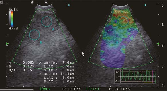
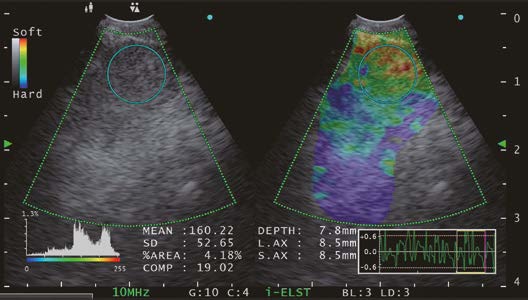
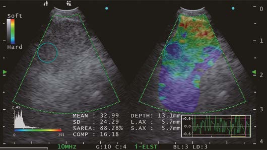
**In strain histogram analysis, lesion stiffness is calculated as mean pixel color values of a single ROI within the target lesion. Values range from 0 to 255 (from blue over green to red). Increasing lesion stiffness causes a decrease in mean histogram values.
EBUS-TBNA is a diagnostic procedure specifically designed for obtaining pathological specimens, where the key emphasis lies in the collection of tissue samples for accurate analysis. Considering the impact 4K and other high resolution imaging technologies bring in a surgical setting, I expect that the higher resolution ultrasound images of EU-ME3 make EBUS-TBNA procedures itself easier to perform, but also expect to develop a further clinical value such as improving safety and improving the positive rate.
- Content Type