For Healthcare Professionals | Interview | LTF-H290
The New LTF-H290
for more Reliable Pleuroscopy

Narayana Health, Mazumdar Shaw Medical Center
Understanding the landscape in medical thoracoscopy
Medical thoracoscopy continues to gain traction as a minimally invasive diagnostic and therapeutic tool for pleural diseases. It remains the preferred approach for investigating undiagnosed exudative pleural effusions, particularly in malignancy and tuberculosis, with a diagnostic yield of up to 95%. Recent trends include the increasing use of semirigid thoracoscopes, cryobiopsies, and narrow-band imaging to enhance diagnostic accuracy. Additionally, its role in therapeutic pleurodesis and selected early empyemas is expanding, complementing VATS in a multidisciplinary approach.
Tuberculous pleuritis remains a leading cause of undiagnosed pleural effusions in high-burden regions, where thoracoscopy offers superior diagnostic yield over pleural fluid analysis. The rising prevalence of malignant pleural effusions (MPEs) and malignant pleural mesothelioma (MPM) has further driven its use for diagnosis and pleurodesis. Advances in optical technology, minimally invasive techniques, and training have also improved accessibility and safety, leading to wider adoption.
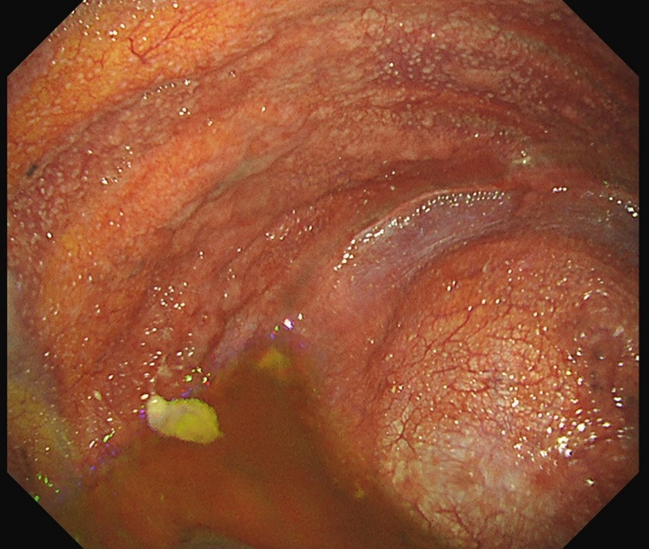
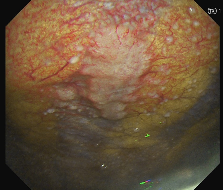
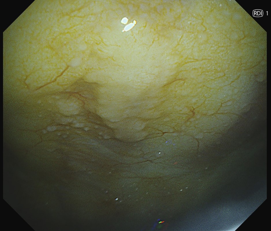
HD Image quality with new EVIS X1 Technology
What was your experience like using the new LTF-H290 in terms of image quality for observation and diagnosis?
The LTF-H290 pleuroscope has significant improvements from LTF-160 with HD imaging, NBI, a wider field of view, and better illumination, enhancing pleural visualization and diagnostic precision. These upgrades improve contrast, detection of subtle lesions, and procedural efficiency, making thoracoscopy more effective for malignancy and inflammatory conditions.
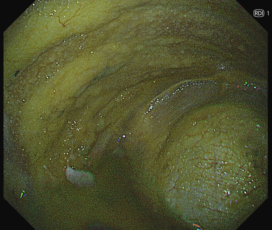
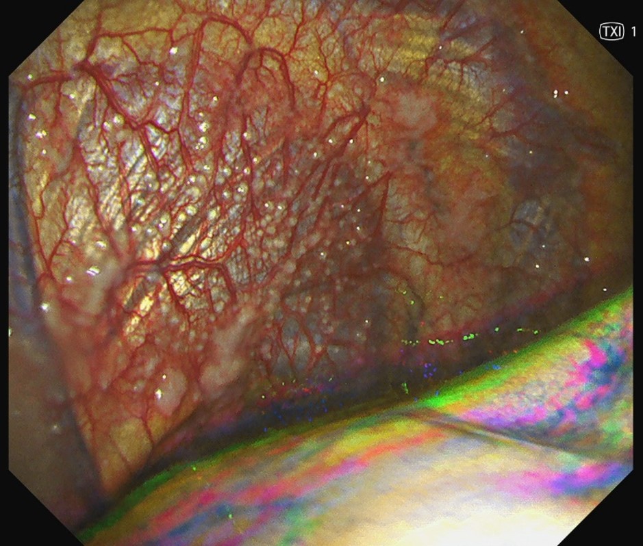
In addition, the use of LTF-H290 together with the new Olympus EVIS X1 CV-1500 processor has new optical technologies that could be useful for procedures. TXI (Texture and Color Enhancement Imaging) improves contrast and texture definition, making subtle pleural abnormalities more distinct, which is particularly useful in identifying early malignant or inflammatory changes and refining biopsy site selection. RDI (Red Dichromatic Imaging), on the other hand, enhances the visualization of blood vessels and bleeding sources, potentially aiding in bleeding control and procedural safety, especially during biopsies and pleurodesis. However, its real-world utility in managing active bleeding requires further exploration, and it currently serves as an additional module to help identify a bleeder than a primary tool for hemostasis.
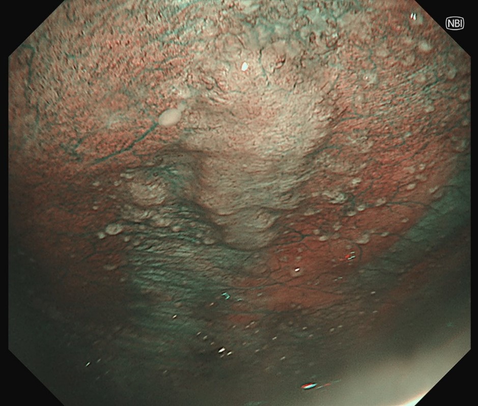
How about the availability of Narrow Band Imaging (NBI) with the use of the LTF-H290, has this improve observation and diagnostic accuracy?
NBI enhances pleural and vascular visualization in medical thoracoscopy, improving contrast and lesion differentiation, especially when white light is inconclusive. It is particularly useful in malignant pleural disease and biopsy site selection, enhancing diagnostic accuracy and reducing repeat procedures, leading to better patient outcomes.
Increased angulation and advanced suction capability
How was your experience with the increased 210° UP angulation of the LTF-H290?
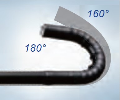
210° UP angulation of the LTF-H290 has greatly improved maneuverability during medical thoracoscopy. It allows better access to difficult-to-reach areas like the diaphragmatic and apical pleura, enhancing pleural examination and targeted biopsy accuracy. This reduces scope manipulation, improving procedural efficiency and patient comfort, making thoracoscopy more effective for diagnosis and treatment.
The increased suction capability with the 3.0mm working channel of the LTF-H290 significantly improves procedural efficiency. It allows for quicker fluid evacuation, maintaining a clearer view of the pleural cavity, especially in cases with heavy exudates or blood. This enhances biopsy precision and overall procedural control, reducing interruptions and improving workflow.
Overall experience and application of LTF-H290
Could you share with us your overall experience with it and is there any key feature of the LTF-H290 that you think could improve medical thoracoscopy procedure moving forward?
The LTF-H290 has significantly enhanced medical thoracoscopy with its HD imaging, NBI, improved angulation (210° UP), and a 3.0mm working channel for better suction. These features provide superior visualization, easier access to difficult areas, and enhanced procedural efficiency.
- Content Type