Introduction
A solitary pulmonary nodule (SPN) is well-defined as an opacity up to 30 mm in diameter that is surrounded by unaltered aerated lung. It is often an incidental finding on chest radiographs and chest CT scans. Sometimes the pathology is already advanced, and in the diagnostic pathway we have found suspected mediastinal lymph nodes by means of CT scans or PET.
Case Report
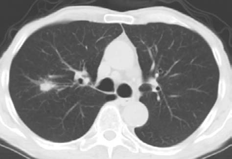
A 71 year-old man, smoker of 48 packs/year, suffered from arterial hypertension and gout, but was in good clinical conditions. During preoperative examinations for right knee replacement surgery, a right upper lobe pulmonary nodule (14 x 18 mm) was identified in a CT scan (image 1). In the CT scan we also noted an increased in short axis right hilar lymph node (11R). A PET-FDG CT scan showed an increased uptake in the nodule (SUV 14) in the 11R lymph node (SUV 8). A head CT scan was negative.
After a Lung Disease Management Team (DMT) meeting, we decided to perform video bronchoscopy helped by fluoroscopy associated with radial-EBUS. Moreover, we suggested the use of linear EBUS for mediastinal staging based on current practice in which a nodule shouldn’t require mediastinal staging but the presence of N1 (11R lymph node) demands it.
Procedure
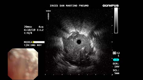
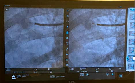
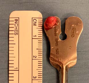
The patient, after signed informed consent, was submitted to a double procedure. After deep sedation with fentanyl and midazolam, we introduced, via the mouth, the ultrathin flexible bronchoscope (Olympus BFMP190F) to the sixth-generation bronchus in the segmentary apical right upper bronchus. Then we introduced the mini radial probe (UM-S20-17S) inside the working channel, which advanced into the branching towards the lesion. We were able to identify a concentric lesion, with a good visibility and distinct margins (image 2). After a fluoroscopy control (image 3), we introduced the dedicated needle (PeriView Flex 21G) used to prepare a first smear. Our cytopathologist performed ROSE and confirmed the right position, because he recognized neoplastic cells. Therefore, we introduced a 1.1 mm cryoprobe and, always under fluoroscopic view, we obtained the first cryo-sample (image 4). This first cryo was used to prepare a slide with the touch imprint (TIC-ROSE) method. The cytopathologist confirmed the malignant cell and formulated a diagnosis of adenocarcinoma.
In the second step we performed a mediastinal staging with an EBUS scope (BF-UC190F). We measured every lymph node beginning from the contralateral node. The only two lymph nodes larger than 5 mm in short axis were 4R and 11R. We conducted three passes in 4R, with ROSE always negative, and two passes in 11R positive for malignant cells.
All the molecular analysys and immunochemistry were negative.
Our patient was evaluated by a multidisciplinary team as T1bN1M0 (stage IIB) and he was submitted to neoadjuvant therapy followed by surgery.
Conclusion
SPN is a common finding in clinical practice, and the importance of its accurate pathological diagnosis is very high.
The BF-MP190F has a large working channel (1.7 mm) despite its small distal end diameter (3.0 mm), allowing the use of different tools.
Extensive sampling is usually undertaken to increase the chance of a diagnosis because it is difficult to assess whether representative tissue has been obtained. Because they use an endobronchial approach through a small working channel, these tools are miniaturized. This generally results in small samples, which are often difficult for the pathologist to interpret. The combination of thin bronchoscopes, lesion guidance and confirmation techniques (fluoroscope and radial EBUS), and sampling methods (needle and cryo) improves the likelihood of diagnosis and therefore the appropriate treatment of patients.
We must encourage the interventional pulmonologist and the DMT, to prioritize the endoscopic approach in the diagnostic pathway of SPN. This facilitates early diagnosis and the staging of suspected lesions in a single process, reducing patient stress and costs.
The Nurse Role in Peripheral Lung Diagnosis
Once the doctor has scheduled the procedure, the nurse organizes the endoscopic suite.
In this case we prepared:
· Monitoring kit
· Fluoroscope
· Ultrathin bronchoscope
· Mini radial probe
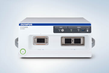
· EBUS
· ROSE kit
In particular, the radial probe must be prepared by evaluating the correct functioning of the probe once attached to the driving unit and subsequent lubrication of the probe to facilitate its movement in the working channel.
The nurse needs to know the patient’s clinical history very well, and consider the possibility of complications based on comorbidity.
Knowledge of sampling tools and proper handling of materials is an essential part of a collaborative healthcare team approach. Nurses play a critical role in ensuring that established hygiene standards are strictly followed throughout the sampling process, thereby minimizing the risk of contamination and infection. By maintaining these standards and applying their technical expertise, nurses help promote safety, accuracy, and overall positive patient outcomes.
Working in a team is very important in an endoscopy suite, both in elective procedures and in emergency ones.
As medical knowledge is constantly growing, technical modifications or changes of the product design, product specifications, accessories and service offerings may be required.
- Content Type