Colorectal Case 1

Prof. Naohisa Yahagi
Cancer Center, Keio University Hospital, Japan
*Prof. Naohisa Yahagi, MD, the authoring physician of this presentation, is a paid consultant to Olympus Corporation of the Americas.
Disclaimer:
- The EVIS X1™ Endoscopy System is not designed for cardiac applications. Other combinations of equipment may cause ventricular fibrillation or seriously affect the cardiac function of the patient. Improper use of endoscopes may result in patient injury, infection, bleeding, and/or perforation. Complete indications, contraindications, warnings, and cautions are available in the Instructions for Use (IFU).
- This site is published by Olympus America Inc., which is solely responsible for its contents and is intended for U.S. audiences only. Not all products are available for sale in all markets.
- This site is intended for Healthcare Professionals. If you are a patient, it is important that you discuss information about the benefits and risks of products with your doctor.
- RDI technology is not intended to replace histopathology sampling as a means of diagnosis.
1) Data on file with Olympus (DC00489968)
2) Uraoka T, Igarashi M. “Development and clinical usefulness of a unique red dichromatic imaging technology in gastrointestinal endoscopy: A narrative review.” Therapeutic Advances in Gastroenterology. 2022;15.
Scope: PCF-H190I
Organ: Rectum
Patient information: M, 70s
Medical history: N/A
RDI™ technology is not intended to replace histopathological sampling as a means of diagnosis.
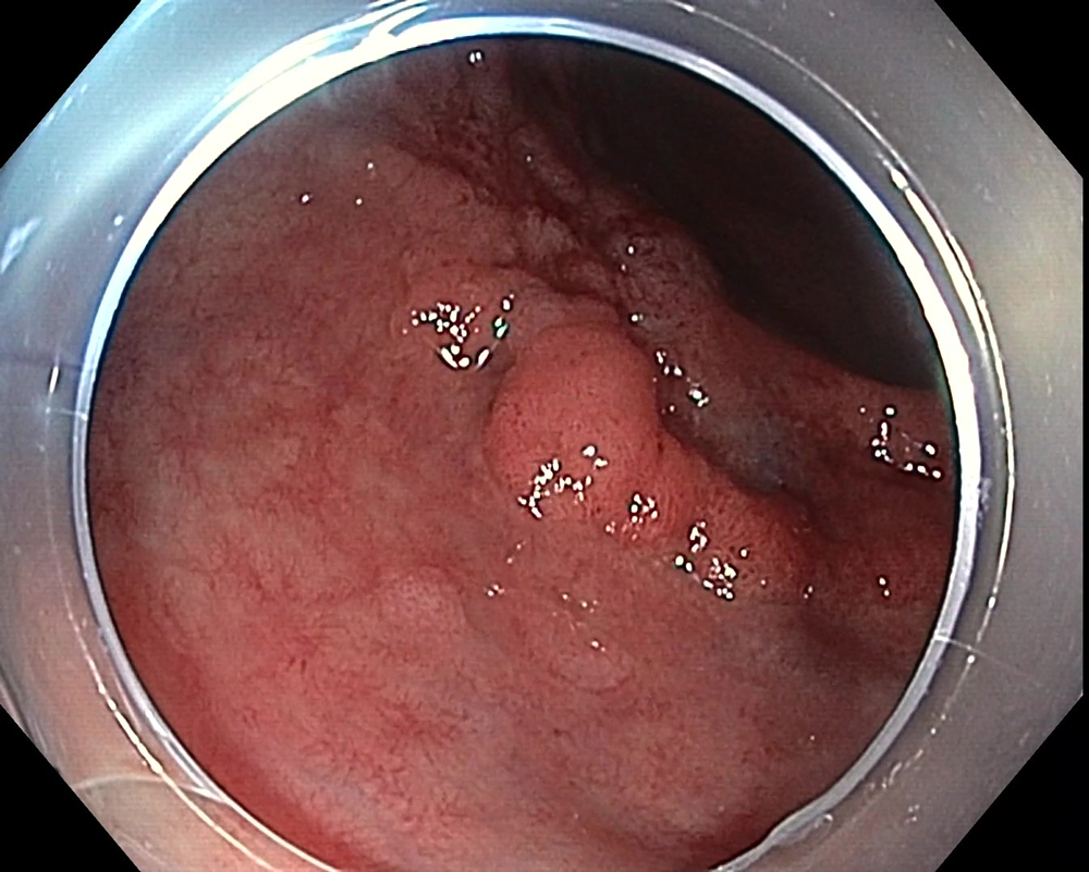
1. WLI Observation
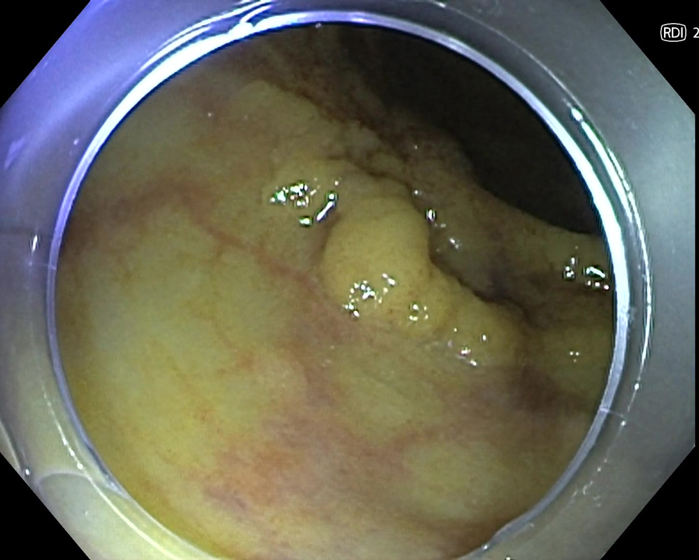
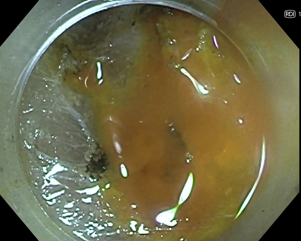
2. RDI Mode 2 observation
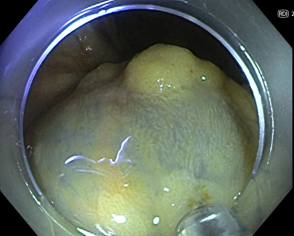
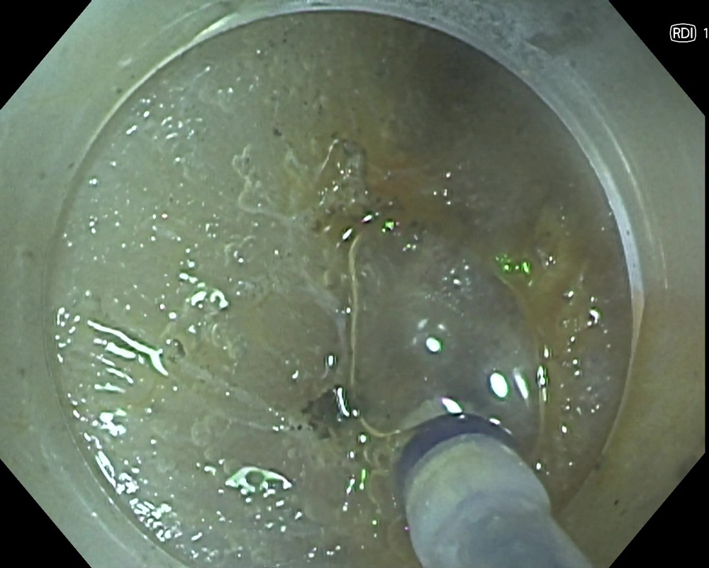
3. Injection under RDI Mode 2

4. Injection under RDI Mode 2
Case Video
5. Dissection
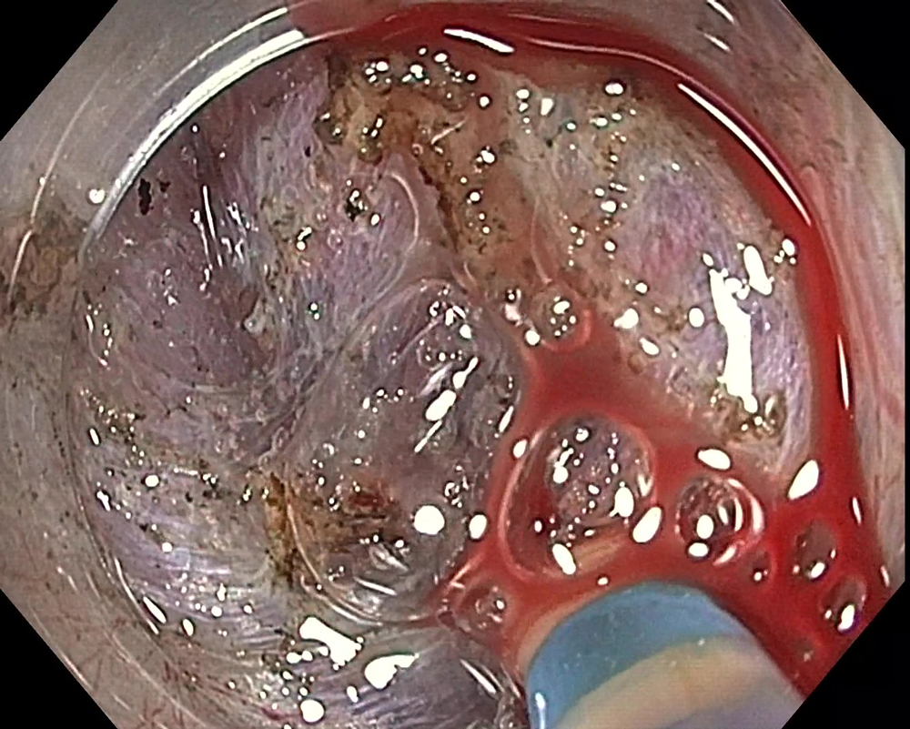
6. Bleeding
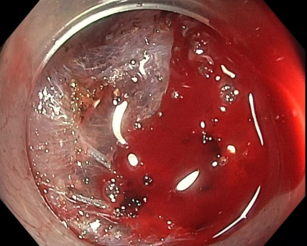
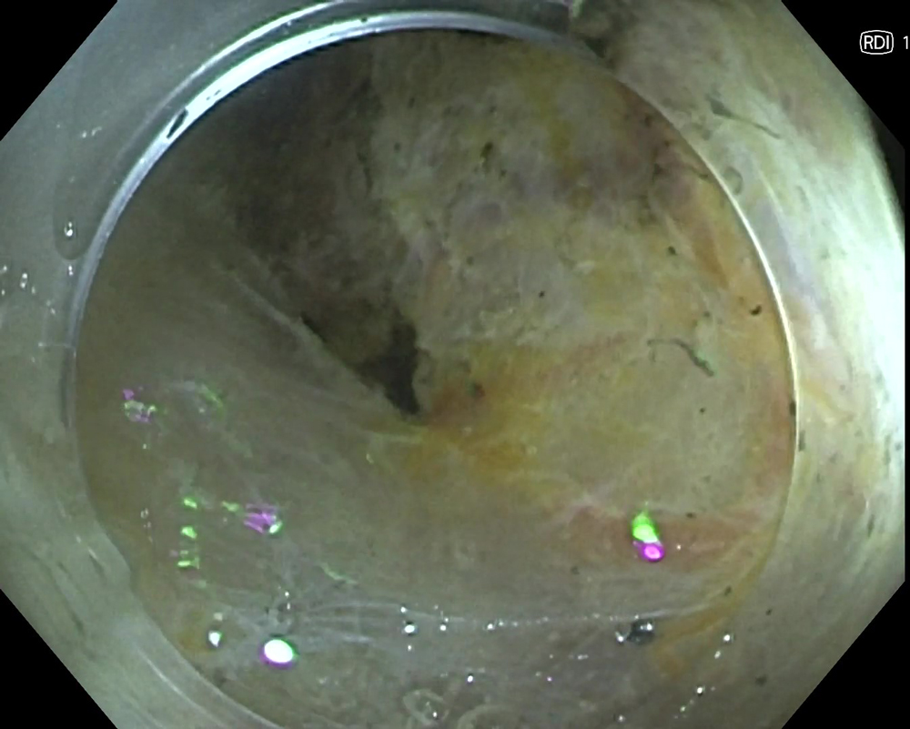
7. RDI Mode 1
8. Hemostasis
9. Completion
Case Video
Overall Comment
Although no studies are yet to verify the effectiveness of RDI technology, in my experience the use of RDI technology may help reduce hemostasis and treatment times. I have experienced the application of RDI technology to support hemostasis of gastrointestinal bleeding and diagnosis or treatment of esophageal varices in my medical center. Additionally, I have been experiencing the benefits of RDI technology use in cases outside of ESD.
I feel strong psychological stress when the entire monitor view turns red due to bleeding. I believe that visualizing hemorrhages in amber color instead of red decreases the sense of urgency and stress of operators towards bleeding, which allows operators to identify the bleeding point easily and perform hemostasis in a composed manner.
I expect that the efficiency and quality of endoscopic diagnosis and treatment are supported using the multiple functions of the EVIS X1™ endoscopy system, including RDI technology.
Disclaimer
The EVIS X1™ Endoscopy System is not designed for cardiac applications. Other combinations of equipment may cause ventricular fibrillation or seriously affect the cardiac function of the patient. Improper use of endoscopes may result in patient injury, infection, bleeding, and/or perforation. Complete indications, contraindications, warnings, and cautions are available in the Instructions for Use (IFU).
This site is published by Olympus America Inc., which is solely responsible for its contents and is intended for U.S. audiences only. Not all products are available for sale in all markets.
This site is intended for Healthcare Professionals. If you are a patient, it is important that you discuss information about the benefits and risks of products with your doctor.
RDI technology is not intended to replace histopathology sampling as a means of diagnosis.
1) Data on file with Olympus (DC00489968)
2) Uraoka T, Igarashi M. “Development and clinical usefulness of a unique red dichromatic imaging technology in gastrointestinal endoscopy: A narrative review.” Therapeutic Advances in Gastroenterology. 2022;15.
- Content Type


