- Home
- Gastroenterology
- Search: EVIS X1
Search: EVIS X1
-
 Add to View
 Colorectal - Imaging AtlasCase 22: Multiple lesions in ascending colon Prof. Dr. Hu Xiao
 -
Add to View
Colorectal - Imaging AtlasCase 31: Multiple lesions in ascending colon Prof. Dr. Hu Xiao
-
Add to View
EVIS X1 Atlas - Imaging AtlasCase 30: Multiple lesions in ascending colon Prof. Dr. Hu Xiao
-
Add to View
Colorectal - Imaging AtlasCases 32: Is+IIa (LST-G) lesion Dr. Hiroaki Ikematsu
-
Add to View
Colorectal - Imaging AtlasCases 33: LST-GM, invasive cancer Prof. Dr. Fatih Aslan
-
Add to View
EVIS X1™ Atlas - Imaging AtlasCases 35: Detection Prof. Yasushi Sano
-
Add to View
EVIS X1™ Atlas - Imaging AtlasCases 36: Detection 0-IIa, SSL with CD Prof. Yasushi Sano
-
Add to View
EVIS X1™ Atlas - Imaging AtlasCase 37: Characterization JNET 2B Prof. Yasushi Sano
-
Add to View
EVIS X1™ Atlas - Imaging AtlasCases 38: Characterization JNET 2B Prof. Yasushi Sano
-
Add to View
EVIS X1 Atlas - Imaging AtlasCases 33: LST-GM, invasive cancer Prof. Dr. Fatih Aslan
-
Add to View
EVIS X1 Atlas - Imaging AtlasCases 32: Is+IIa (LST-G) lesion Dr. Hiroaki Ikematsu
-
 Add to View
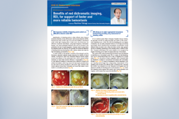
ESD - ReferenceBenefits of red dichromatic imaging, RDI, for support of faster and more reliable hemostasis
Add to View
ESD - ReferenceBenefits of red dichromatic imaging, RDI, for support of faster and more reliable hemostasis -
Add to View
ESD - ReferenceBenefits of red dichromatic imaging, RDI, for support of faster and more reliable hemostasis
-
 Add to View
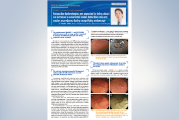
Colonoscopy - ReferenceEVIS X1 Endoscopist Interview
Add to View
Colonoscopy - ReferenceEVIS X1 Endoscopist Interview -
Add to View
Colonoscopy - ReferenceEVIS X1 Endoscopist Interview
-
 Add to View
Colonoscopy - Technology InformationX1 Elevating the Standard of Endoscopy
Add to View
Colonoscopy - Technology InformationX1 Elevating the Standard of Endoscopy -
Add to View
Upper GI Endoscopy - Technology InformationX1 Elevating the Standard of Endoscopy
-
 Add to View
ESD - Video - ProcedureThe use of RDI™ technology during gastric ESD
Add to View
ESD - Video - ProcedureThe use of RDI™ technology during gastric ESD -
 Add to View
ESD - Video - ProcedureThe use of RDI during gastric ESD
Add to View
ESD - Video - ProcedureThe use of RDI during gastric ESD -
Add to View
Colorectal - Imaging AtlasCase 11: Colonic polyp (Tubular adenoma) Dr. Supakij Khomvilai
-
Add to View
Colorectal - Imaging AtlasCase 11: Colonic polyp (Tubular adenoma) Dr. Supakij Khomvilai
-
Add to View
EVIS X1 Atlas - Imaging AtlasCase 11: Colonic polyp (Tubular adenoma) Dr. Supakij Khomvilai
-
Add to View
Colorectal - Imaging AtlasCase 8: Colonic polyp (Tubular adenom) Dr. Supakij Khomvilai
-
Add to View
Colorectal - Imaging AtlasCase 8: Colonic polyp (Tubular adenoma) Dr. Supakij Khomvilai
-
Add to View
EVIS X1 Atlas - Imaging AtlasCase 8: Colonic polyp (Tubular adenom) Dr. Supakij Khomvilai
-
Add to View
Colorectal - Imaging AtlasCase 9: Colonic polyp (Sessile Serrated Lesion) Dr. Supakij Khomvilai
-
Add to View
Colorectal - Imaging AtlasCase 9: Colonic polyp (Sessile Serrated Lesion) Dr. Supakij Khomvilai
-
Add to View
EVIS X1 Atlas - Imaging AtlasCase 9: Colonic polyp (Sessile Serrated Lesion) Dr. Supakij Khomvilai
-
Add to View
Colorectal - Imaging AtlasCase 7: Colonic polyp (Well differentiate adenocarcinoma with shallow submucosal invasion) Dr. Supakij Khomvilai
-
Add to View
Colorectal - Imaging AtlasCase 7: Colonic polyp (Well differentiate adenocarcinoma with shallow submucosal invasion) Dr. Supakij Khomvilai
-
Add to View
EVIS X1 Atlas - Imaging AtlasCase 7: Colonic polyp (Well differentiate adenocarcinoma with shallow submucosal invasion) Dr. Supakij Khomvilai
-
Add to View
Colorectal - Imaging AtlasCase 10: Colonic polyp (Tubular adenoma) Dr. Supakij Khomvilai
-
Add to View
Colorectal - Imaging AtlasCase 10: Colonic polyp (Tubular adenoma) Dr. Supakij Khomvilai
-
Add to View
EVIS X1 Atlas - Imaging AtlasCase 10: Colonic polyp (Tubular adenoma) Dr. Supakij Khomvilai
-
Add to View
Colorectal - Imaging AtlasCase 6: Colonic polyp (Tubular Adenoma) Dr. Supakij Khomvilai
-
Add to View
Colorectal - Imaging AtlasCase 6: Colonic polyp (Tubular Adenoma) Dr. Supakij Khomvilai
-
Add to View
EVIS X1 Atlas - Imaging AtlasCase 6: Colonic polyp (Tubular Adenoma) Dr. Supakij Khomvilai
-
Add to View
Colorectal - Imaging AtlasCase 19: 0-Is (LST-mixed type), 22mm, JNET 2A with fern-like pits Prof. Yasushi Sano
-
Add to View
Colorectal - Imaging AtlasCase 17: 0-Is (LST-mixed type), 22mm, JNET 2A with fern-like pits Prof. Yasushi Sano
-
Add to View
Colorectal - Imaging AtlasCase 20: 0-Is (LST-mixed type), 22mm, JNET 2A with fern-like pits Prof. Yasushi Sano
-
Add to View
EVIS X1 Atlas - Imaging AtlasCase 20: 0-Is (LST-mixed type), 22mm, JNET 2A with fern-like pits Prof. Yasushi Sano
-
Add to View
Colorectal - Imaging AtlasCase 18: 0-IIa (LST-non-granular type), 25mm, JNET 2A+2B Prof. Yasushi Sano
-
Add to View
Colorectal - Imaging AtlasCase 21: 0-IIa (LST-non-granular type), 25mm, JNET 2A+2B Prof. Yasushi Sano
-
Add to View
EVIS X1 Atlas - Imaging AtlasCase 21: 0-IIa (LST-non-granular type), 25mm, JNET 2A+2B Prof. Yasushi Sano
-
Add to View
Colorectal - Imaging AtlasCase 24: A large anal submucosal lesion Dr. Shiaw-Hooi Ho
-
Add to View
EVIS X1 Atlas - Imaging AtlasCase 24: A large anal submucosal lesion Dr. Shiaw-Hooi Ho
-
Add to View
Colorectal - Imaging AtlasCase 25: Crohn’s Ileo-colitis Dr. Shiaw-Hooi Ho
-
Add to View
EVIS X1 Atlas - Imaging AtlasCase 25: Crohn’s Ileo-colitis Dr. Shiaw-Hooi Ho
-
Add to View
Colorectal - Imaging AtlasCase 30: Rectal ESD Prof. Naohisa Yahagi
-
Add to View
EVIS X1 Atlas - Imaging AtlasCase 31: Rectal ESD Prof. Naohisa Yahagi
-
Add to View
Colorectal - Imaging AtlasCase 29: Multifocal Anal Squamous Intraepithelial Lesions (ASIL) with low and high-grade dysplasia Mr. Peter Borch-Johnsen
-
Add to View
EVIS X1 Atlas - Imaging AtlasCase 29: Multifocal Anal Squamous Intraepithelial Lesions (ASIL) with low and high-grade dysplasia Mr. Peter Borch-Johnsen
-
Add to View
Colorectal - Imaging AtlasCase 28: Pedunculated polyp Mr. Peter Borch-Johnsen
-
Add to View
EVIS X1 Atlas - Imaging AtlasCase 28: Pedunculated polyp Mr. Peter Borch-Johnsen
-
Add to View
Colorectal - Imaging AtlasCase 21: Intramucosal Carcinoma (High-grade Dysplasia) Prof. Yoji Takeuchi
-
Add to View
Colorectal - Imaging AtlasCase 27: Intramucosal Carcinoma (High-grade Dysplasia) Prof. Yoji Takeuchi
-
Add to View
EVIS X1 Atlas - Imaging AtlasCase 27: Intramucosal Carcinoma Prof. Yoji Takeuchi
-
Add to View
Colorectal - Imaging AtlasCase 26: Large 40mm splenic flexure NG-LST Dr. Shiaw-Hooi Ho
-
Add to View
EVIS X1 Atlas - Imaging AtlasCase 26: Large 40mm splenic flexure NG-LST Dr. Shiaw-Hooi Ho
-
Add to View
Colorectal - Imaging AtlasCase 20: Large non-polypoid rectal tumor Prof. Han-Mo Chiu
-
Add to View
Colorectal - Imaging AtlasCase 23: Large non-polypoid rectal tumor Prof. Han-Mo Chiu
-
Add to View
EVIS X1 Atlas - Imaging AtlasCase 23: Large non-polypoid rectal tumor Prof. Han-Mo Chiu
-
Add to View
Colorectal - Imaging AtlasCase 19: 0-Is (submucosal invasive carcinoma), 11mm, JNET 2B Prof. Yasushi Sano
-
Add to View
Colorectal - Imaging AtlasCase 22: 0-Is (submucosal invasive carcinoma), 11mm, JNET 2B Prof. Yasushi Sano
-
Add to View
EVIS X1 Atlas - Imaging AtlasCase 22: 0-Is (submucosal invasive carcinoma), 11mm, JNET 2B Prof. Yasushi Sano
-
Add to View
Colorectal - Imaging AtlasCase 16: LST-NG-PD (Pseudo-depressed type), pT1a colonic cancer Dr. Ho Dang Quy Dung
-
Add to View
Colorectal - Imaging AtlasCase 19: LST-NG-PD, pT1a colonic cancer Dr. Ho Dang Quy Dung
-
Add to View
EVIS X1 Atlas - Imaging AtlasCase 19: LST-NG-PD (Pseudo-depressed type), pT1a colonic cancer Dr. Ho Dang Quy Dung
-
Add to View
Colorectal - Imaging AtlasCase 12: Sessile Serrated Lesion (SSL) - JNET1 Dr. Serhii Polishchuk
-
Add to View
Colorectal - Imaging AtlasCase 13: Sessile Serrated Lesion (SSL) - JNET1 Dr. Serhii Polishchuk
-
Add to View
EVIS X1 Atlas - Imaging AtlasCase 13: Sessile Serrated Lesion (SSL) - JNET1 Dr. Serhii Polishchuk
-
Add to View
Colorectal - Imaging AtlasCase 13: Tubular adenoma with LGD - JNET2A Dr. Serhii Polishchuk
-
Add to View
Colorectal - Imaging AtlasCase 14: Tubular adenoma with LGD - JNET2A Dr. Serhii Polishchuk
-
Add to View
EVIS X1 Atlas - Imaging AtlasCase 14: Tubular adenoma with LGD - JNET2A Dr. Serhii Polishchuk
-
Add to View
Colorectal - Imaging AtlasCase 14: Tubular adenoma with HGD - JNET2B Dr. Serhii Polishchuk
-
Add to View
Colorectal - Imaging AtlasCase 15: Tubular adenoma with HGD - JNET2B Dr. Serhii Polishchuk
-
Add to View
EVIS X1 Atlas - Imaging AtlasCase 15: Tubular adenoma with HGD - JNET2B Dr. Serhii Polishchuk
-
Add to View
Colorectal - Imaging AtlasCase 15: SM deep invasive cancer of colon - JNET3 Dr. Serhii Polishchuk
-
Add to View
Colorectal - Imaging AtlasCase 16: SM deep invasive cancer of colon - JNET3 Dr. Serhii Polishchuk
-
Add to View
EVIS X1 Atlas - Imaging AtlasCase 16: SM deep invasive cancer of colon - JNET3 Dr. Serhii Polishchuk
-
Add to View
Colorectal - Imaging AtlasCase 17: Tubular adenoma with LGD - JNET2A Dr. Serhii Polishchuk
-
Add to View
EVIS X1 Atlas - Imaging AtlasCase 17: Tubular adenoma with LGD - JNET2A Dr. Serhii Polishchuk
-
Add to View
Colorectal - Imaging AtlasCase 18: Tubular adenoma with LGD - JNET2A Dr. Serhii Polishchuk
-
Add to View
EVIS X1 Atlas - Imaging AtlasCase 18: Tubular adenoma with LGD - JNET2A Dr. Serhii Polishchuk
-
Add to View
Colorectal - Imaging AtlasCase 12: JNET2a Colonic Polyp A/Prof. Stephen Tsao
-
Add to View
EVIS X1 Atlas - Imaging AtlasCase 12: JNET2a Colonic Polyp A/Prof. Stephen Tsao
-
 Add to View
Gastroenterology - Imaging AtlasCase 5: Low grade dysplasia (Duodenum) Dr. Jae Young Jang
Add to View
Gastroenterology - Imaging AtlasCase 5: Low grade dysplasia (Duodenum) Dr. Jae Young Jang -
Add to View
Gastroenterology - Imaging AtlasCase 5: Low grade dysplasia (Duodenum) Dr. Jae Young Jang
-
Add to View
EVIS X1™ Atlas - Imaging AtlasCase 9: Duodenal second part tubular adenoma with high grade dysplasia Prof. Dr. Fatih Aslan
-
Add to View
Gastroenterology - Imaging AtlasCase 9: Duodenal second part tubular adenoma with high grade dysplasia Prof. Dr. Fatih Aslan
-
Add to View
Gastric - Imaging AtlasCase 12: High grade of dysplasia Dr. Ashutosh Mohapatra
-
Add to View
Gastroenterology - Imaging AtlasCase 11: High grade of dysplasia Dr. Ashutosh Mohapatra
-
Add to View
EVIS X1™ Atlas - Imaging AtlasCase 10: High grade of dysplasia Dr. Ashutosh Mohapatra
-
Add to View
EVIS X1 Atlas - Imaging AtlasCase 6: High grade of dysplasia Dr. Ashutosh Mohapatra
-
Add to View
EVIS X1 Atlas - Imaging AtlasCase 14: High grade of dysplasia Dr. Ashutosh Mohapatra
-
Add to View
Gastroenterology - Imaging AtlasCase 9: High grade of dysplasia Dr. Ashutosh Mohapatra
-
Add to View
Gastric - Imaging AtlasCase 9: Duodenal second part tubular adenoma with high grade dysplasia Prof. Dr. Fatih Aslan
-
Add to View
Gastric - Imaging AtlasCase 5: Low grade dysplasia (Duodenum) Dr. Jae Young Jang
-
 Add to View
Gastroenterology - Imaging AtlasCase 1: Multiple deep ulcers with increased friability in the jejunum Dr. Mohan Ramchandani
Add to View
Gastroenterology - Imaging AtlasCase 1: Multiple deep ulcers with increased friability in the jejunum Dr. Mohan Ramchandani -
Add to View
EVIS X1 Atlas - Imaging AtlasCase 1: Multiple deep ulcers with increased friability in the jejunum Dr. Mohan Ramchandani
-
 Add to View
Colonoscopy - Technology InformationEndoscopy CAD System OIP-1 ENDO-AID : Intelligent Support for Future Endoscopies
Add to View
Colonoscopy - Technology InformationEndoscopy CAD System OIP-1 ENDO-AID : Intelligent Support for Future Endoscopies -
 Add to View
EVIS X1™ Technology - Technology InformationEDOF™ technology: The Power to See Things Clearer
Add to View
EVIS X1™ Technology - Technology InformationEDOF™ technology: The Power to See Things Clearer -
Add to View
EVIS X1™ Technology - Technology InformationEDOF: The Phenomenon of Full Focus
-
Add to View
Colonoscopy - Technology InformationEDOF: The Phenomenon of Full Focus
-
 Add to View
EVIS X1™ Technology - Technology InformationNBI™ Technology with EVIS X1™ Endoscopy System
Add to View
EVIS X1™ Technology - Technology InformationNBI™ Technology with EVIS X1™ Endoscopy System -
 Add to View
EVIS X1™ Technology - Technology InformationRDI™ Technology (Red Dichromatic Imaging)
Add to View
EVIS X1™ Technology - Technology InformationRDI™ Technology (Red Dichromatic Imaging) -
Add to View
EVIS X1™ Technology - Technology InformationRDI (Red Dichromatic Imaging) technology
-
Add to View
Colonoscopy - Technology InformationRDI (Red Dichromatic Imaging) technology
-
 Add to View
EVIS X1™ Technology - Technology InformationTXI™ Technology
Add to View
EVIS X1™ Technology - Technology InformationTXI™ Technology -
Add to View
ESD - Technology InformationRDI (Red Dichromatic Imaging) technology
-
Add to View
EVIS X1™ Technology - Technology InformationNBI technology with EVIS X1
-
Add to View
Colonoscopy - Technology InformationNBI technology with EVIS X1
-
Add to View
EVIS X1™ Technology - Technology InformationTXI technology - The New White Light -
-
Add to View
Colonoscopy - Technology InformationTXI technology - The New White Light -
-
Add to View
EVIS X1 Atlas - Technology InformationNBI technology
-
Add to View
Upper GI Endoscopy - Technology InformationNBI technology with EVIS X1
- Home
- Gastroenterology
- Search: EVIS X1