Gastric case 12

Takashi Kawai, MD, PhD
Department of Gastroenterological Endoscopy
Tokyo Medical University Hospital, Japan
Scope : GIF-1200N / EVIS X1
Case : Early gastric cancer (por2>sig>tub2, 0-IIc, pT1a(M), UL(-), ly0, v0)
Organ : From the angulus to the lesser curvature of the antrum
Patient Information : M, 50s
Medical History : H. pylori eliminated
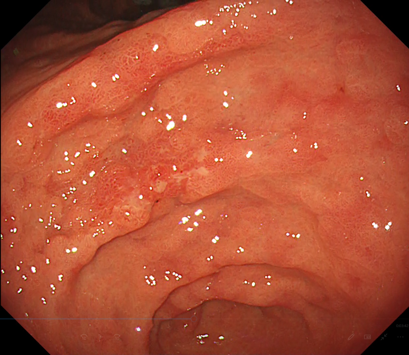
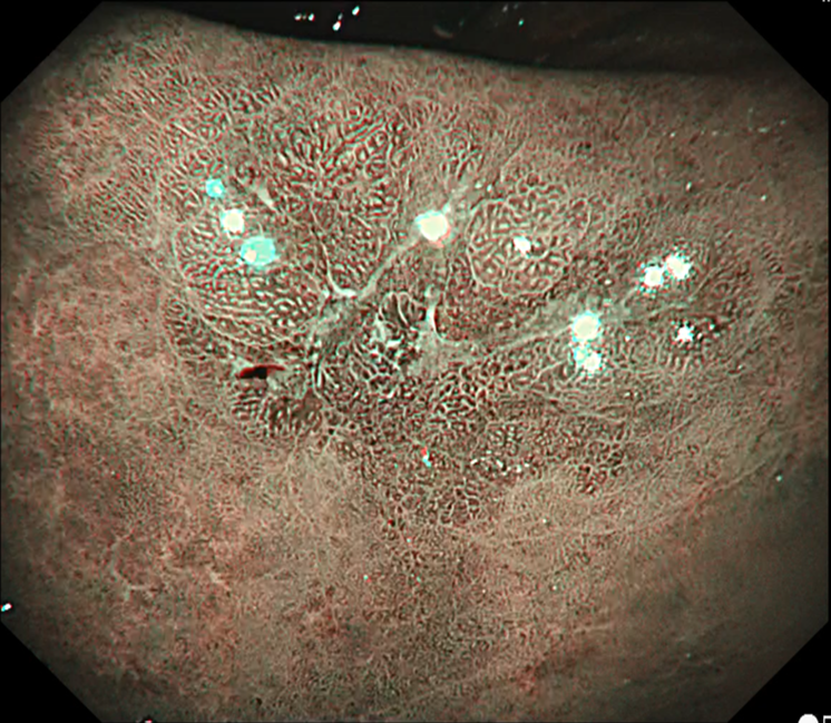
WLI
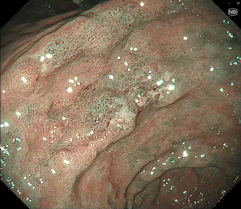
NBI
Overall comment
Under white light, the lesion appeared to be depressed and had a clear boundary with the surrounding tissue. Patchy reddening and an irregular uneven surface suggested a diagnosis of gastric cancer. Narrow Band Imaging (NBI) clarified the demarcation lines and revealed patchy brownish areas. The lesion exhibited an irregular uneven micro-surface structure with a non-structural surface in the depression in the center of the lesion. Thanks to the Brightness Adjustment Imaging with Maintenance of Contrast (BAI-MAC) function, it was possible to clearly observe all the way to the margin of the lesion.
Scope : GIF-1200N / CV290
Case : Early gastric cancer (tub1, 0-IIa+IIc, pT1a(M), ly0, v0)
Organ : Lesser curvature of the angulus
Patient Information : M, 70s
Medical History : H. pylori eliminated
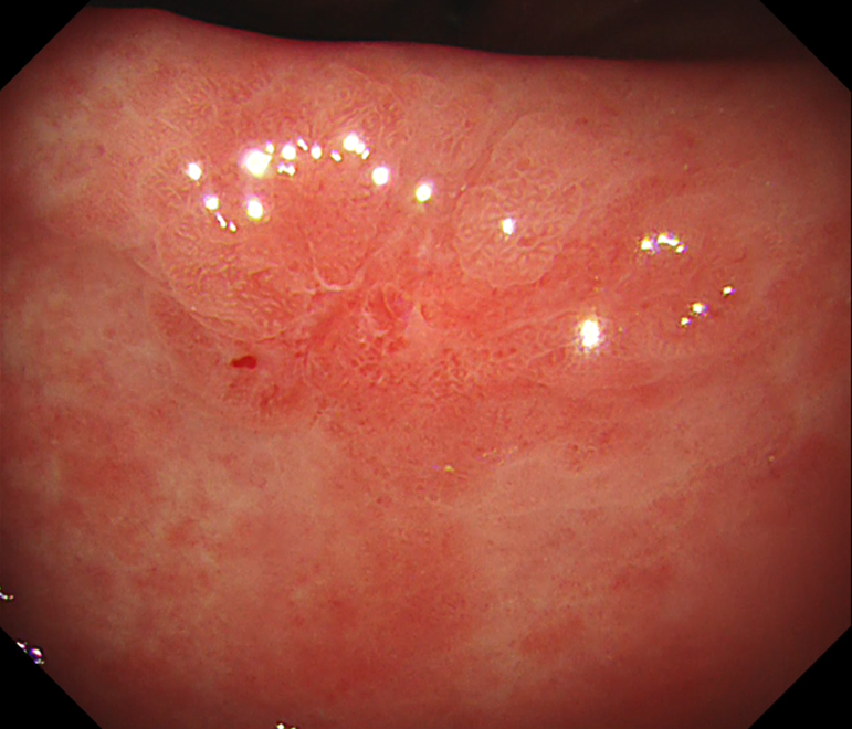
WLI
NBI
Overall comment
Under white light, the lesion appeared to be depressed in the center and had a clear boundary with the surrounding tissue. Patchy reddening and uneven irregularity in the depression were recognized, leading to a diagnosis of gastric cancer. Narrow Band Imaging (NBI) brought the demarcation lines into clear view. The intervening parts were enlarged non-uniformly and irregular surface pattern was observed in the depression in the center of the lesion.
* Specifications, design and accessories are subject to change without any notice or obligation on the part of the manufacturer
Takashi Kawai, MD, PhD
- Content Type