Colorectal Case 4
.jpg)
Prof. Yoji Takeuchi
Disclaimer:
- NBI™, BAI-MAC™ and TXI™ Technologies are not intended to replace histopathological sampling as a means of diagnosis
- The positions and statements made herein by Prof. Takeuchi are based on Prof. Takeuchi’s experiences, thoughts and opinions. As with any product, results may vary, and the techniques, instruments, and settings can vary from facility to facility. The content hereof should not be considered as a substitute for carefully reading all applicable labeling, including the Instructions for Use. Please thoroughly review the relevant user manual(s) for instructions, risks, warnings, and cautions. Techniques, instruments, and setting can vary from facility to facility. It is the clinician’s decision and responsibility in each clinical situation to decide which products, modes, medications, applications, and settings to use.
- The EVIS X1™ endoscopy system is not designed for cardiac applications. Other combinations of equipment may cause ventricular fibrillation or seriously affect the cardiac function of the patient. Improper use of endoscopes may result in patient injury, infection, bleeding, and/or perforation. Complete indications, contraindications, warnings, and cautions are available in the Instructions for Use (IFU)
1)Data on file with Olympus (DC00489968)
2)Data on file with Olympus (DC00479164, DC0047877)
3)Data on file with Olympus (DC00510434 & DC00567392)
Scope: CF-EZ1500DI
Case: Early rectal cancer, SM deep invasion carcinoma
Organ: Lower rectum
Patient information: F, 50s
Medical history: Nothing particular
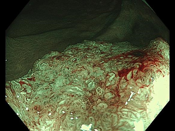
1. White Light Imaging (WLI)
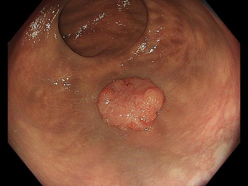
When observed with non-magnifying WLI, the entire image is in focus and looks bright and clear. A 12-mm diameter sessile elevated lesion (0-Is) is recognized in the lower rectum.
2. TXI™ technology
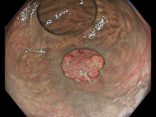
When observed with non-magnifying TXI™ technology, the contrast between red and white and surface irregularities become clear. The depressed surface looks clear, emphasizing the irregular morphology more than WLI1.
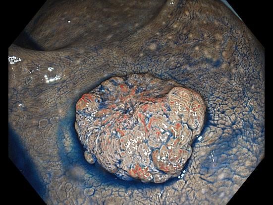
3. NBI™ technology non-magnifying observation
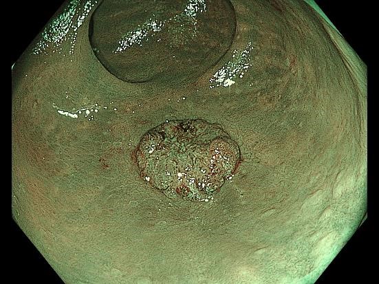
While the surface structure and vascular pattern in the margins of the lesion can be recognized even with relatively distant observation, they appear irregular and unclear in the center of the lesion. This lesion may be interpreted as Type 2 or 3 in the NICE classification, depending on the doctor.
4. WLI non-magnifying chromoendoscopy (with indigo carmine staining)
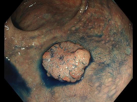
Pooling of indigo carmine in the grooves emphasizes the surface structure. The difference between the relatively inconspicuous irregularities in the margins and the dense yet unclear irregularities in the center is recognized more clearly.
5. TXI™ technology non-magnifying chromoendoscopy (with indigo carmine staining)
Color contrast between red, blue, and white are emphasized more than under WLI. The difference in tones enhances the surface structure1.
6. NBI™ technology non-magnifying observation (close-up observation)
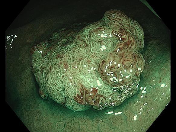
Bringing the scope tip close to the lesion makes it possible to clearly recognize surface microstructures and microvascular patterns without magnification. The image is in focus in both near and far areas3, making it easier to tell which part of the entire image requires further magnifying examination.
7. NBI™ technology magnifying chromoendoscopy (in Near Focus mode)
Disappearance of the surface structure and disruption of the microvascular network are recognized. The lesion can be classified as type 3 in the JNET classification. The image is mostly in focus at both near and far points, making it possible to comprehend the characteristics of the lesion in certain regions1.
Case video
Overall Comment
[Summary for this case]
This case involved a deeply invasive, elevated-type carcinoma in the lower rectum. In a distant view under non-magnified observation using WLI, TXI™ technology, NBI™ technology, and chromoendoscopy, the entire lesion appeared bright and in focus due to BAI-MAC™ technology. This clarity made it easy to assess the lesion’s irregular morphology, raising suspicion of invasive carcinoma. With TXI™ technology observation, surface structures became even more distinct after the application of indigo carmine.
Even without magnification, in normal focus mode, rough surface structures and vascular patterns were clearly visible during close-up NBI™ technology observation. This allowed for the targeted selection of areas requiring more detailed examination with magnified observation. Unlike conventional magnified observation, which requires precise, incremental focus adjustments, the Near Focus mode enabled quick switching with a simple button press2. Additionally, EDOF™ technology made it easy to capture sharp, in-focus images, which could contribute to the broader adoption of magnifying endoscopy.
Colon tumors often present as elevated lesions, which pose a challenge in maintaining a consistent focus due to variations in the distance between the lens and the lesion. However, EDOF™ technology is designed to keep both near and far points in focus, providing clear, edge-to-edge imaging3. Elevated lesions in the colon, particularly invasive carcinomas, tend to be fragile and prone to bleeding upon contact, which can obstruct further observation.
Magnified observation in the upper digestive tract is primarily used for histological assessment and to determine the extent of lesions. This requires a detailed examination of surface and vascular structures at full zoom. In contrast, pinpoint invasion is uncommon in the colon, so magnified observation for invasion diagnosis focuses on assessing irregularities and disruptions in surface structures over a limited area rather than analyzing each structure individually.
The Near Focus mode does not provide the same level of magnification as conventional magnified observation. However, it allows for a relatively wide field of view while maintaining a safe distance between the lens and the lesion. Because there is no direct contact with the lesion, the risk of bleeding is minimized. This enables high-quality diagnostic imaging, which supports informed decision-making when determining a treatment strategy.
Additionally, in esophagogastroduodenoscopy, lesion detection and detailed examination are often performed on separate days since the procedure only requires fasting. In colonoscopy, however, treatment is frequently performed immediately after lesion detection, as bowel preparation is burdensome for patients. The EZ1500 endoscope is particularly well suited for colorectal diagnosis, as its advanced TXI™ technology enhances lesion detection, while the bright NBI™ technology and seamless transition between close-up and magnified observation facilitate accurate assessment.
* Specifications, design and accessories are subject to change without any notice or obligation on the part of the manufacturer.
- Keyword
- Content Type


