Colorectal Case 7

Dr. Supakij Khomvilai
Hospital of Chulalongkorn University, Thailand
Disclaimer:
- NBI™ and BAI-MAC™ technologies are not intended to replace histopathological sampling as a means of diagnosis
- The positions and statements made herein by Dr. Supakij Khomvilai are based on Dr. Supakij Khomvilai’s experiences, thoughts and opinions. As with any product, results may vary, and the techniques, instruments, and settings can vary from facility to facility. The content hereof should not be considered as a substitute for carefully reading all applicable labeling, including the Instructions for Use. Please thoroughly review the relevant user manual(s) for instructions, risks, warnings, and cautions. Techniques, instruments, and setting can vary from facility to facility. It is the clinician’s decision and responsibility in each clinical situation to decide which products, modes, medications, applications, and settings to use.
- The EVIS X1™ endoscopy system is not designed for cardiac applications. Other combinations of equipment may cause ventricular fibrillation or seriously affect the cardiac function of the patient. Improper use of endoscopes may result in patient injury, infection, bleeding, and/or perforation. Complete indications, contraindications, warnings, and cautions are available in the Instructions for Use (IFU)
1) Data held on file with Olympus (K192793) as of 07/17/2020
Procedure Information
Scope: CF-EZ1500DL
Case Findings: Colonic polyp. Well differentiate adenocarcinoma with shallow submucosal invasion (668 micron from muscularis mucosa )
Patient information: F, 70s
Medical history: Screening for Colorectal cancer
1. White light observation
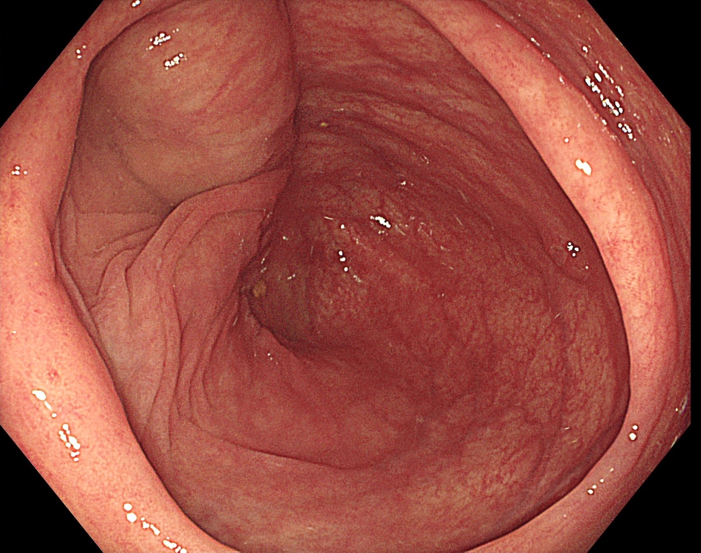
Bright, white light with 4K image showed the details of cecum where the abnormality is clearly visible.
Enhancement : A8
NBI Mode : NA
TXI Mode : NA
RDI Mode : NA
BAI-MAC : On
2. White light close observation
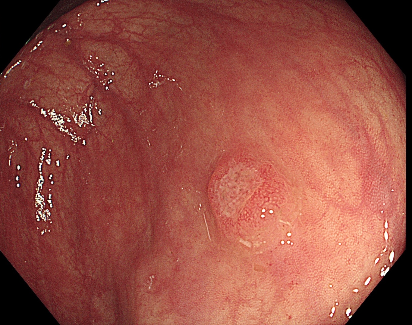
Closer look at the lesion we can see depressed area in the middle of the lesion. EDOF™ (extended depth of field) technology colonoscope showed details of surface which need more information.
Enhancement : A8
NBI Mode : NA
TXI Mode : NA
RDI Mode : NA
BAI-MAC : On
3. White light with Near Focus
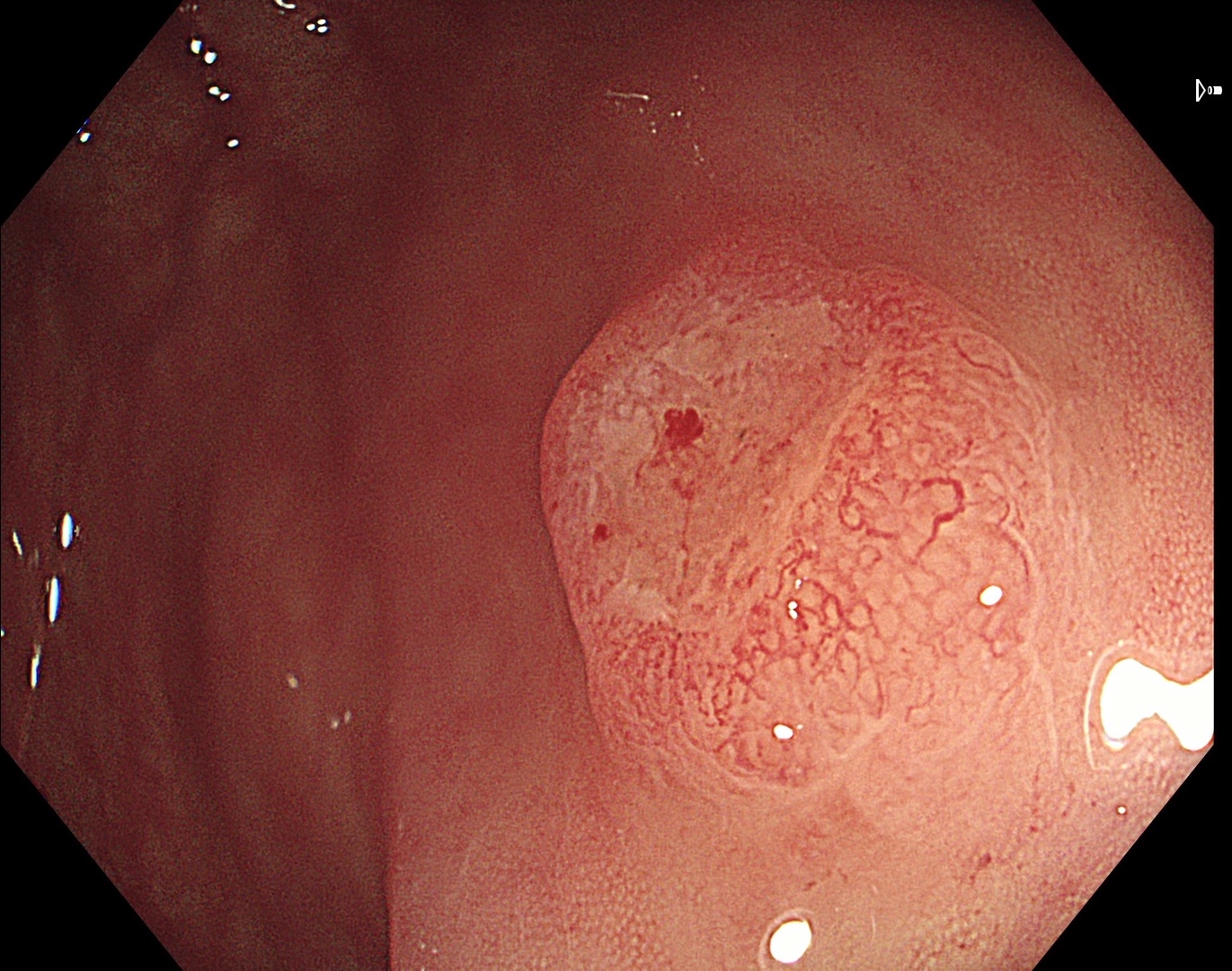
Near focus image showed details of surface which might be cancerous lesion.
Enhancement : A8
NBI Mode : NA
TXI Mode : NA
RDI Mode : NA
BAI-MAC : On
4. NBI™ technology observation
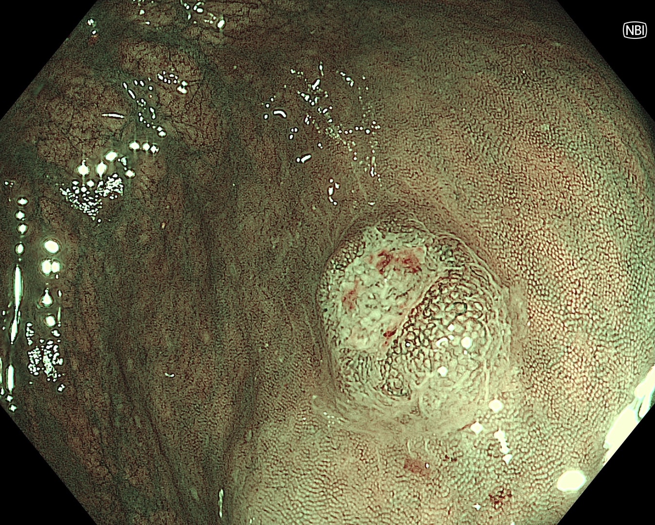
NBI™ technology enhances visibility of surface and vascular pattern.1
Enhancement : B8
NBI Mode : 3
TXI Mode : NA
RDI Mode : NA
BAI-MAC : On
* Specifications, design and accessories are subject to change without any notice or obligation on the part of the manufacturer
Case 6: Colonic polyp (Tubular Adenoma)
Dr. Supakij Khomvilai Case 8: Colonic polyp (Tubular adenom)
Dr. Supakij Khomvilai
Dr. Supakij Khomvilai Case 8: Colonic polyp (Tubular adenom)
Dr. Supakij Khomvilai
- Keyword
- Content Type


