Colorectal Case 15

Dr. Serhii Polishchuk
Gastrointestinal endoscopist at LLC “GASTROCENTER” Olymed
Trainer at NGO “EndoAcademy”, Ukraine
Disclaimer:
NBI™ and TXI™ Technologies are not intended to replace histopathological sampling as a means of diagnosis.
The positions and statements made herein by Dr. Serhii Polishchuk are based on Dr. Polishchuk experiences, thoughts and opinions. As with any product, results may vary, and the techniques, instruments, and settings can vary from facility to facility. The content hereof should not be considered as a substitute for carefully reading all applicable labeling, including the Instructions for Use. Please thoroughly review the relevant user manual(s) for instructions, risks, warnings, and cautions. Techniques, instruments, and setting can vary from facility to facility. It is the clinician’s decision and responsibility in each clinical situation to decide which products, modes, medications, applications, and settings to use.
The EVIS X1™ endoscopy system is not designed for cardiac applications. Other combinations of equipment may cause ventricular fibrillation or seriously affect the cardiac function of the patient. Improper use of endoscopes may result in patient injury, infection, bleeding, and/or perforation. Complete indications, contraindications, warnings, and cautions are available in the Instructions for Use (IFU)
Scope: CF-EZ1500DL
Organ: Colon (sigmoid colon)
Patient information: F, 50s, come for screening colonoscopy with FIT positive test
Medical history: No family history of CRC. No alcohol abuse, no smoking
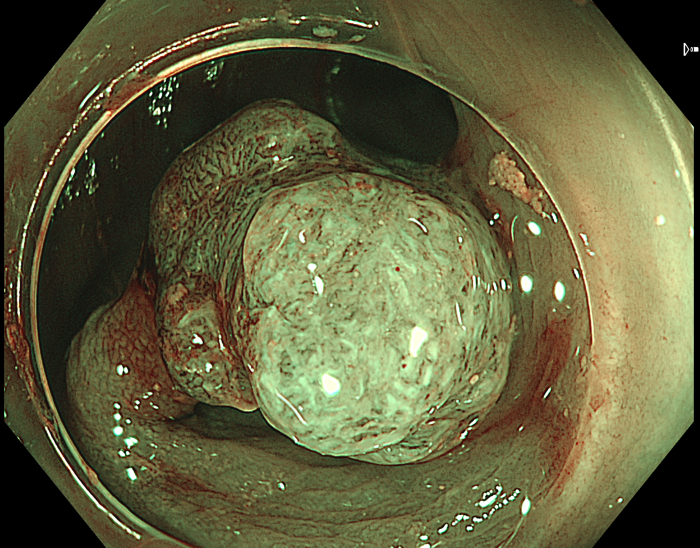
1. Tubular adenoma with HGD
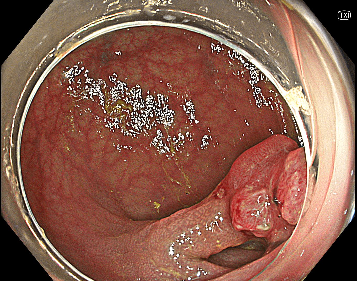
Texture and Color Enhancement Imaging (TXI): Mode 1
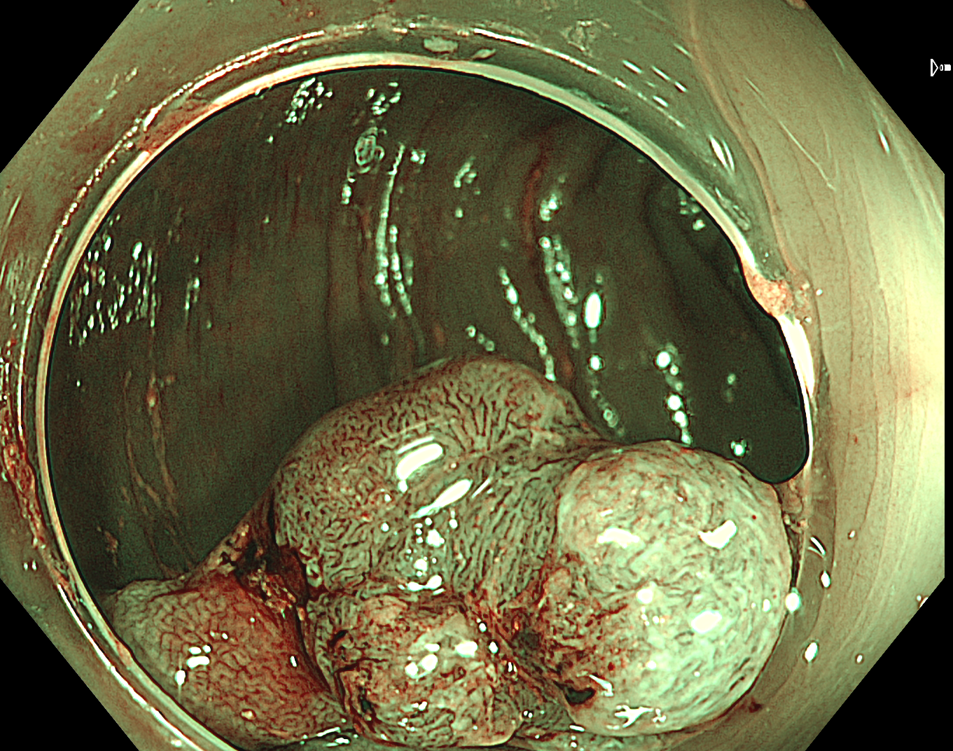
2. Tubular adenoma with HGD
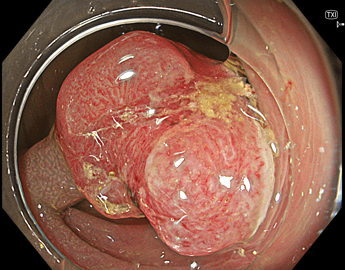
Texture and Color Enhancement Imaging (TXI): Mode 1
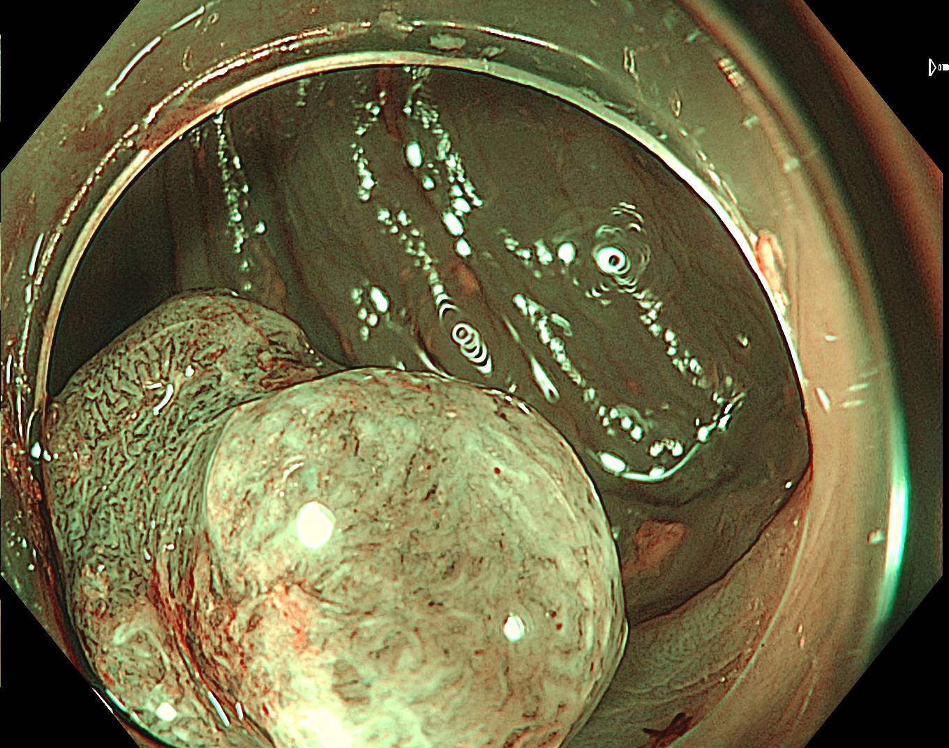
3. Tubular adenoma with HGD
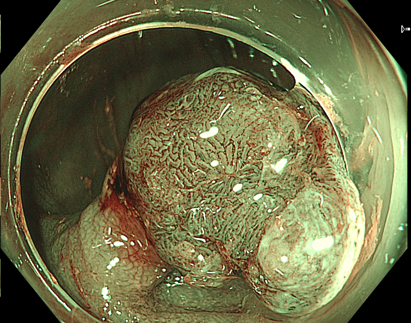
NBI Color Mode 3
Enhancement: A5
4. Tubular adenoma with HGD
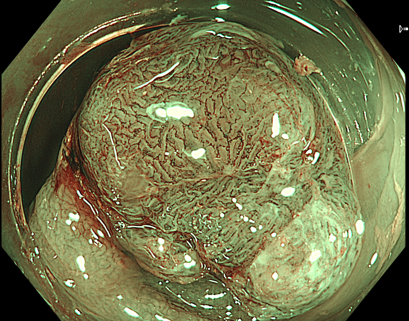
NBI Color Mode 3
Enhancement: A5
5. Tubular adenoma with HGD
NBI Color Mode 3
Enhancement: A5
6. Tubular adenoma with HGD
NBI Color Mode 3
Enhancement: A5
7. Tubular adenoma with HGD
NBI Color Mode 3
Enhancement: A5
Case Video
Tubular adenoma with HGD - JNET2B
Overall Comment
The final optical histology prediction in this case was:
Location: sigmoid colon
Size: ~12mm
Morphology (Paris classification): 0-Іp
Pit pattern: Kudo classification IIIL+Vi
JNET classification: JNET2B (vessel pattern – irregular caliber of vessels and irregular distribution of vessels; irregular surface pattern)
Optical histology prediction: Tubular adenoma with HGD
Treatment: hot snare polypectomy (HSP)
Pathology report: Tubular adenoma with HGD (ICD-O code: 8211/2). No signs of cytological dysplasia in resection margins (complete removal)
* Specifications, design and accessories are subject to change without any notice or obligation on the part of the manufacturer
Dr. Serhii Polishchuk Case 19: LST-NG-PD (Pseudo-depressed type), pT1a colonic cancer
Dr. Ho Dang Quy Dung
- Keyword
- Content Type


