Colorectal Case 25

Dr. Shiaw-Hooi Ho
Associate Professor of Medicine at the Department of Medicine,
Universiti Malaya, Malaysia
Disclaimer:
NBI™ and TXI™ technologies are not intended to replace histopathological sampling as a means of diagnosis
The positions and statements made herein by Dr. Ho Shiaw Hooi are based on Dr. Ho Shiaw Hooi’s experiences, thoughts and opinions. As with any product, results may vary, and the techniques, instruments, and settings can vary from facility to facility. The content hereof should not be considered as a substitute for carefully reading all applicable labeling, including the Instructions for Use. Please thoroughly review the relevant user manual(s) for instructions, risks, warnings, and cautions. Techniques, instruments, and setting can vary from facility to facility. It is the clinician’s decision and responsibility in each clinical situation to decide which products, modes, medications, applications, and settings to use.
The EVIS X1™ endoscopy system is not designed for cardiac applications. Other combinations of equipment may cause ventricular fibrillation or seriously affect the cardiac function of the patient. Improper use of endoscopes may result in patient injury, infection, bleeding, and/or perforation. Complete indications, contraindications, warnings, and cautions are available in the Instructions for Use (IFU)
1) Data on file with Olympus (DC00489968)
Scope: PCF-H190DL
Organ: Terminal ileum & colon
Patient information: F, 20s
Medical history: Chronic diarrhea for about 1 ½ years prior to consultation; no background medical illness in the past
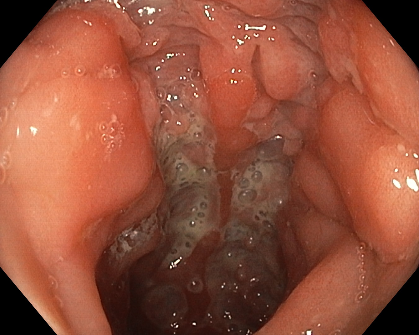
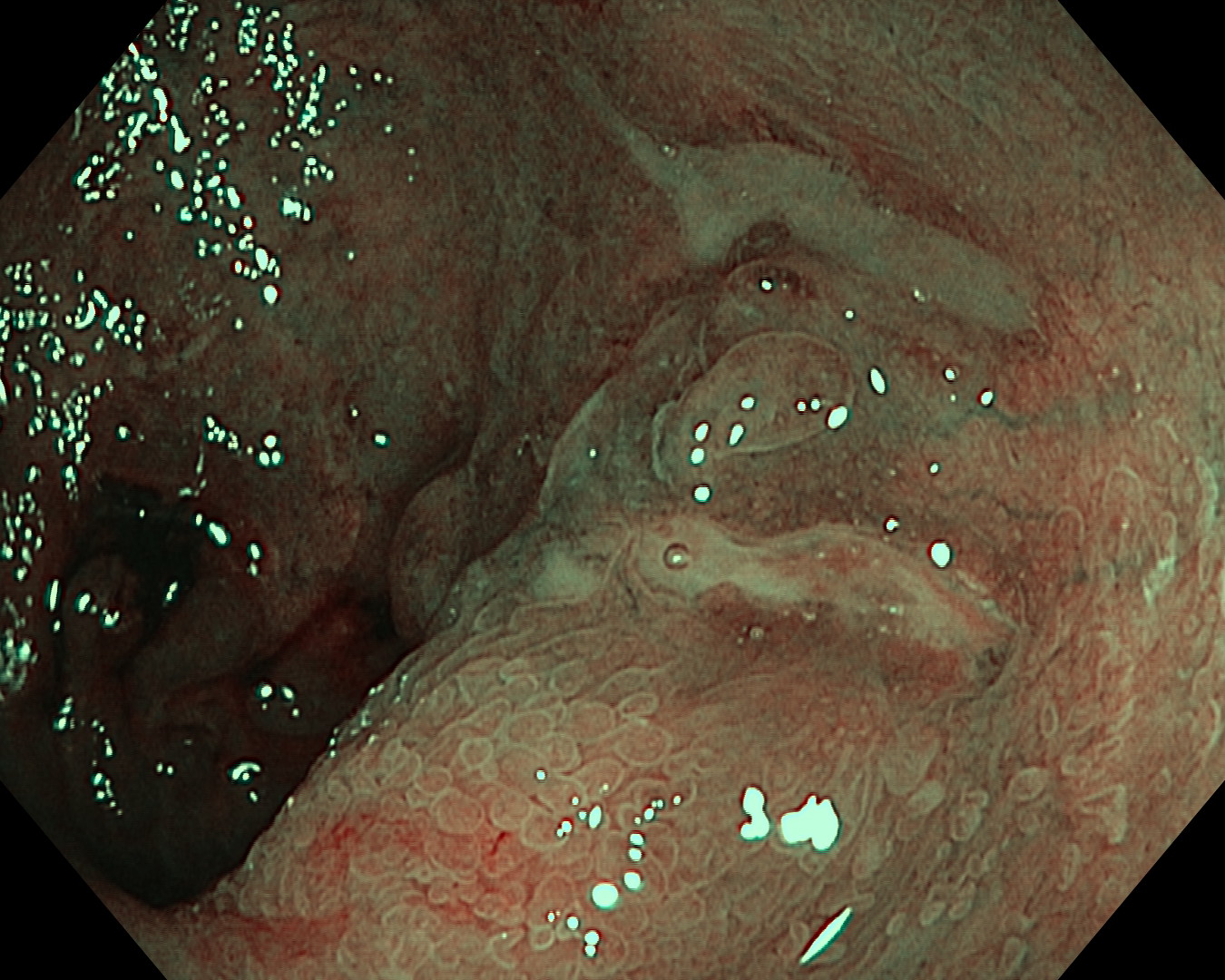
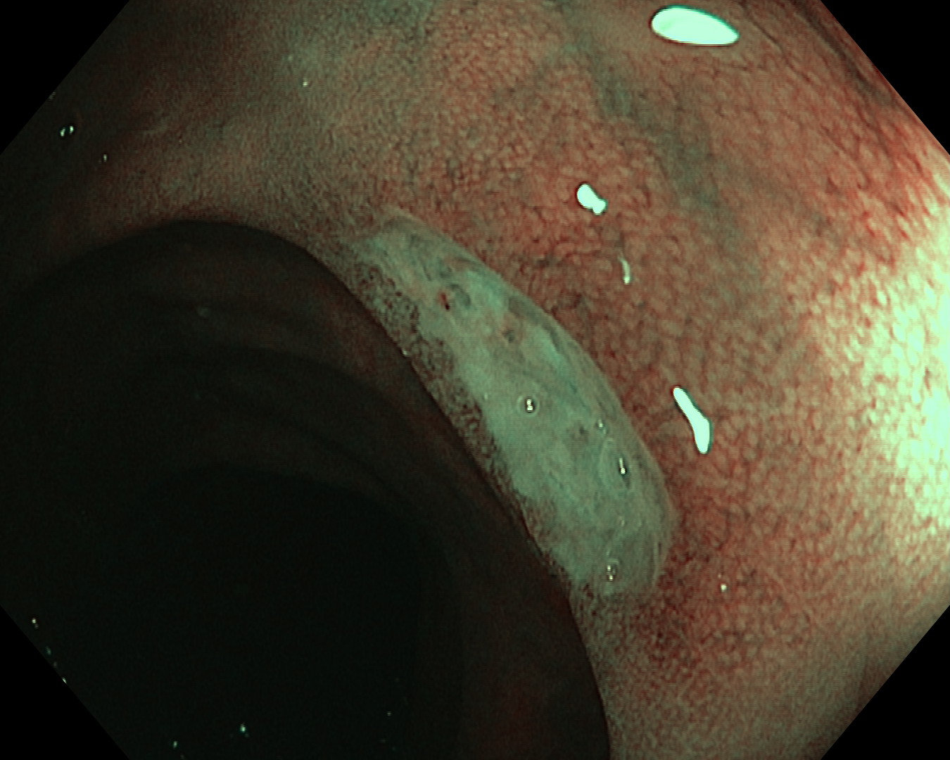
1. WLI Observation
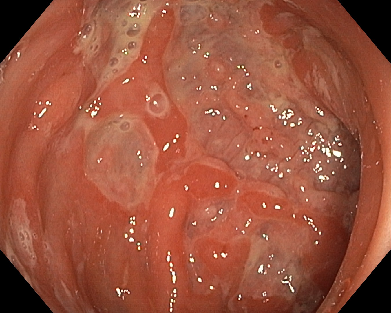
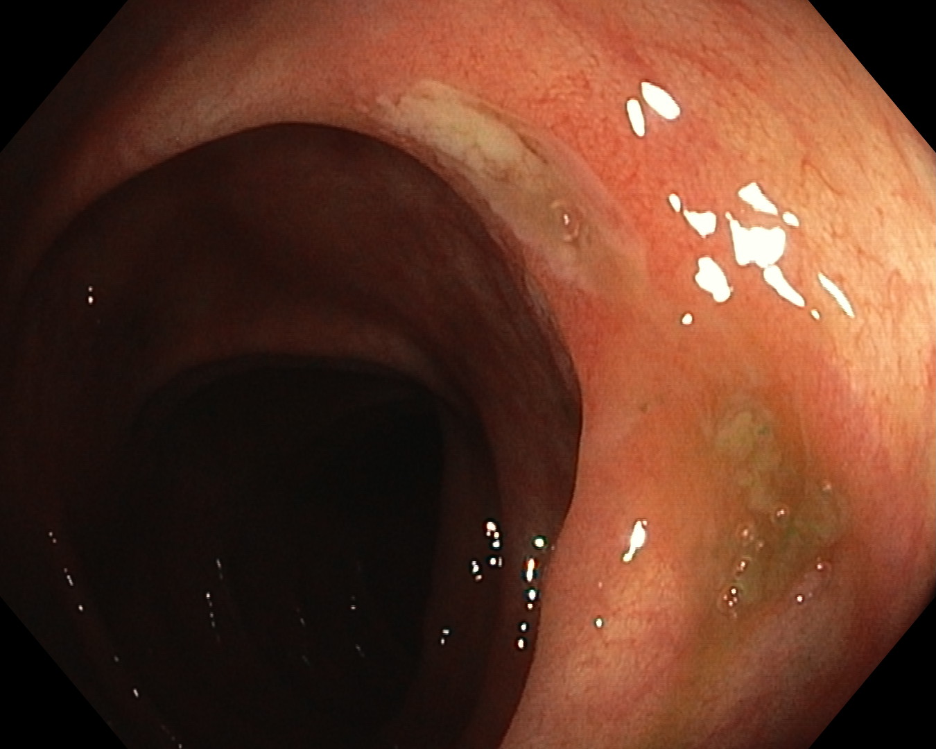
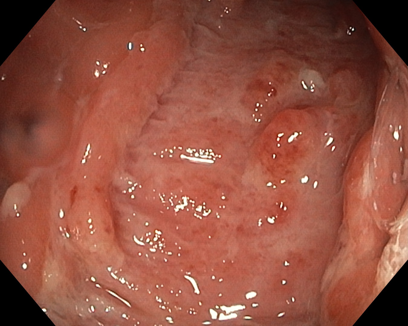
2. WLI Observation
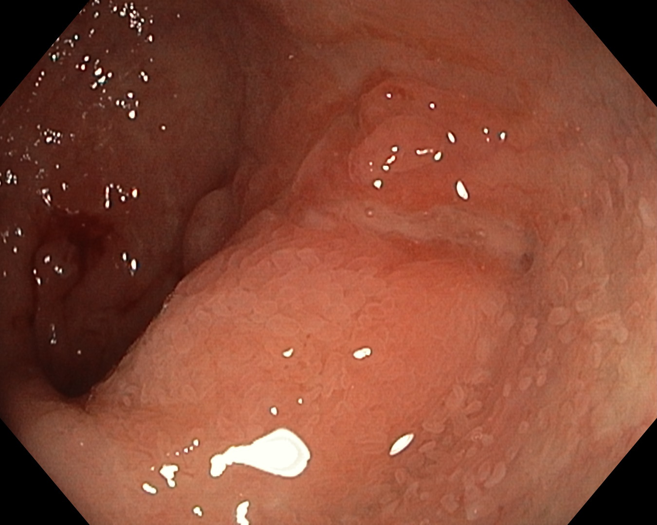
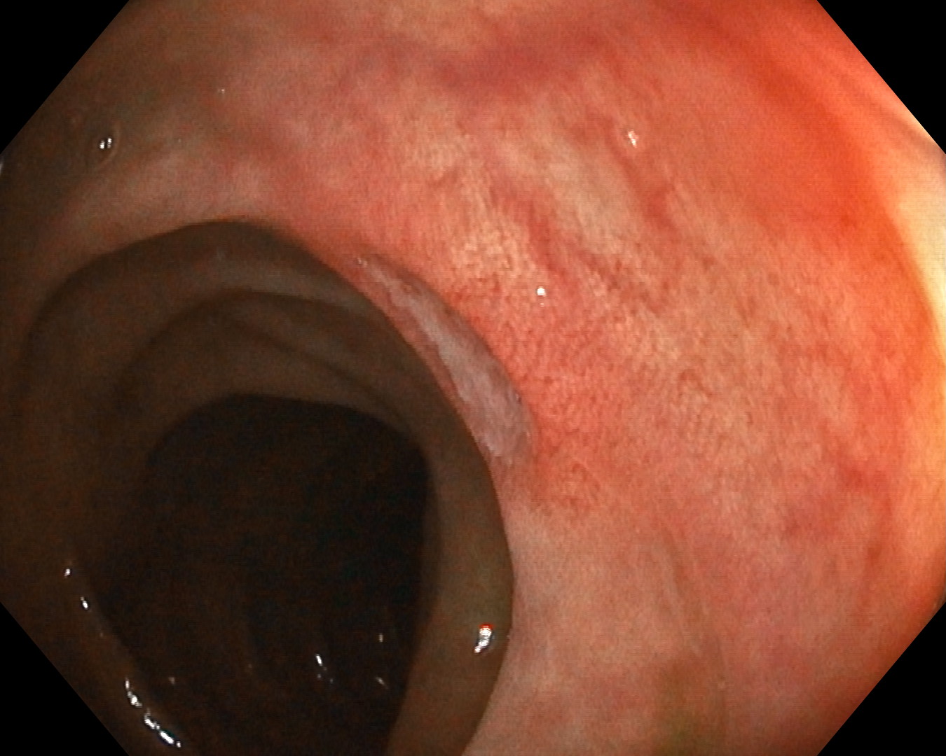
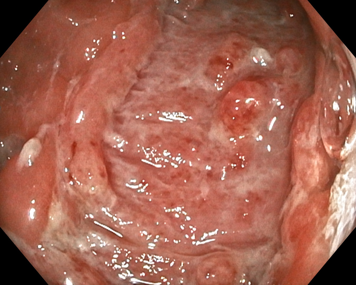
3. WLI Observation
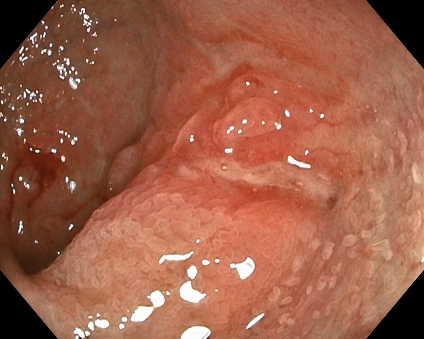
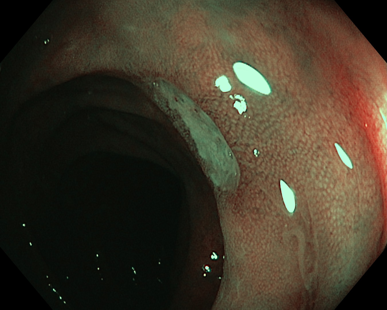
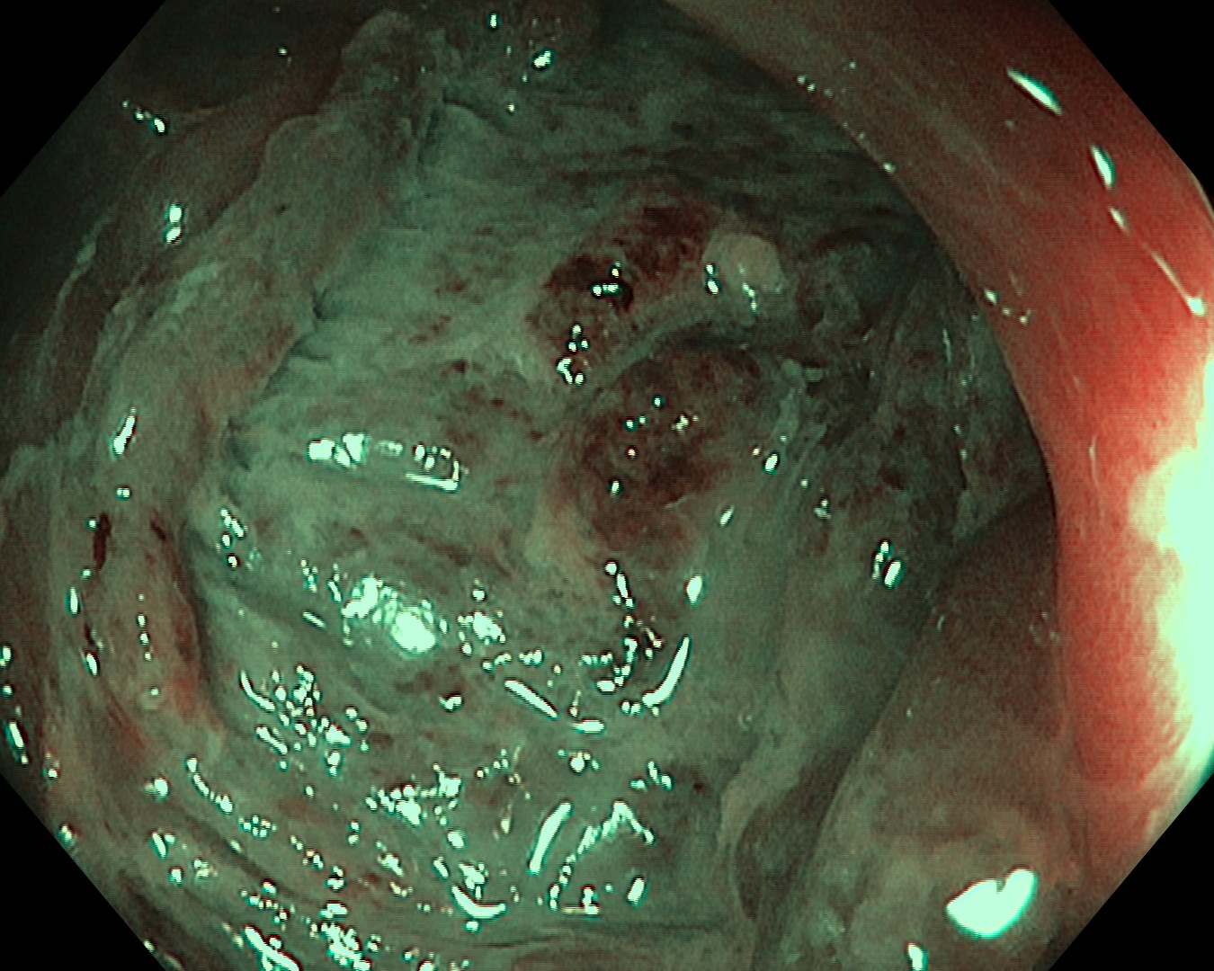
4. TXI™ technology Observation
5. NBI™ technology Observation
6. WLI Observation
7. TXI™ technology Observation
8. NBI™ technology Observation
9. NBI™ technology Observation
10. WLI Observation
11. TXI™ technology Observation
12. NBI™ technology Observation
Case Video
Overall Comment
This young female patient presented with chronic diarrhea for 1 ½ years prior to her consultation in this hospital. The endoscopic findings were classical of typical Crohn’s ileo-colitis: deep longitudinal ulcer with skip lesions and cobblestone appearance. She was started on steroid, azathioprine and was planned for biologic therapy.
* Specifications, design and accessories are subject to change without any notice or obligation on the part of the manufacturer
Dr. Shiaw-Hooi Ho Case 26: Large 40mm splenic flexure NG-LST
Dr. Shiaw-Hooi Ho
- Content Type


