Dr. V. R. Pattabhi Raman / Royal Care Hospital, India
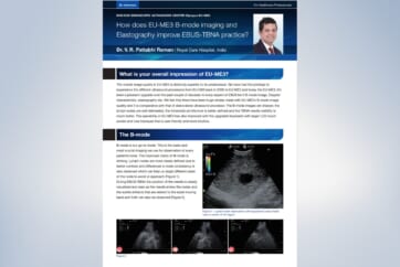
What is your overall impression of EU-ME3?
The overall image quality in EU-ME3 is distinctly superior to its predecessor. We have had the privilege to experience the different ultrasound processors from EU-C60 back in 2008 to EU-ME2 and today the EU-ME3. It’s been a pleasant upgrade over the past couple of decades in every aspect of EBUS be it B-mode image, Doppler characteristic, elastography etc. We feel that there have been huge strides made with EU-ME3’s B-mode image quality and it is comparative with that of stand-alone ultrasound processor. The B-mode images are sharper, the lymph nodes are well delineated, the intranodal architecture is better defined and the TBNA needle visibility is much better. The operability in EU-ME3 has also improved with the upgraded keyboard with larger LCD touch screen and new trackpad that is user friendly and more intuitive.
The B-mode
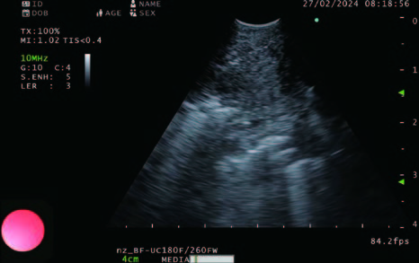
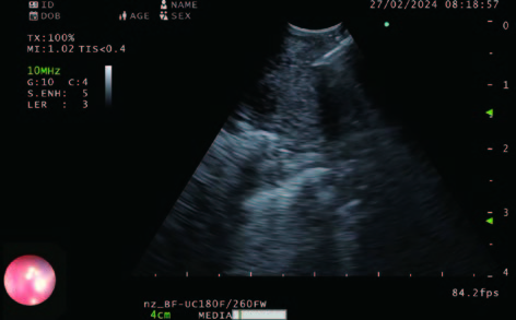
B-mode is our go-to mode. This is the basic and most crucial imaging we use for observation of every patient’s node. The improved clarity of B-mode is striking. Lymph nodes are more clearly defined due to better contrast and differences in node consistency is also observed which can help us target different areas of the node to avoid or approach (Figure 1).
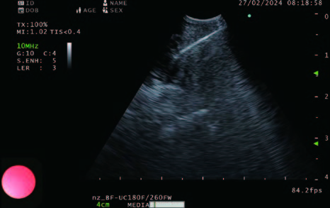
During EBUS-TBNA the position of the needle is clearly visualized and seen as the needle enters the nodes and the subtle artifacts that are related to the stylet moving back and forth can also be observed (Figure 2).
Figure 2
Flow mode in EU-ME3
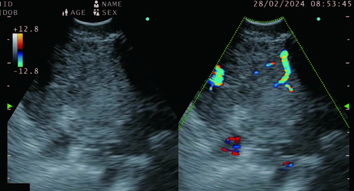
The doppler in the new processor comes in three modes. The color flow, power flow and the H-flow mode. The contrast seems better and the intranodal vessels are visualized clearly. We are also able to have the doppler super imposed and corresponding B-mode side by side and thus the poor visibility that we used to have with the previous processors when the doppler was on as the needle enters the node is no longer an issue (Figure 3).
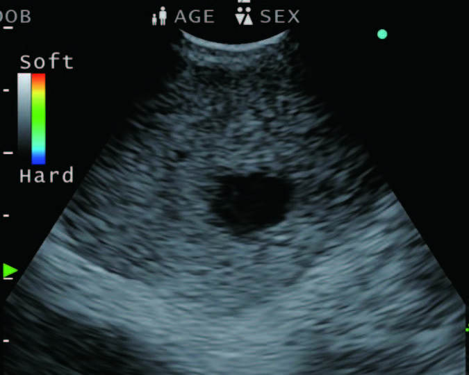
Elastography
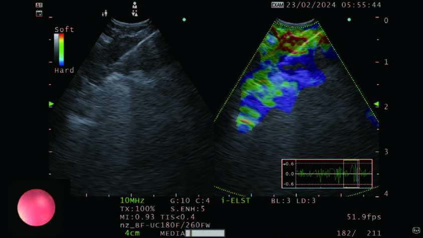
Elastography is a recent addition that can assess the hardness of the lymph node during EBUS. There is a theoretical advantage when one can assess the nature of the node before sampling it and to decide whether sampling would be useful, and even better if there are different areas of the nodes that need to be sampled to improve the outcome of the puncture. While the previous version of this processor had this capability, the EU-ME3 is newly equipped with “i-ELST” mode, that enables stable ELST images even with weak pulsation. The option of being able to view a B-mode and Elastography or Doppler in the same frame as adjacent pictures (see Figure 4) helps us target the desired area of interest in the node.
Ergonomics
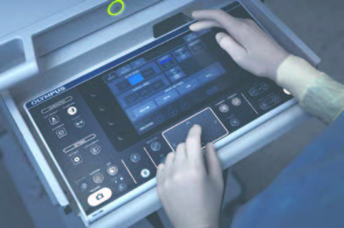
Rollers and mouse belong to technology of yesteryears and in keeping up with the trend the keyboard in EU-ME3 has a trackpad which is practical as it is very intuitive to use, easy to complete the measurements and the addition of all the mediastinal lymph node stations as a pre-programmed list is a welcome addition as all these small nuances help reduce the scope time which in turn helps us to improve patient comfort without compromising on the data that is being generated. The shorter scope time in the patient will eventually lead to better overall lifetime of the scope thanks to reduced wear and tear.
- Content Type