Disclaimer:
The techniques and clinical opinions presented in this material reflect the personal experience and professional judgment of the healthcare professional and do not necessarily represent the views of Olympus. This material is intended for healthcare professionals only. This material is not intended to substitute the Instructions for Use (IFU). Users should always refer to the applicable IFU and use Olympus products in accordance with the approved indications and local regulatory requirements. The healthcare professional presenting this material has provided this complimentary service for the benefit of clinical education.
# POA 2025 Global Live Surgery: Precise EBUS-Cryobiopsy by Prof. Gu Ye’s Team – Advanced Endoscopy Enabling High‑Value Clinical Care
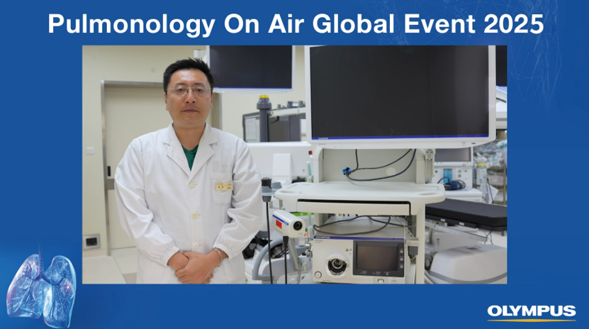
On December 5, 2025, the Pulmonology on Air (POA) global respiratory online academic platform successfully held its international live symposium. Prof. Gu Ye’s team from Shanghai Pulmonary Hospital presented a live clinical case focusing on advanced lung cancer, demonstrating a standardized, integrated procedure of EBUS‑guided mediastinal lymph node cryobiopsy combined with primary lung lesion cryobiopsy. This live session highlighted how state‑of‑the‑art endoscopic equipment enables precise lesion localization and safe, high‑quality tissue acquisition, delivering a reproducible, high‑standard workflow for interventional pulmonologists worldwide.
1. Clinical Background
A male patient in his sixties with a 1‑year history of pulmonary opacity presented with persistent cough, yellow sputum, and recent worsening with hemoptysis.
Imaging revealed:
– A definite malignant lesion in the right lower lobe;
– Significant enlargement of mediastinal lymph nodes (station 7 and station 11), highly suspicious for metastasis;
– Right pleural effusion and osteolytic destruction of the left 9th rib, consistent with advanced malignant lung disease.
The primary clinical objectives were:
– To obtain sufficient, high‑quality viable tumor tissue for histopathological diagnosis and molecular testing;
– To achieve accurate oncologic staging to guide subsequent therapy;
– To perform minimally invasive procedures with optimal safety and low complication rates.
2. Detailed Surgical Procedure
(1) Multi‑Modal Imaging Evaluation Using the EVIS X1 System
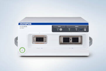
The procedure began with endoscopic airway inspection using the EVIS X1 (CV‑1500) system.
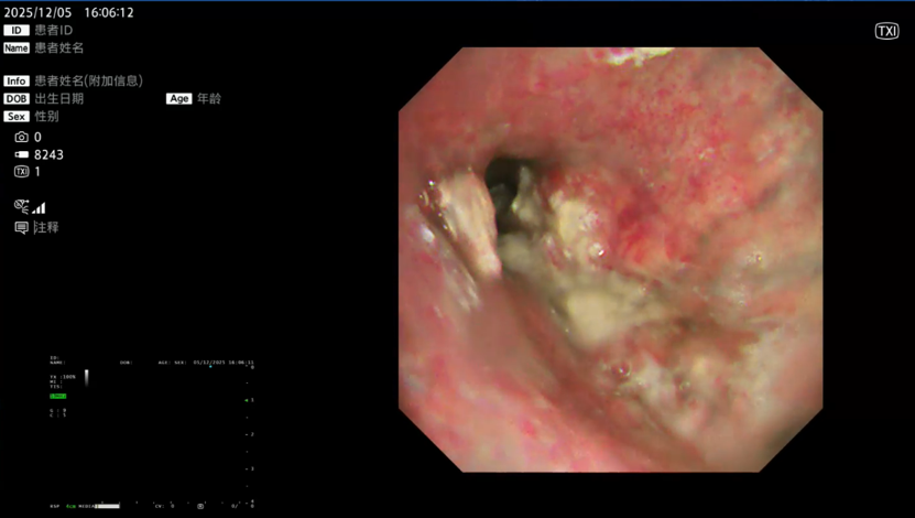
– TXI mode clearly delineated mucosal texture, structural layers, and vascular distribution to define tumor boundaries.
– NBI mode enhanced visualization of microvascular abnormalities, identifying regions of high tumor activity for targeted biopsy.
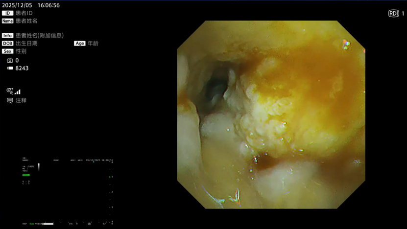
– RDI mode identified potential bleeding sites in advance, supporting proactive hemostasis.
Combined multi‑modal imaging reliably distinguished necrotic tissue from viable tumor, helping to minimize non-target sampling and supporting a “see‑first, operate‑precisely” strategy.
(2) EBUS Localization with BF‑UC290F Ultrasound Bronchoscope
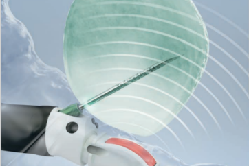
The BF‑UC290F EBUS bronchoscope was introduced for real‑time mediastinal sonographic evaluation. It clearly identified enlarged station 7 and station 11 lymph nodes, delineated size, echotexture, margins, and adjacent vascular structures, and allowed safe trajectory planning while avoiding vessels.
With a thinner distal tip, shorter ultrasound probe, and 20° forward view (closer to conventional bronchoscopy and free of ultrasound probe obstruction), the device enabled smooth insertion and stable, safe manipulation.
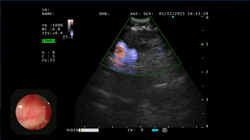
(3) Targeted Transbronchial Needle Aspiration (TBNA) with ViziShot 2–22G Needle
Under continuous ultrasound monitoring, the ViziShot 2–22G cutting needle was advanced. Its sharp cutting tip penetrated lymph node capsules and fibrous tissue smoothly. Each target node was sampled 3 times to ensure adequate cellular yield.
The needle’s enhanced flexibility allowed access to deep, technically challenging locations such as station 11 nodes. The green distal sheath provided high visibility under bronchoscopic view, further improving procedural safety.
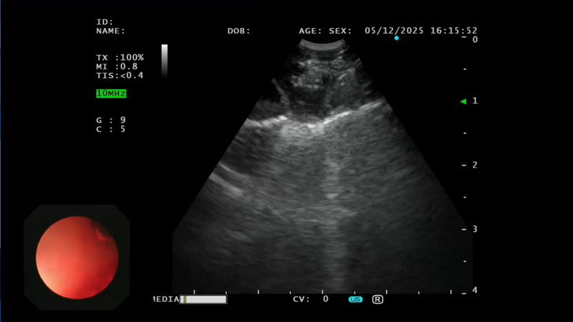
(4) Ultrasound ‑Guided Cryobiopsy for High‑Quality Tissue Acquisition
To obtain large, intact, histology‑ready tissue cores required for pathological and molecular analysis, cryobiopsy was performed.
A 1.1 mm cryoprobe was inserted through the working channel. Under combined endoscopic and ultrasound visualization, the probe tip was precisely positioned on the target lymph node area. Freezing was activated to attach tissue to the probe; the probe was then withdrawn gently to retrieve large, continuous tissue strips. This step ensured specimens were sufficient in quantity and well‑preserved in architecture.
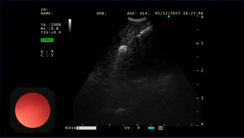
(5) Post‑Procedural Assessment and Management
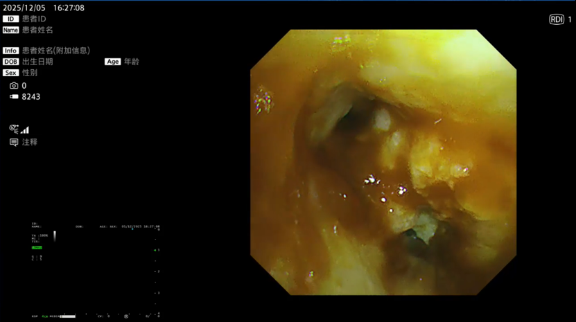
A standard bronchoscope was reinserted to inspect the entire airway. Secretions and mild oozing were aspirated.
Hemostasis was confirmed under RDI mode, with no active bleeding or airway injury detected. The procedure was concluded safely. The entire workflow was minimally invasive, well‑organized, and well‑tolerated by the patient.
3. Clinical Value of Core Equipment
– EVIS X1 (CV‑1500)
Multi‑modal imaging improves tumor identification, reduces non‑target biopsy, predicts bleeding risk, and increases diagnostic yield and procedural safety.
– BF‑UC290F EBUS Bronchoscope
Enables precise mediastinal lymph node mapping, safe vascular avoidance, and smooth navigation, improving the accuracy of lung cancer staging.
– ViziShot 2–22G Needle
Delivers reliable access to deep or difficult lymph nodes, ensures smooth penetration, and enhances visibility and success rates in challenging sampling.
4. Clinical Takeaways
This POA live procedure embodied a fully visualized, precise, and minimally invasive approach to advanced lung cancer evaluation. It delivered four key clinical advantages:
- Higher diagnostic accuracy: Tissue quality may contribute to improved histopathological and molecular testing success rates.
- More precise staging: Complete lymph node sampling strengthens the reliability of oncologic staging.
- Greater efficiency: Diagnosis and staging are completed in a single procedure, shortening the diagnostic timeline.
- Improved safety: Real‑time visualization reduces risks of bleeding, perforation, and other complications.
The standardized approach developed by Prof. Gu Ye’s team at Shanghai Pulmonary Hospital establishes a reproducible, reference model for interventional pulmonology in advanced lung cancer. It underscores China’s growing role in bronchoscopic precision diagnosis and care.
*The above section 3&4 represents clinical experience from expert physicians from Prof. Gu Ye’s Team. Actual results may vary depending on patient‑specific conditions and operator proficiency.
- Content Type