Hospital: Professor, PGIMER Chandigarh Hospital
Disclaimer:
- NBI™ and TXI™ Technologies are not intended to replace histopathological sampling as a means of diagnosis
- The positions and statements made herein by Dr. Agarwal are based on Dr. Agarwal’s experiences, thoughts and opinions. As with any product, results may vary, and the techniques, instruments, and settings can vary from facility to facility. The content hereof should not be considered as a substitute for carefully reading all applicable labeling, including the Instructions for Use. Please thoroughly review the relevant user manual(s) for instructions, risks, warnings, and cautions. Techniques, instruments, and setting can vary from facility to facility. It is the clinician’s decision and responsibility in each clinical situation to decide which products, modes, medications, applications, and settings to use.
- The EVIS X1™ endoscopy system is not designed for cardiac applications. Other combinations of equipment may cause ventricular fibrillation or seriously affect the cardiac function of the patient. Improper use of endoscopes may result in patient injury, infection, bleeding, and/or perforation. Complete indications, contraindications, warnings, and cautions are available in the Instructions for Use (IFU)
- Dr Agarwal, the authoring physician(s) of this presentation, are/ is a paid consultant(s) to Olympus Corporation of the Americas.
Scope: BF-1TH1100
Patient information: Male, 71 years old
Medical history: Not Significant
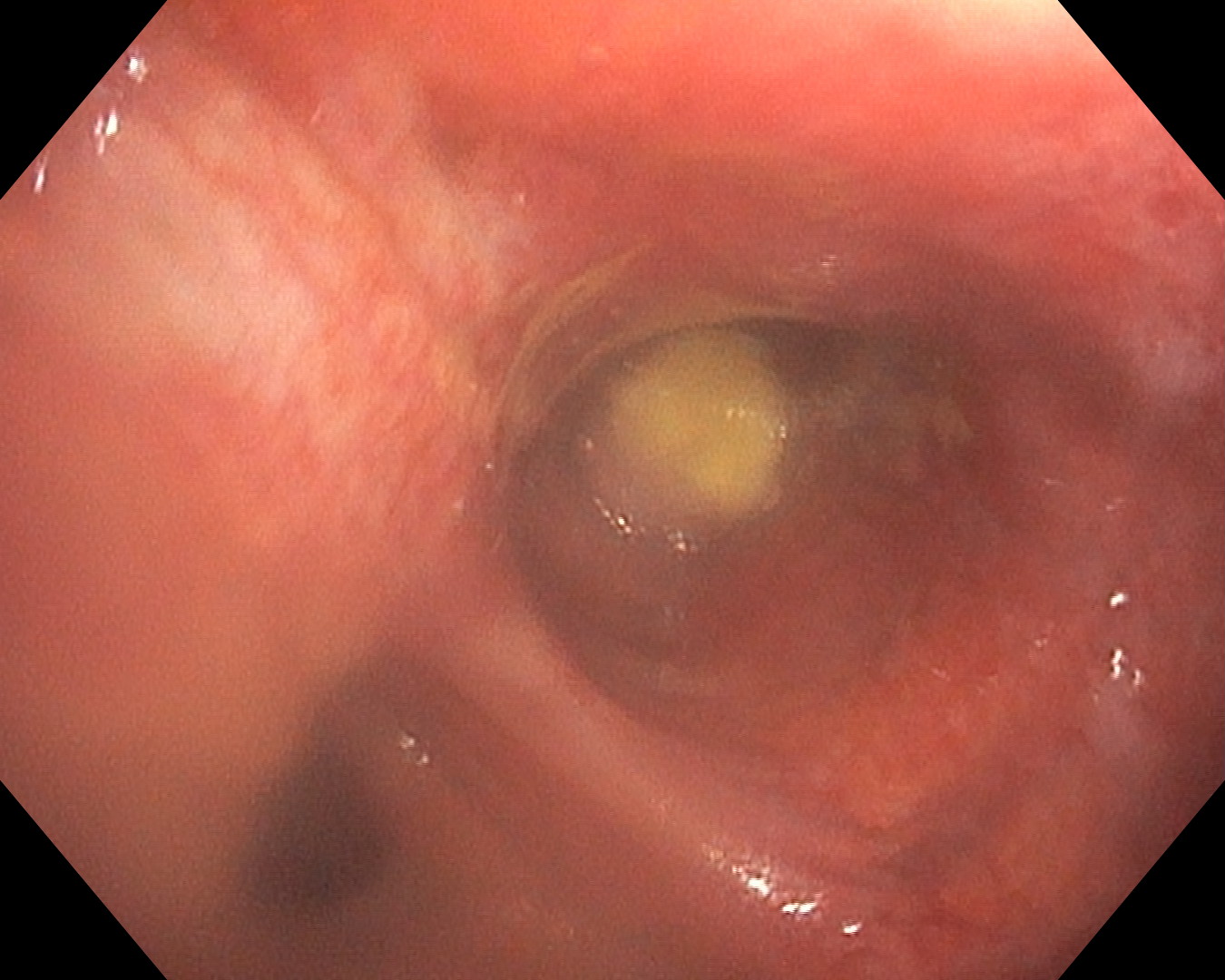
1. WLI
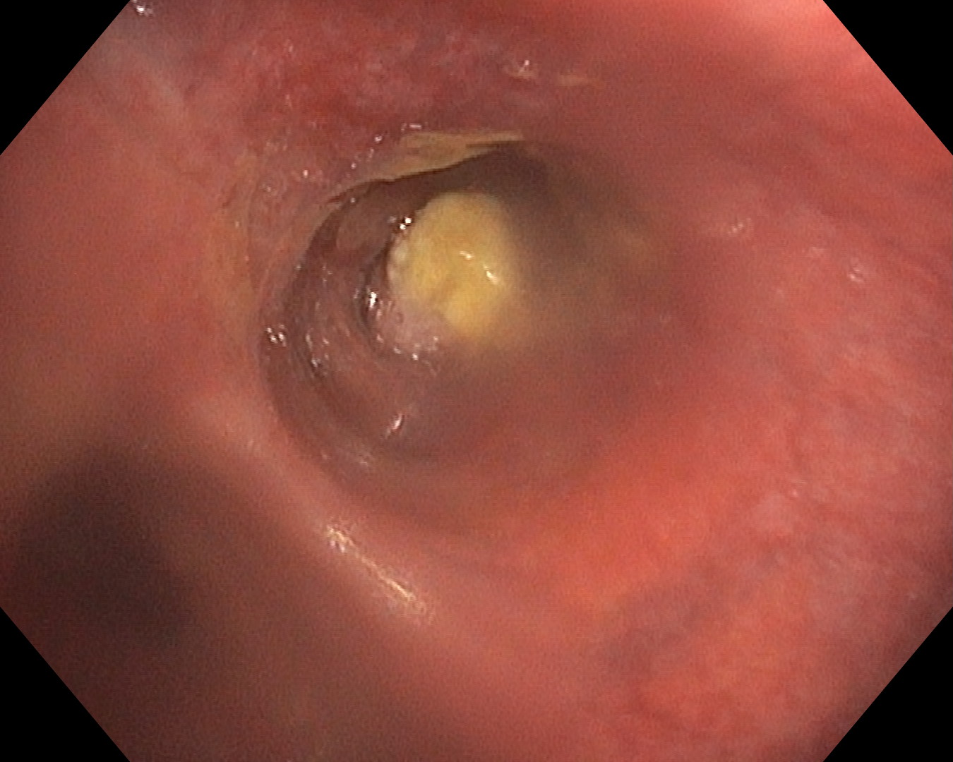
Lingula opening occluded by necrotic growth.
2. WLI +TXI™ Technology
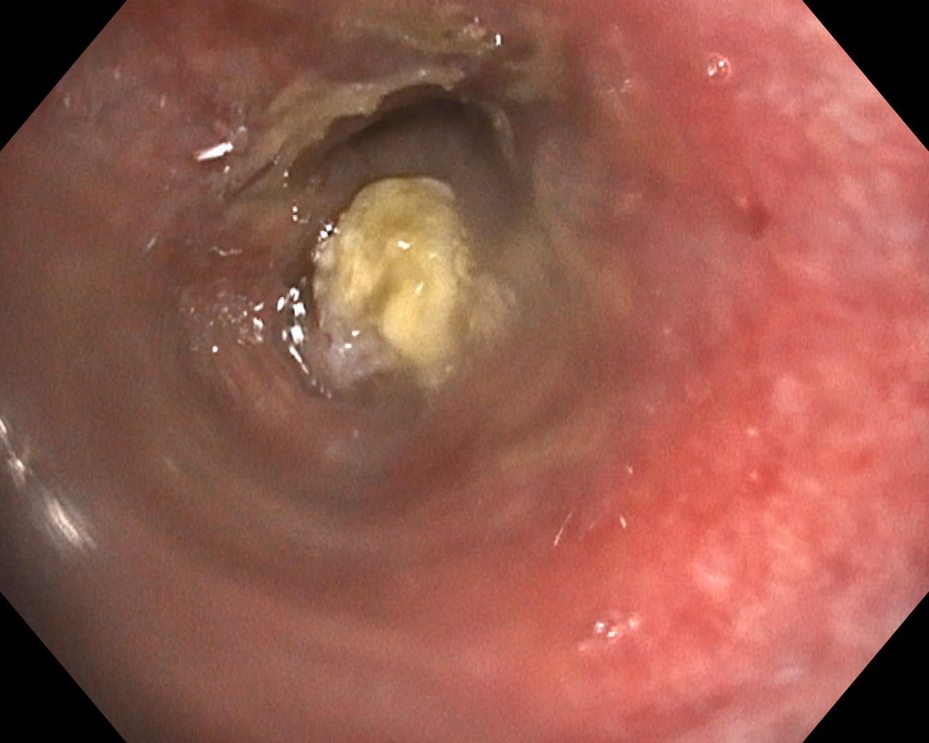
Surrounding erythema and edema being visualized better with TXI™ Technology imaging, along with necrotic growth occluding the opening of lingula
3. WLI
Surrounding vascularity not clearly visible with WLI.
4. WLI + NBI™ Technology
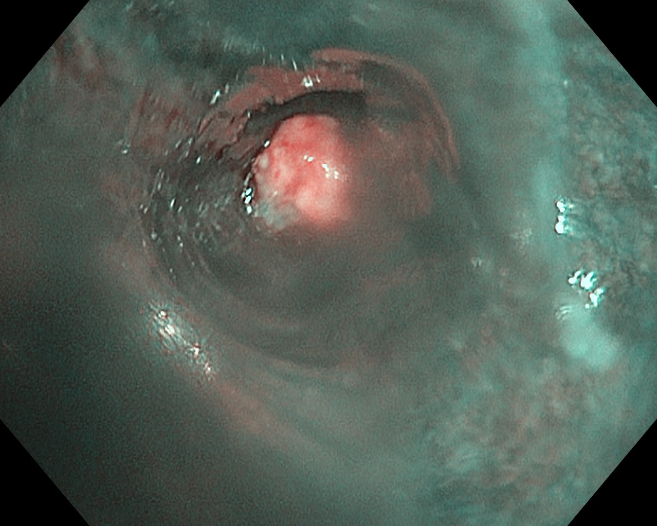
Surrounding vascularity is better visualized with NBI™ Technology.
Overall Comment
This 71-year-old gentleman, presented with dry cough and progressive breathlessness of 2 months duration. He was a reformed smoker with smoking index of 400. CT thorax showed left upper lobe segmental collapse and consolidation. Bronchoscopy was done to check for airway tumors. In flexible bronchoscopy, we observed necrotic growth occluding the opening of lingula with surrounding erythema and edema.
- Keyword
- Content Type