Author: Daisuke Himeji, MD, PhD
Miyazaki Prefectural Miyazaki Hospital, Japan
Endoscopy Center, Department of Internal Medicine, Department of Medical Informatics
Disclaimer:
- TXI™ and RDI™ Technologies are not intended to replace histopathological sampling as a means of diagnosis
- The positions and statements made herein by Dr. Himeji’s are based on Dr. Himeji’s experiences, thoughts and opinions. As with any product, results may vary, and the techniques, instruments, and settings can vary from facility to facility. The content hereof should not be considered as a substitute for carefully reading all applicable labeling, including the Instructions for Use. Please thoroughly review the relevant user manual(s) for instructions, risks, warnings, and cautions. Techniques, instruments, and setting can vary from facility to facility. It is the clinician’s decision and responsibility in each clinical situation to decide which products, modes, medications, applications, and settings to use.
- The BF-H1200 used in this case is not available in the US market at this time nor is there an established time for its release. The safety and effectiveness of this product and or the use of these products has not yet been established in the United States market.
- The EVIS X1™ endoscopy system is not designed for cardiac applications. Other combinations of equipment may cause ventricular fibrillation or seriously affect the cardiac function of the patient. Improper use of endoscopes may result in patient injury, infection, bleeding, and/or perforation. Complete indications, contraindications, warnings, and cautions are available in the Instructions for Use (IFU)
- Dr Himeji, the authoring physician(s) of this presentation, are/ is a paid consultant(s) to Olympus Corporation.
Scope: BF-H1200
Patient information: Female in her 50s
Medical history:
Right breast cancer was diagnosed 11 years ago and mastectomy was performed. Eight years later, it recurred and metastasized to the lung. A right lower lobectomy was performed, followed by the administration of various chemotherapies. In a recent chest CT performed as part of post-operative monitoring, a possible tumor was detected in the right main bronchus. The patient was consequently referred to us.
1. Observation from the lower trachea (WLI)
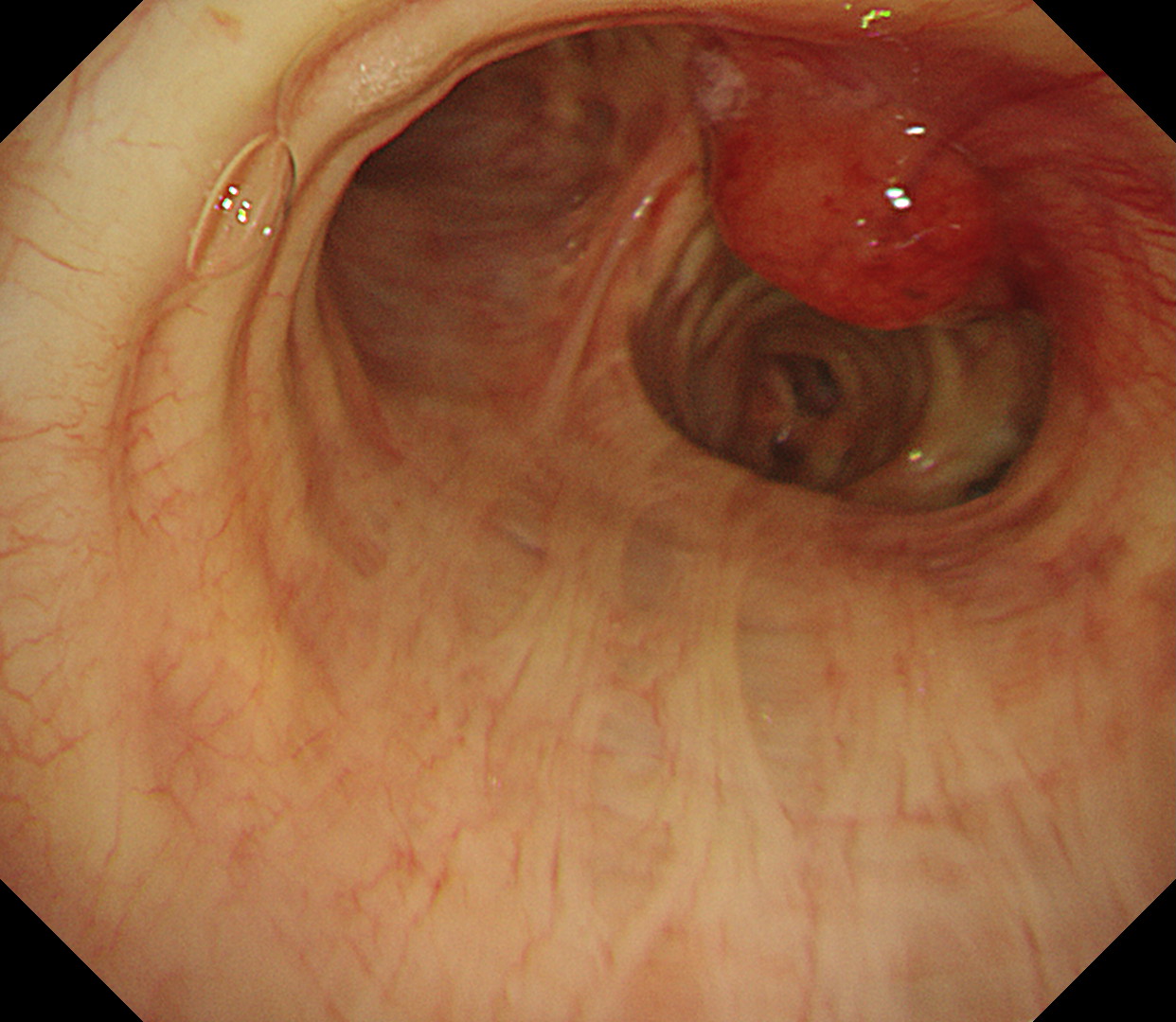
A tumorous lesion with a wide base is recognized near the cartilage of the right main bronchus.
2. Observation from the lower trachea (TXI™ Technology)
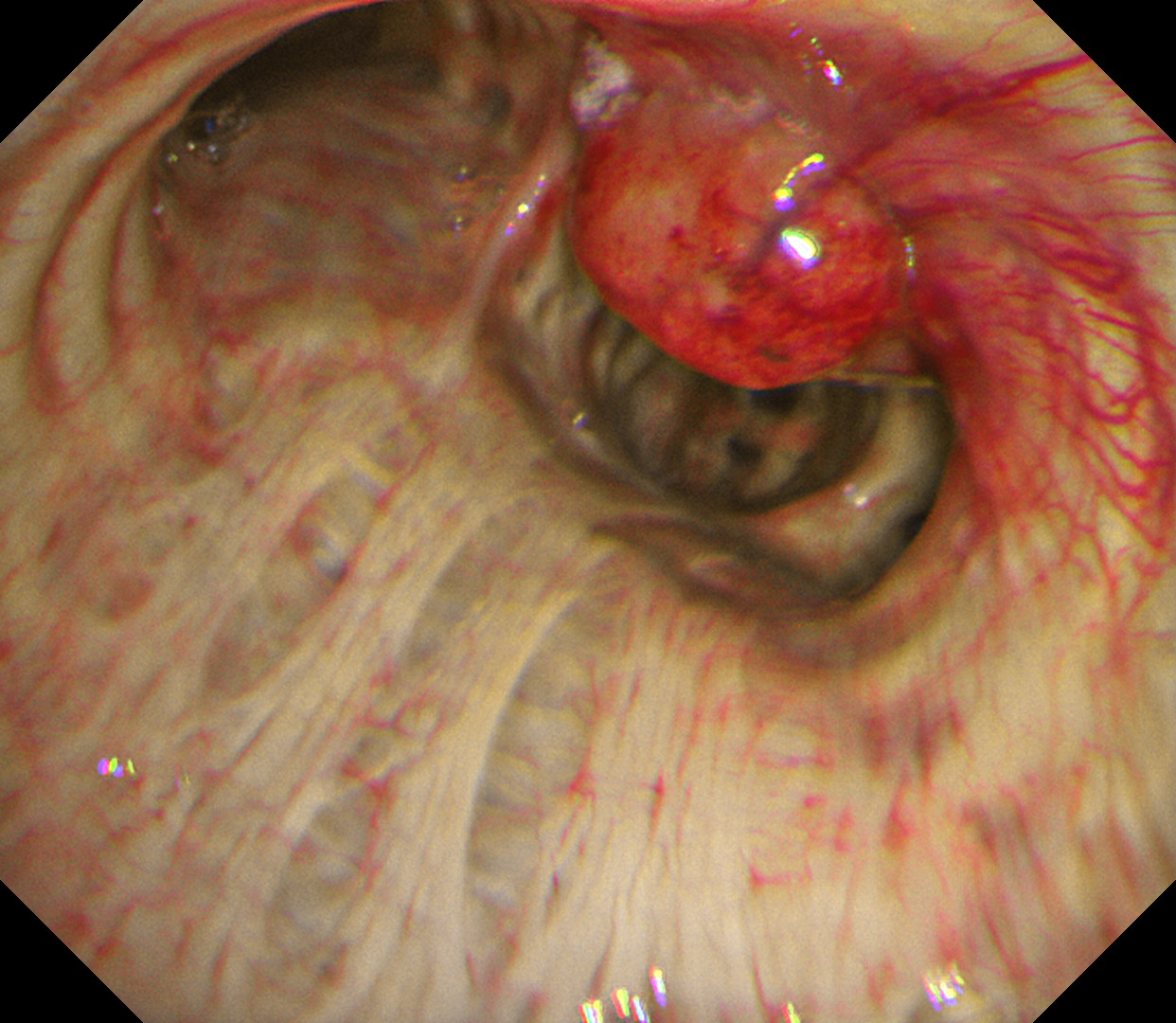
When observed with Texture and Color Enhancement Imaging (TXI™ Technology), vascular properties are enhanced, revealing a network of abnormal vessels on the surface of the tumor as well as longitudinal mural thickening of the tumor base.
3. Observation from the lower trachea (RDI™ Technology)
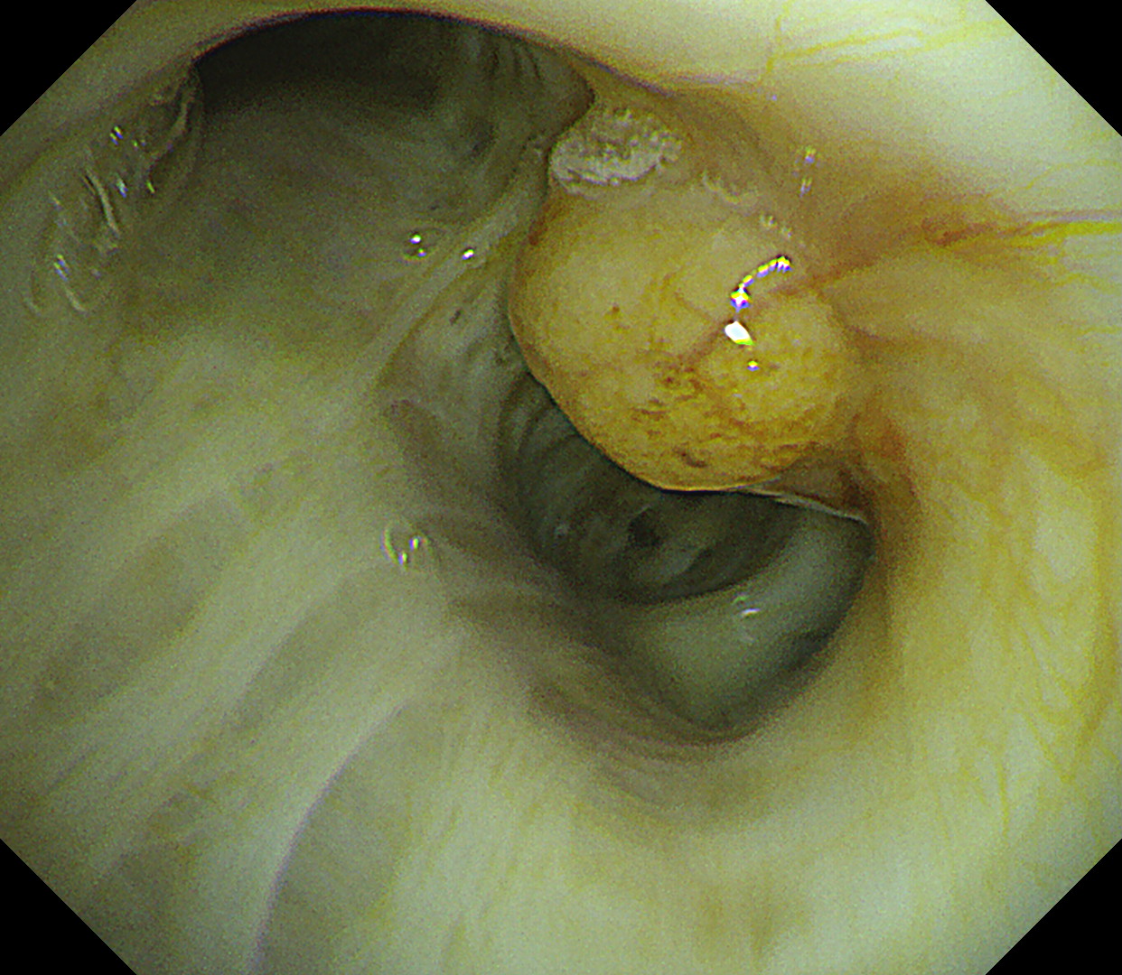
When observed with Red Dichromatic Imaging (RDI™ Technology), no active bleeding is recognized.
4. Observation from the lower trachea (close-up)
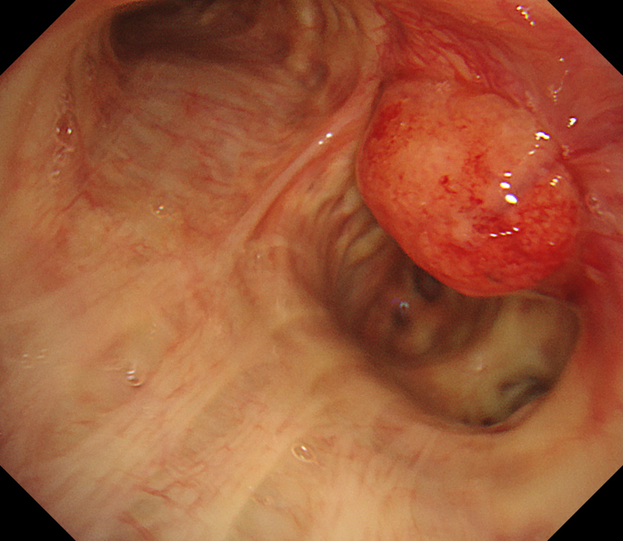
In close-up observation, a tumorous lesion with a wide base is recognized near the cartilage of the right main bronchus.
5. Observation from the lower trachea (close-up)
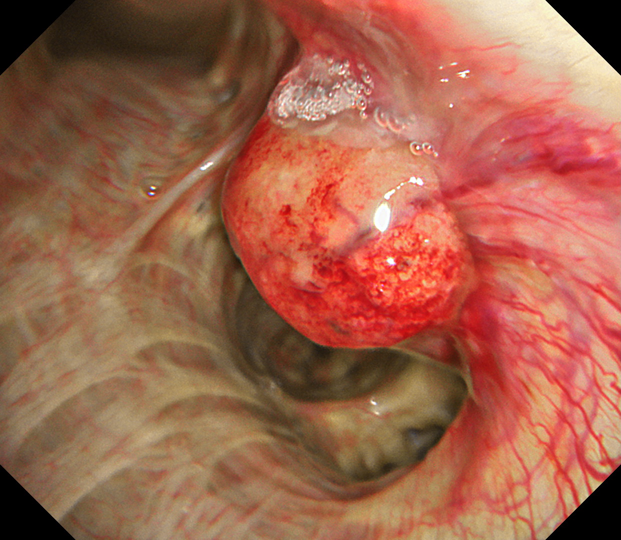
In close-up observation, vascular properties are enhanced, revealing a network of abnormal vessels on the surface of the tumor as well as longitudinal mural thickening of the tumor base.
Case video
In comparison with conventional white light imaging, the TXI™ Technology mode noticeably enhances the properties and distribution of vessels on the surface of the tumor, as well as the characteristics of the longitudinal wall, thereby providing images that facilitate assessment of the degree of infiltration of the tumor into the bronchial wall. This video also shows images of bronchial intervention. It is easy to see the bleeding point of the tumor in the RDI™ Technology mode. The bleeding point is cauterized with argon plasma coagulation for hemostasis.
Pathological Findings
Diagnosis of metastatic lung cancer (breast cancer)
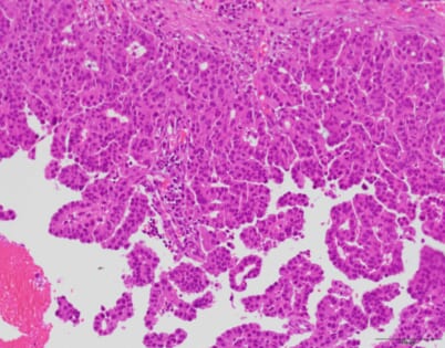
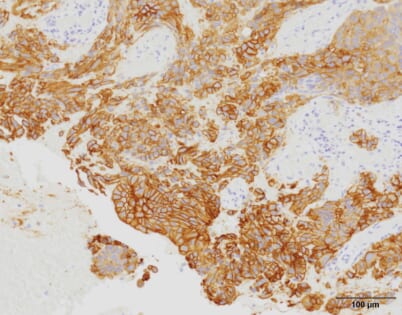
Tumor cells feature a sheet-like architecture with a honeycomb or solid pattern, part of which also shows papillary growth. Some disruption is visible, as well. The tumor cells are HER2 positive, so it is suspected that the breast cancer has metastasized to the lung (bronchial metastasis).
Overall Comment
We performed a bronchoscopy in order to determine an appropriate interventional strategy. While the image quality of white light images was extremely clear, using the TXI™ Technology mode enhanced the vascular properties, highlighting the network of vessels on the tumor surface and the thickening of the longitudinal wall, thereby facilitating comprehension of the characteristics of the lesion and the extent of infiltration. To prevent occlusion of the right main bronchus, we resected the tumor using rigid bronchoscopy. During the procedure, we performed bronchoscopy to observe the lesion and were able to clearly observe active bleeding from the resected site in the RDI™ Technology mode.
Co- author
Department of Internal Medicine, Miyazaki Prefectural Miyazaki Hospital
Dr. Gen-ichi Tanaka, Dr. Ryoichi Matsumoto, Dr. Ritsuya Shiiba
Department of Surgery, Miyazaki Prefectural Miyazaki Hospital
Dr. Kiichiro Beppu, Dr. Seiichi Odate
Department of Anatomic Pathology, Miyazaki Prefectural Miyazaki Hospital
Dr. Kousuke Marutsuka, Dr. Murasaki Aman
* Specifications, design and accessories are subject to change without any notice or obligation on the part of the manufacturer
- Content Type