Fumihiro Asano, MD, PhD
Department of Pulmonary Medicine,
Gifu Prefectural General Medical Center
Disclaimer:
- TXI™ and RDI™ Technologies are not intended to replace histopathological sampling as a means of diagnosis
- The positions and statements made herein by Dr. Asano, are based on Dr. Asano’s experiences, thoughts and opinions. As with any product, results may vary, and the techniques, instruments, and settings can vary from facility to facility. The content hereof should not be considered as a substitute for carefully reading all applicable labeling, including the Instructions for Use. Please thoroughly review the relevant user manual(s) for instructions, risks, warnings, and cautions. Techniques, instruments, and setting can vary from facility to facility. It is the clinician’s decision and responsibility in each clinical situation to decide which products, modes, medications, applications, and settings to use.
- The BF-1TH1200 used in this case is not available in the US market at this time nor is there an established time for its release. The safety and effectiveness of this product and or the use of these products has not yet been established in the United States market.
- The EVIS X1™ endoscopy system is not designed for cardiac applications. Other combinations of equipment may cause ventricular fibrillation or seriously affect the cardiac function of the patient. Improper use of endoscopes may result in patient injury, infection, bleeding, and/or perforation. Complete indications, contraindications, warnings, and cautions are available in the Instructions for Use (IFU)
- Dr Asano, the authoring physician(s) of this presentation, are/ is a paid consultant(s) to Olympus Corporation
Scope: BF-1TH1200
Case:Tumor in the right upper lobe
Location: Right upper lobe bronchus
Patient information: Male, 70 years old
Medical history:Chest CT during his health checkup revealed a mass shadow in the right pulmonary hilum, and he was referred to our institution.
1. Tumor in the right intermediate trunk
(WLI)
2. Tumor in the right intermediate trunk
(TXI™ Technology)
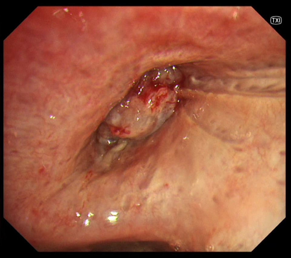
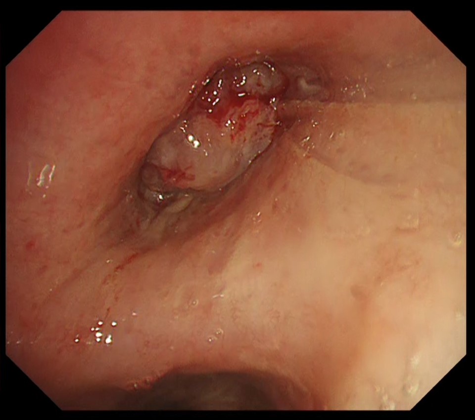
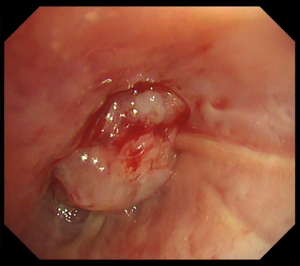
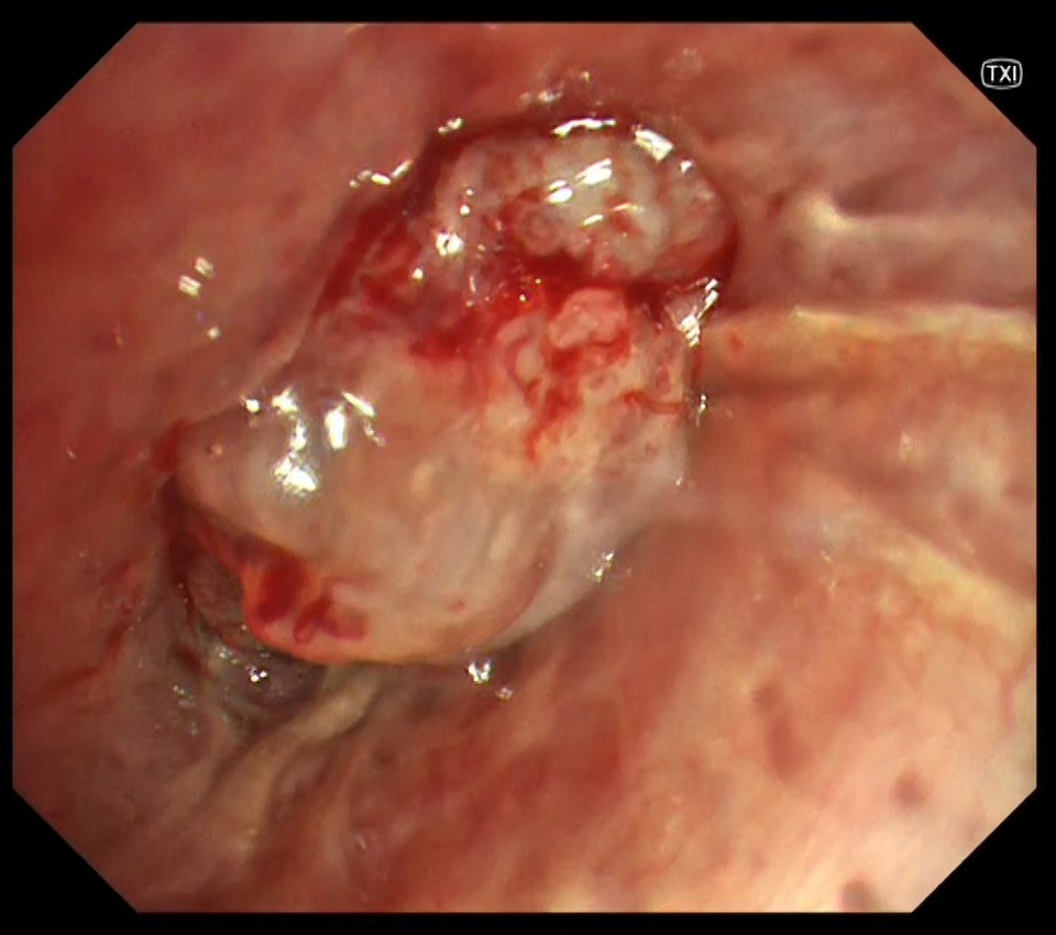
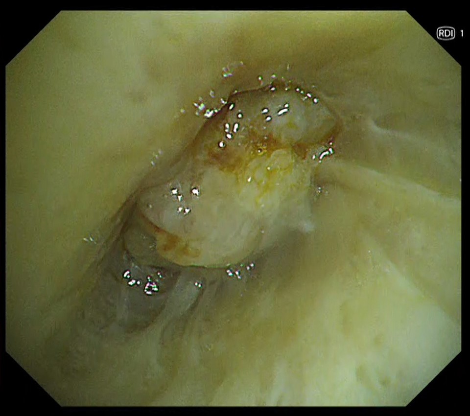
3. Close-up of the tumor in the right upper lobe
(WLI)
4. Close-up of the tumor in the right upper lobe
(TXI™ Technology)
5. Close-up of the tumor in the right upper lobe
(RDI™ Technology)
Case Video
Pathological Findings
- Lung squamous cell carcinoma (diagnosed with TBB of the right upper lobe tumor and EBUS-TBNA of metastasized mediastinal lymph node)
- Proliferation of lung squamous cell carcinoma with a tendency to keratinization is observed.
Overall Comment
This was a case of squamous cell carcinoma of the lung (cT1N2M0 stage, IIIA). Bronchoscopy showed that the advanced peripheral squamous cell carcinoma partially progressed towards the central bronchi in a polypoid fashion. The convergence of longitudinal folds suggested that the upper right lobe bronchus was narrowed in a pointed fashion and the lesion extended around the bronchus. In TXI™ technology, structures such as longitudinal folds and fine vascular networks on the mucosa were more enhanced and could be observed more clearly than with white light.
- Content Type