Tips on Colorectal ESD Techniques
Dr. Tanaka
The preparation should be as good as possible in order to reserve a good field of view during the ESD procedure and to reduce the risk of leakage of fecal fluid into the abdominal cavity in case of perforation.
Dr. Yahagi
We put the patient on a low-residue diet + laxative on the previous day, and administer 2 liters of Niflec* (oral lavage solution)*+ 10 ml of Gascon drops (defoaming agent)* on the day of the treatment.
Dr.Tamegai
Preparation is stricter than for an ordinary colonoscopy. We put the patients on a low-residue diet whenever possible before the treatment, and administer a larger amount of intestinal cleaning solution (Niflec©, etc.) than usual.
Dr.Tanaka
Basically, we do not use sedation for colorectal ESD because the patient has to change position frequently.
Dr.Yahagi
We rarely use sedation for rectal ESD. When the patient feels a strong sense of distension in the deep part of the colon or is experiencing a lot of anxiety, we apply Opystan© (pethidine hydrochloride)* and add diazepam as required.
Dr.Tamegai
We use 10 mg diazepam at the beginning and maintain the sedation by intravenous injection of 35 mg Opystan© by 1/2 ampule. The total amount of Opystan© is variable depending on the procedure time, but we prepare 2 ampules of Opystan© before each procedure.
and what precautions do you have to take for local injections?
Dr.Tanaka
We mix a small amount of indigo carmine with sodium hyaluronate (Mucoup©*) We also use Glyceol© (concentrated glycerin fructose)* when the condition is favourable. We begin local injection with Glyceol© to create a space in the submucosa, and then proceed to local injection of MucoUp©. This aims at avoidance of local injection inside the muscle layer.
Dr.Yahagi
Basically, we use Glyceol© (200 ml) + Bosmin (epinephrine)* (0.2 ml) + indigo carmine (0.2 to 0.4 ml).
The dose of indigo carmine is basically low in order to improve the visual recognition of blood vessels. It is increased when fibrosis is present. With difficult cases, we add a local injection of MucoUp© + indigo carmine after tissue lifting using Glyceol©.
Dr.Tamegai
We generally use a stock solution of Glyceol© or MucoUp© according to the situation. We apply local injection from the mucosal side in the initial stage of incision and dissection, and then inject the solution directly into the submucosa after the dissection has advanced. Care is taken to avoid deep injection because extension of the submucosa would become difficult if the liquid is injected into the proper muscle layer.
1. What do you use if there is bleeding during ESD, and how do you use it?
Dr.Tanaka
Minor bleeding can be stopped by coagulation using the distal end of the knife, but arterial bleeding should be treated with a haemostatic forceps (Soft Coagulation at 50 W). The colon wall is thin and delayed perforation may occur if a wide area of the muscle layer is grasped. As precisely a.s possible, only the blood vessel should be grasped (at a single point). If it is necessary to grasp the muscle layer, try to grasp only the inner circular muscle, and lift the muscle layer slightly toward the centre of the lumen of the intestinal tract before activation.
The most important thing with the colon and rectum is to take care to avoid excessive activation.
* May not be available in your area.
Dr.Yahagi
Provided that the submucosa is sufficiently intact, bleeding from a small blood vessel can be stopped by coagulation using the distal end of the knife. When the bleeding is spurting or the muscle layer is exposed, bleeding should be stopped using haemostatic forceps. When doing this, a scope with a water jet function is very useful
Dr.Tamegai
We treat bleeding at the oozing level with coagulation using the distal end of the Hook Knife, and use the Coagrasper to deal with spurting bleeding.
2. What device do you use to treat the blood vessels at the ulcer floor after ESD, and what is the objective of treatment?
Dr.Tanaka
We use a haemostatic forceps (Soft Coag., 50 W). We also treat the oozing area and the cut the end of the spurting artery using coagulation by grasping with a haemostatic forceps.
Dr.Yahagi
We apply coagulation using the Coagrasper to spurting bleeding or a thicker blood vessel. We apply the current till spurting stops and the tissue becomes white. Care is taken not to burn the tissue too much.
Dr.Tamegai
We treat thick exposed blood vessels using a short clip, and treat other small blood vessels with light coagulation using the Coagrasper or APC. The point of blood vessel treatment is to treat only the visible blood vessels from the ulcer floor, while leaving other vessels alone.
1.What measures do you take to prevent perforation?
Dr.Tanaka
We use sodium hyaluronate (MucoUp) in an injection solution and reposition the patient so that the injection solution can be pooled in the submucosal layer. Also, we take care not to contact the distal end of the Hook Knife with the muscle layer. When dissecting mucosa with the knife, we perform dissection using a gentle stroking technique on the submucosa, taking care not to touch the muscle layer and avoiding pressing the knife. We also take care not to risk ESD in regions where the scope is difficult to manoeuvre.
Dr.Yahagi
After making sure that the tissue is sufficiently lifted, we perform incision and dissection while checking the knife tip under the endoscopic view. Maintaining the field of view and keeping the tissue lifted are critical. Make sure that the knife contacts the tissue gently.
Dr.Tamegai
To avoid operating blindly, we try to incise and dissect only where we can observe endoscopically. Since the lesions with the highest risk of perforation in colorectal ESD are those accompanied with fibrosis in the submucosa, we believe it important to take measures according to the cause, type and severity of fibrosis.
(Also see Q6 on Page 15.)
2.What kinds of countermeasures do you take if you end up with a perforation?
Dr.Tanaka
We treat small perforations with short clips. If the perforation is large, we also use a long clip sometimes.
Dr.Yahagi
Our first choice is a short clip (HX-610-090S), but we select the clip size and jaw angle according to the perforation size. When we find a perforation, we immediately clip it to prevent leakage of fecal fluid into the abdominal cavity. When releasing the clip, we exhaust air and close it gently (in order not to tear the muscle layer).
Dr.Tamegai
I have only experienced a small perforation, so I closed it with a clip. However, if the perforation is large, I believe it would be ideal to perform clipping in the same way as after EMR, that is, attaching indwelling clips to the edge of the perforation to make a spindle shape, then suture it by attaching clips from the easiest-to-clip position on the edge toward the centre. The clips we would use for this purpose are the HX-610-135 and HX-610-090L.
3. What kinds of countermeasures do you take if closure using cUps is difficult?
Dr.Tanaka
With a very small perforation, we exhaust air as much as possible, put the patient in a position so that the fecal fluid will not contact the perforation area, and administer an antibiotic. However, since perforative peritonitis is unavoidable if a perforation cannot be closed, it is important to prepare for emergency surgery by holding close communication with a surgeon. Perforation in a region without the serosa presents a high
risk of formation of abscess on the retroperitonium or around the rectum.
Dr.Yahagi
We perform closure using an indwelling snare + clips if possible. If this is difficult, we perform surgery.
Dr.Tamegai
I haven’t had this problem so I cannot be specific, but I believe that a possible endoscopic approach would be to draw the greater omenta or soft tissues with negative pressure and close the opening with clips.
However, as there is no established theory on what countermeasure to take when closure is difficult, so it may be appropriate to consider emergency surgery as the first choice.
4.What kind of equipment should we have on hand for use in the ease of perforation?
Dr.Tanaka
Clips for use in closure of perforations are indispensable. In addition, if a CO2 insufflator is available, it is possible to reduce the intestinal pressure and therefore reduce the risk of leakage of fecal fluid into the abdominal cavity.
Dr.Yahagi
A set of clips, a CO2 insufflator and antibiotic are necessary.
Dr.Tamegai
An abdominal ultrasound system should also be available in case deaeration is required.
5.What precautions do you take in postprocedural management of patients who have suffered a perforation?
Dr.Tanaka
We manage the respiration and circulation dynamics in order not to overlook the abdominal observations (symptoms of peritonitis). It is also necessary not to miss the timing of emergency surgery in order to avoid hazardous consequences such as sepsis. Special care is required with elderly patients because they do not always present inflammation reactions or leukocytosis.
Dr.Yahagi
Abstinence from food and drink is necessary. Fever can be reduced with continuous drip infusion and antibiotics. The patient should be closely observed until the inflammation reaction begins to decrease. Surgery should also be considered in the case of fever above 39°C, abdominal pain, or muscular defense.
Dr.Tamegai
It is important to determine whether or not immediate emergency surgery is necessary based on the position and degree of perforation, the degree of contamination. and the effectiveness of clip closure. Consultation with the surgical team and measures in accordance with the risk management regulations are also necessary. If conservative management is possible, special attention should be paid to the fluctuation of vitals, the presence of signs such as muscular defense in abdominal observation, the presence of an abscess in CT or echo
observation, and any changes in inflammation observed in peripheral blood and biochemical examinations.
Dr.Tanaka
To make it easier to extend the fibrotic area of the submucosa, we start the incision and advance dissection from outside the fibrotic area. To improve visibility of the submucosa we inject it with indigo carmine. Dissection should be performed precisely, cautiously and gently using the Hook Knife. Do not risk proceeding in a situation where the submucosa cannot be recognised visually because advanced fibrosis is mixed into the muscle layer. In this case, you may have to consider stopping the procedure.
Dr.Yahagi
Stay away from such a lesion if you are a beginner. Apply sufficient local injection, begin dissection from a point that’s away from the fibrotic area, and enter the submucosa together with a distal attachment to ensure the direct endoscopic view of the scar area. Inject a sufficient amount of sodium hyaluronate around the scarred area to lift up the lesion as much as possible.
Dr.Tamegai
Submucosal fibrosis can be categorised into two types: 1) non-cancerous fibrosis with unknown cause, which is associated with treatment procedures such as local injection, biopsy and EMR as well as with inflammation and intestinal peristalsis; and, 2) fibrosis accompanying SM invasion of cancer. Non-cancerous fibrosis presents in endoscopic observation as a trabecular white tone with light fibrosis, a cingulated white tone with medium fibrosis, and a screen-type overall view with advanced fibrosis. Fibrosis accompanying cancer invasion appears white to brown, which may be the invading cancer cells, and has a large number of abnormal blood vessels. We applied ESD to 28 cases involving submucosal fibrosis, performing en-bloc resection on 21 cases and piecemeal
resection on 5 cases. Resection in the last 2 cases was stopped before completion. In the cases in which ESD was Dr. Tl.mtgal completed, the factors that enabled resection were: I} the injection solution succeeded in infiltrating the fibrous tissue stroma; 2) the dissection line could be set because the proper muscle layer position was identified from the normal or loose area around the fibrosis; and, 3) the distal end of the Hook Knife was able to enter the fibrotic tissue. On the other band, we found that advanced screen-type fibrosis and advanced cancer invasion cases cannot be used as standard indications because local injection is not possible and there is no space for the Hook Knife to enter. Submucosal fibrosis can only be dissected when the boundary between the proper muscle layer
and the submucosal layer can be identified and there is space for the knife to enter. In the operative field of view, it should be possible to identify the incision line by observing the fibrotic section and the lines of the proper muscle layer and the submucosal layer on both sides of the fibrotic section.
Usefulness of Carbon Dioxide (C02) Yutaka Saito, National Cancer Center Hospital
Carbon dioxide (CO2) is absorbed in the intes1inaltract more quickly than air, and reports from Europe and North America have demonstrated its safety and usefulness in reduction of patient discomfort. In addition, while insufflation of normal air sometimes produces feeling of distension and discomfort for about haH a day after completion of ltte procedure, CO2 gas reduces postprocedural abdominal discomfort.
Usefulness and safety of CO2 insufflation in colorectal ESD
ESD sometimes lasts for many hours and patient discomfort due to excessive Insufflation presents problems. We conducted colorectal ESD treatment with ordinary air Insufflation and 002 Insufflation, compared ltte results of two groups, and recognised a significant decrease In the usage amount of the sedative agent In the CO2 group, without observing a r1se In the blood CO2 concentration. Later. we also measured the change In Pte CO2 value over time, and found that the average peak value was 56 mmHg. The rise over 60 mmHg Is said to Induce arrhythmia, but there were only a few cases and they were only transient. Although the risk of causing CO2 narcosis is extremely low in colorectal examination under conscious sedation, the use of CO2 should be avoided In patients with chronic obstructive pulmonary disease and ser1ous heart disease. At present, we are able to safely conduct colorectal ESD under CO2 Insufflation using only oxygen concentration and electrocardiogram monitoring, except for patients with contraindications.
Reducing the Risk of accidental symptoms in colorectal ESD
Even in the case of perforation, CO2 can be absorbed quickly so the risk of subcutaneous emphysema can be prevented or reduced. CO2 may also be useful In prevention of the abdominal compartment syndrome due to Insufflation. In addition, while air embolisms have been reported as an occasional complication In endoscopic examinations, CO2’s rapid absorption can be expected to reduce the risk of this complication.
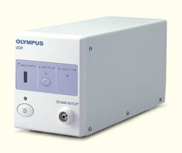
Usefulness of the UCR
The Olympus UCR Is capable of supplying stable CO2 flow without complicated setup. Even if the operator makes a mistake in valve opening or closing, the UCR prevents rapid flow of CO2 into the Intestinal tract. Although It is more expensive than other gas regulators, it is very useful from the viewpoint of risk management.
- Content Type