Advancing the Dimensions of Endosonography
Improved Ultrasound Imaging
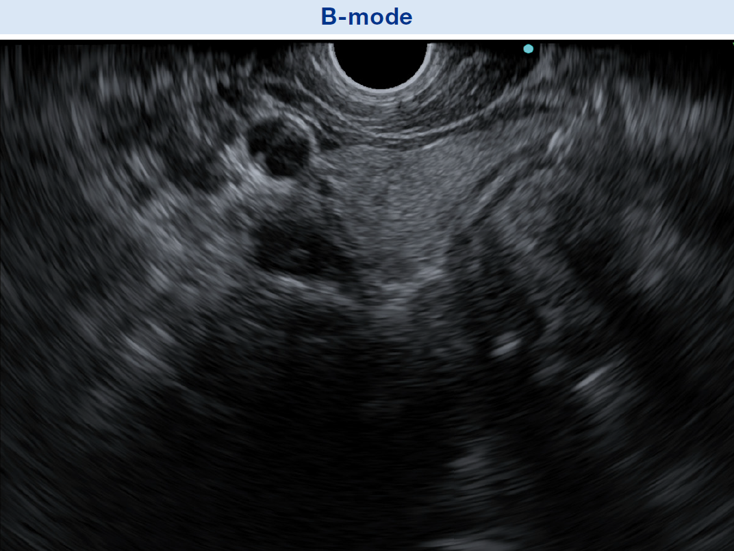
Enhanced B-mode
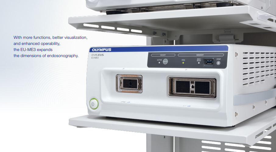
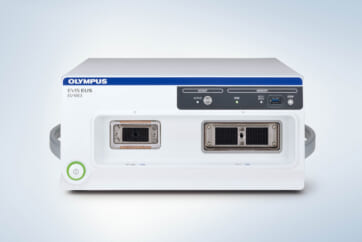
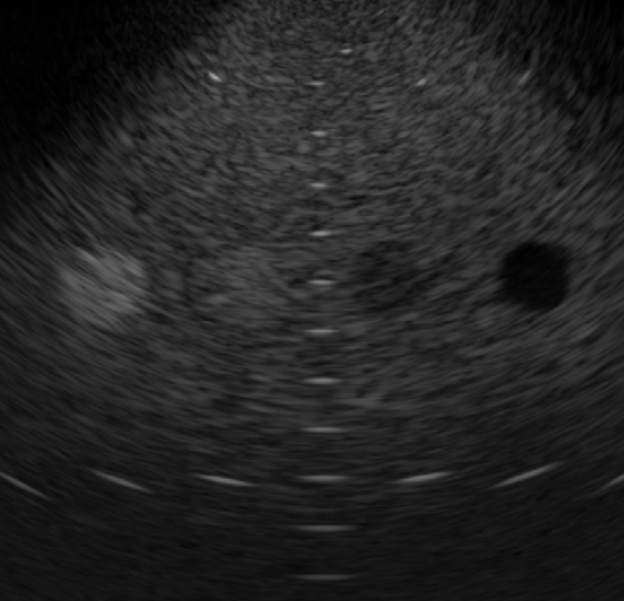
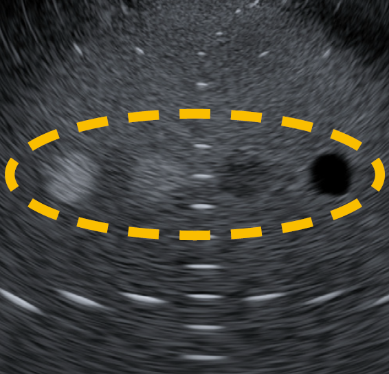
The EU-ME3 provides outstanding image quality and functionality – compatible to a high-end ultrasound center – in a compact body. B-mode image quality has been substantially enhanced compared to our conventional processor (EU-ME2).

Improved Ultrasound Imaging
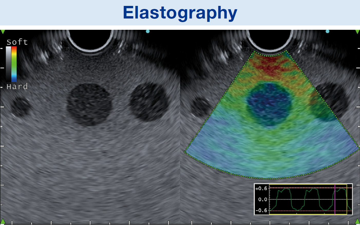
Improved Elastography
The EU-ME3 features an elastography function which visualizes the amount of strain in the tissue (tissue stiffness) during compression and retraction, making it possible to obtain more information about tissue properties.
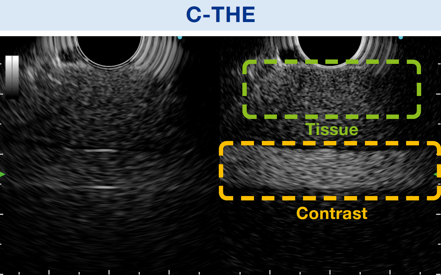
Contrast Harmonic Echo (CHE)
Contrast Harmonic Echo (CHE) images harmonic components from ultrasound contrast agents. The newly added C-THE mode images signals from biological tissue and the contrast.
Tissue Harmonic Echo (THE)
When ultrasound waves are propagated through tissue, distortion is produced and harmonic components are generated. The Tissue Harmonic Echo (THE) mode uses these components to build an image of the targeted area, providing a more detailed granular depiction. Advantages of harmonic imaging include improved resolution, improved signal-to-noise ratio, and fewer artifacts.
Improved Ultrasound Imaging
Doppler Modes
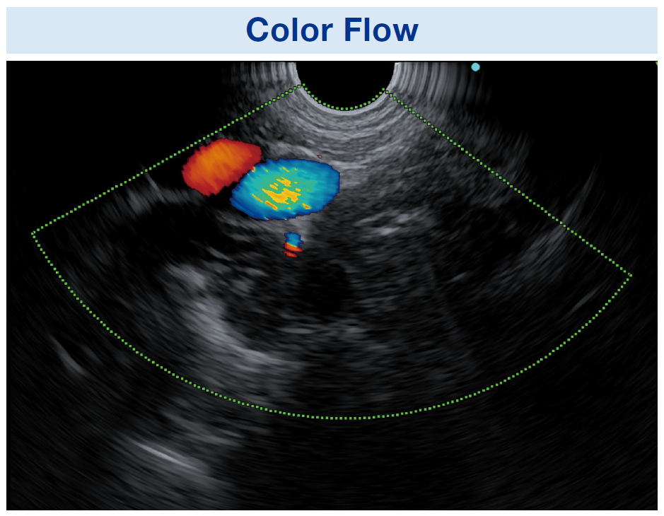
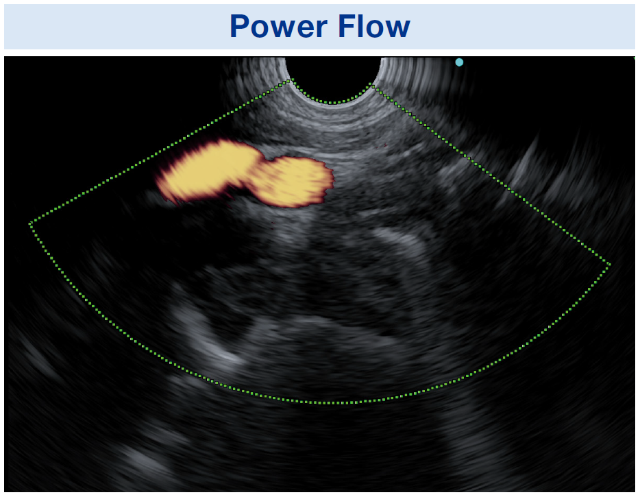
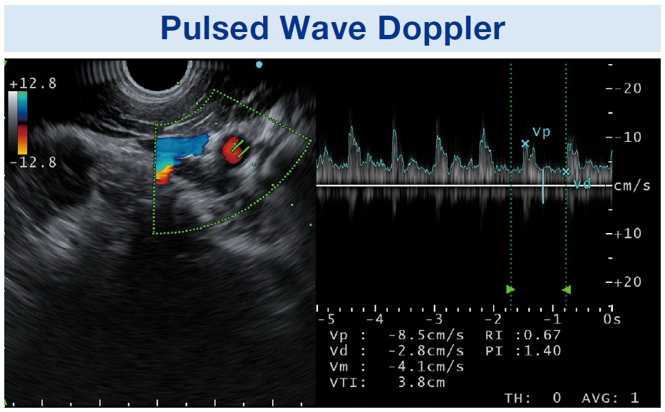
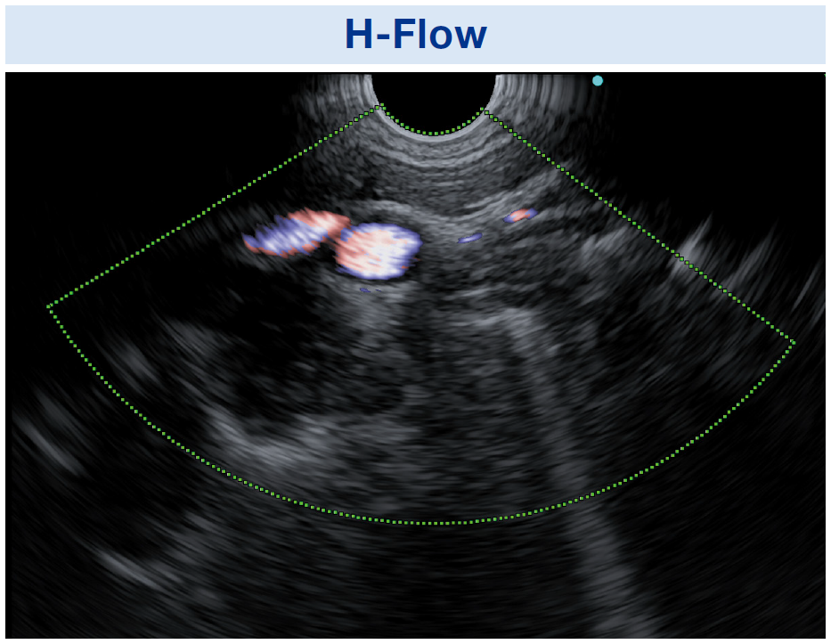
The EU-ME3 offers three basic Doppler modes to distinguish blood flow more clearly – Color Flow, Power Flow, and Pulsed Wave Doppler (PWD). Doppler modes can be used to support safer procedures, benefitting both the patient and the physician. In addition to the three basic Doppler modes, the EU-ME3 also features H-Flow. H-Flow is a more sensitive Doppler mode that shows directional blood flow with less blooming. It is especially useful for imaging small vessels around the tip of the echoendoscope.
Designed for Enhanced Usability
Shear Wave Quantification (SWQ)
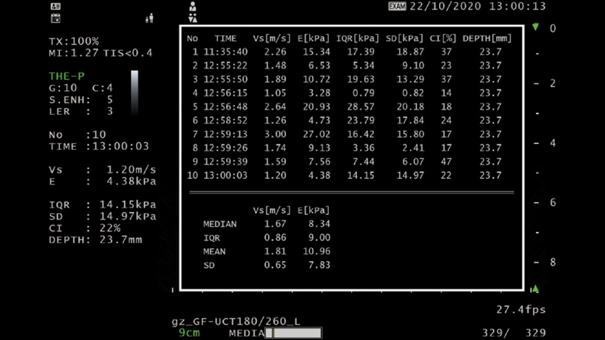
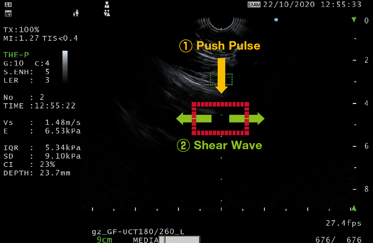
SWQ provides an absolute value of tissue stiffness within a region of interest. It performs this quantitative tissue assessment by calculating the propagation velocity of shear waves, generated from a push-pulse.
Elastography (i-ELST)
i-ELST is a new technology incorporated into the EU-ME3 that makes it easier to display elastic images, even when displacement due to pulsation is modest.
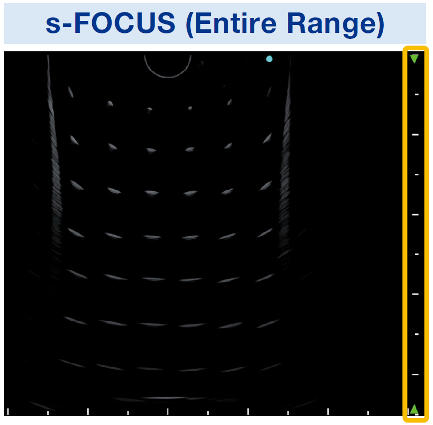
s-FOCUS
The EU-ME3 is equipped with an s-FOCUS mode that reduces the change in resolution with distance from the ultrasound transducer surface. s-FOCUS eliminates the need to manually adjust the focal zones during the procedure.
Designed for Enhanced Usability
Keyboard Usability
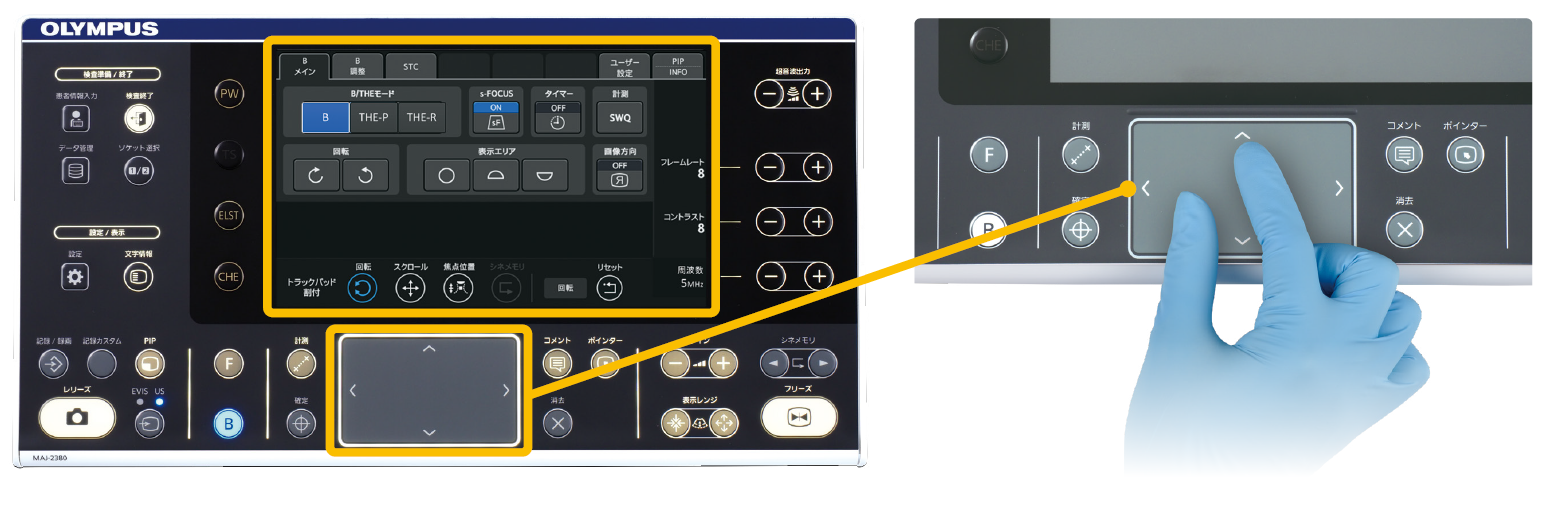
The keyboard was designed with a simple layout in mind and includes a user-friendly built-in touch panel, LED backlit keys and a trackpad for ease of use and cleaning. The larger LCD touch panel allows for a greater range of functions to be displayed at one time.
Ease of Targeting
The position and size of the Doppler region of interest (ROI) can be conveniently adjusted with a trackpad or buttons on the touch panel.
Designed for Enhanced Usability
Wide Range of Compatibility
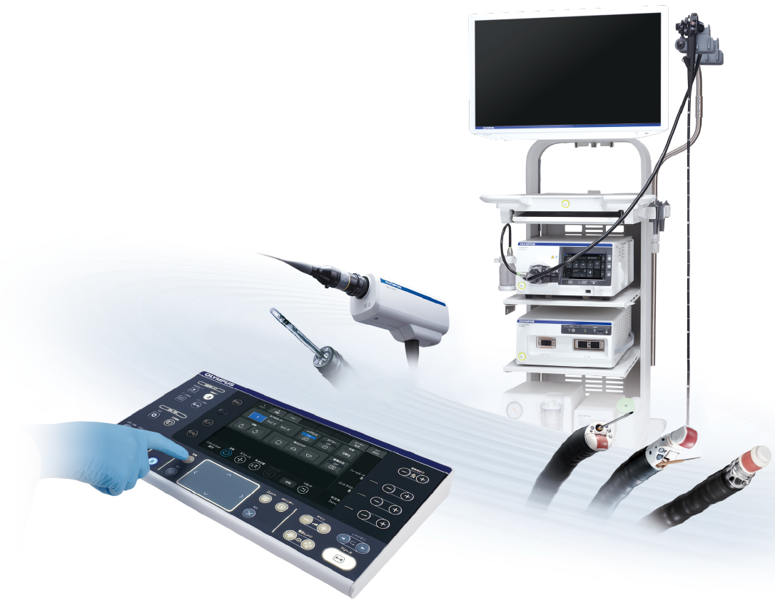
Integrating both electronic and mechanical scanning technologies, the EUME3 is compatible with echoendoscopes and miniature probes, creating a total endosonography solution for a full range of applications.
Customizable Features
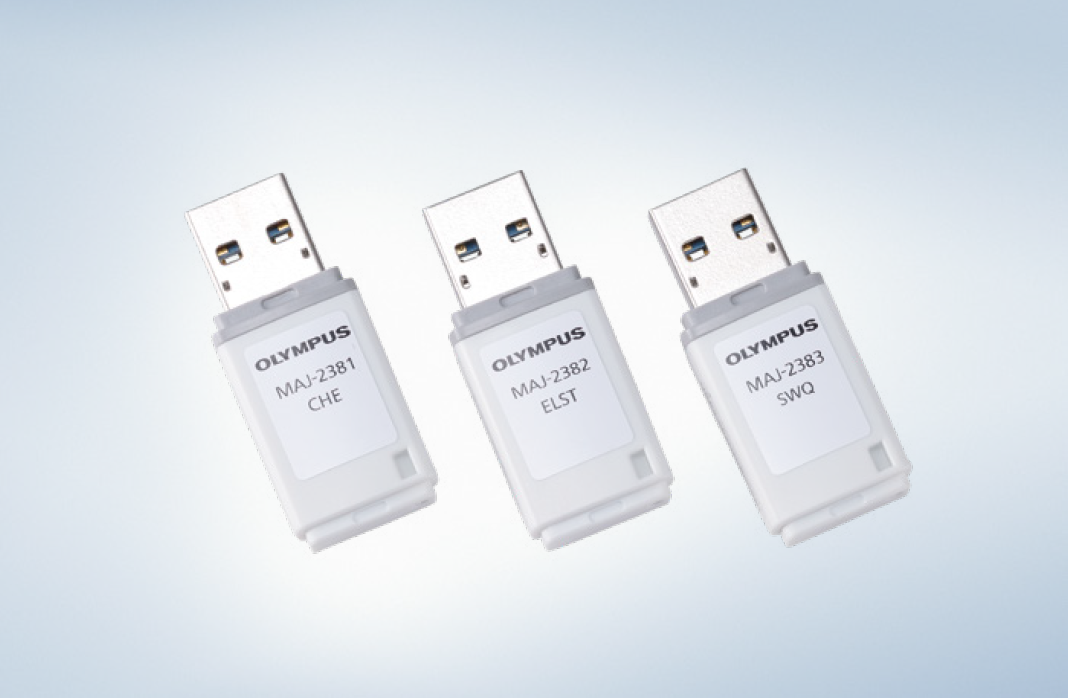
Software options are available to meet the needs of any facility. Because the functions are optional, you can select and add the necessary functions according to your needs and budget.
- Content Type

