Dr. Thawee Ratanachu-ek
RAJAVITHI HOSPITAL, Thailand
What are your initial thoughts after using EU-ME3?
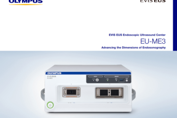
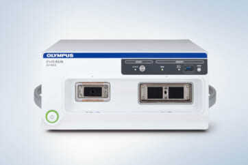
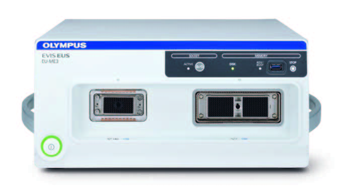
In the market, there are two types of ultrasound processor for endoscopic ultrasound. The first is large standalone type ultrasound processor separated from the endoscopy trolley. The second is a compact box-type ultrasound processor that can be incorporated within the endoscopy trolley (One cart system). Generally, the first one provides better image quality when comparing with the second. Surprisingly, the new Olympus ultrasound processor, namely EU-ME3 provides high quality ultrasound image which is not inferior to the standalone type ultrasound processor.
What do you think is the benefit of B-mode image of EU-ME3?
I have the opportunity to do several cases with EU-ME3.
Case number 1
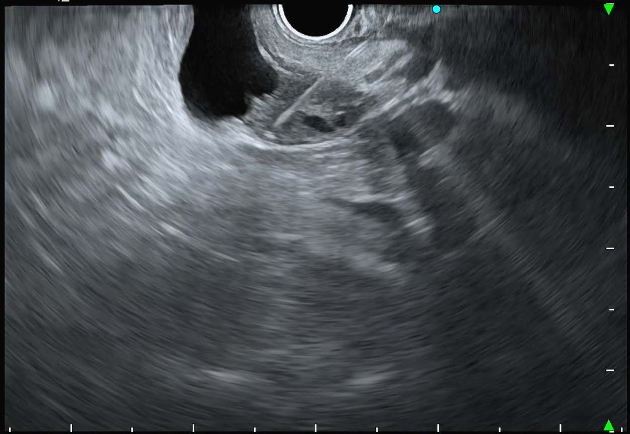
72-year-old male patient with asymptomatic pancreatic lesion detected by trans-abdominal ultrasound and solid mass on CT. EUS with EU-ME3 found solid cystic mass (cystic component 13×12 mm and solid component 17×13 mm) and fine needle biopsy was performed. The pathology diagnosis confirmed neuroendocrine cell proliferation.
Case number 2
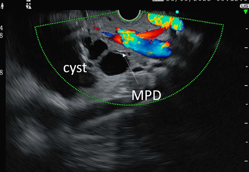
36-year-old female patient present with abdominal discomfort and transabdominal ultrasound and MRI/MRCP found 4.3 cm pancreatic cyst at head of pancreas with suspected connection to main pancreatic duct. EUS with EU-ME3 was performed and found 4.2 cm non enhanced multiloculated with microcystic cyst without clearly seen pancreatic duct connection. Straw color fluid was obtained from 22-gauge fine needle aspiration and subsequent fluid analysis found low CEA and low amylase.
Serous cystadenoma neoplasm (SCN) was diagnosis.
EU-ME3 provides homogeneous smooth high quality scanning ultrasound images on B-mode without any artifact and interference in default tissue harmonic setting, and new full range focus function (s-FOCUS) avoiding manually adjust which clearly defines both solid and cystic abnormality from normal structure in all deep level of images. With high resolution B-mode, small calcifications, ductal system and vessels are also clearly identified.
How do you think about functionality and versatility of EU-ME3?
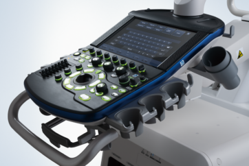
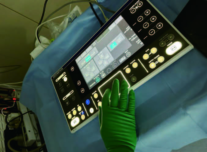
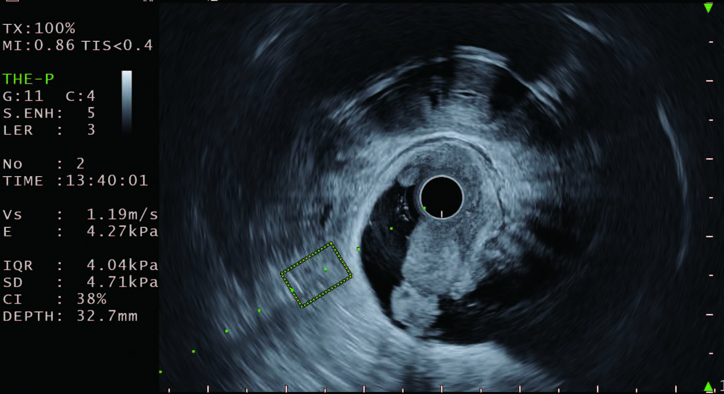
There are standard functions such as color flow, power flow, pulsed wave doppler, tissue elastography, contrast harmonic echo and several new innovations including the new elastography, shear wave measurement namely “Shear Wave Quantification (SWQ)” and user-friendly trackpad that provides smooth and precise targeting and allows swift review of ultrasound images.
Furthermore, the larger LCD touch screen panel, and additional functions such as allocation of FLOW and US record start/stop on scope switches in combination with EVIS X1 system makes it easy for a seamless workflow.

How does new system improve clinical practice?
In my cases, I found the EU-ME3 endoscopic ultrasound center is a user-friendly EUS system. It provides high resolution B-mode images integrated with several additional functions with clinical benefits which, as I already mentioned, makes EUS practices more valuable. Because of well visualized FNA needle, it is easy to perform therapeutic interventional procedures to acquire maximum sample yields. This one trolley compact EUS processor allows more space in endoscopy room to perform interventional procedures combined with several devices. In my view, EU-ME3 is the latest advanced technology worth to have in your centers providing EUS services.
- Content Type