Masayuki Kitano MD, PhD
Wakayama Medical University Hospital
Japan
D-THI
Differential Tissue Harmonic Imaging (D-THI) delivers both high resolution and deep penetration, enabling consistent image quality from superficial to deep regions.
By utilizing dual-frequency transmission, it expands the effective bandwidth compared to conventional THI, improving visualization of deeper structures while maintaining excellent resolution.
This contributes to clearer lesion delineation and enhances overall diagnostic confidence
- Frequency (Routine): 8.5 MHz (standard for routine exams)
- Penetration mode: 7.5 MHz (for improved deep penetration)
- Resolution mode: 11 MHz (for high resolution superficial imaging)
- Operational tip: switch to D7.5 for deep structures and D11 for fine superficial detail
Case 1 D-THI Normal Pancreas
D-THI mode enables clear visualization of the pancreatic head from transgastric view , even up to the region near Major Papillar, which is traditionally difficult to assess with conventional imaging.
Case 2 D-THI Chronic Pancreatitis
In cases such as chronic pancreatitis with strong attenuation, D-THI mode clearly demonstrates key pathological features including echogenic stranding, lobularity, and continuous visualization of the pancreatic duct
SMI
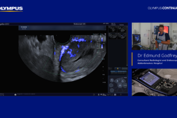
Superb Micro-vascular Imaging(SMI) enables highly sensitive visualization of microvascular flow, supporting more precise lesion assessment.
By reducing noise and suppressing motion artifacts, SMI preserves true low-velocity blood flow signals that are often difficult to detect with conventional Doppler techniques.
With mSMI (monochrome mode) highlighting flow signals and cSMI (color-coded) overlaying them on B-mode images, it provides both detailed vascular information and anatomical context, improving diagnostic accuracy.
-
ROI placement: position over target using the trackball
-
Frequency: 7.5 MHz (routine evaluation)
-
Display option: use mSMI to suppress B mode background and emphasize flow when needed
-
Operational tip: use SMI to detect low velocity microvascular flow; switch from Advanced Dynamic Flow (ADF) to SMI to expand vascular detail
Case 1 SMI Neuroendocrine Tumor
SMI (mSMI) reveals very low velocity intratumoral flow that ADF cannot, suppressing background and highlighting microvascularity for superior visualization.
Case 2 SMI Gastric GIST
SMI detects primary blood flows and branched, irregular, vascular structures not visible on ADF; identification of these irregular flows aided the GIST diagnosis.
Case 3 SMI Neuroendocrine Tumor Small Lesion
High resolution B mode identifies a ~13 mm hypoechoic lesion; switching to SMI shows blood flow signals entering the tumor, supporting the high vascularity characteristic of PNET.
Case 4 SMI Ethanol Injection
SMI visualizes needle and injected ethanol inflow in real time. SMI’s flow emphasis confirms injection that grayscale alone may not clearly show.
SWE
Shear Wave Elastography (SWE) allows both quantitative and visual assessment of tissue stiffness across the entire region of interest.
Unlike conventional point-based measurement, SWE provides stiffness values along with color mapping, enabling a more comprehensive evaluation.
In addition, the Propagation Map enhances measurement reliability by visualizing shear wave behavior, leading to more consistent and reproducible assessments.
-
ROI placement: position ROI over tissue to be measured using the trackball
-
ARFI push: apply ARFI push pulse after ROI is set to generate shear waves
-
ROI size: adjust by application (example: 5 mm diameter for pancreatic parenchyma
Case 1 SWE GIST
SWE using Aplio i800 EUS enables reliable stiffness measurement even in gastric subepithelial tumors, which are traditionally considered difficult to assess with shear wave measurement.
Case 2 SWE Early Chronic Pancreatitis
SWE provides quantitative assessment of pancreatic tissue stiffness, enabling detection of increased stiffness suggestive of mild or chronic pancreatitis.
- Content Type