Disclaimer
The techniques and clinical opinions presented in this material reflect the personal experience and professional judgment of the healthcare professional and do not necessarily represent the views of Olympus. This material is intended for healthcare professionals only. Users should always refer to the applicable Instructions for Use (IFU) and use Olympus products in accordance with the approved indications and local regulatory requirements. The healthcare professional presenting this material has been engaged by Olympus and compensated at fair market value for their services.
Colorectal Case 23

Prof. Yunho Jung
Department of Internal Medicine, Division of Gastroenterology,
Soonchunhyang University College of Medicine,
Cheonan, Republic of Korea
Scope: CF-EZ1500DI
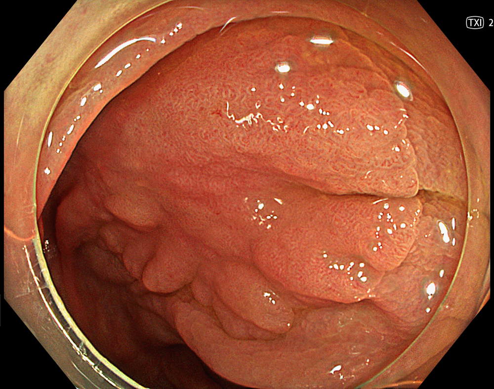
Case: A 30-mm non-granular laterally spreading tumor (LST-NG-FE) located at the hepatic flexure.
Organ: Colon
Patient Information: M, 80s
Medical History: No significant medical history
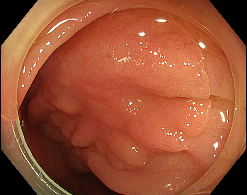
1. LST-NG in WLI
Enhancement : A8
NBI Mode : NA
TXI Mode : NA
RDI Mode : NA
BAI-MAC : On
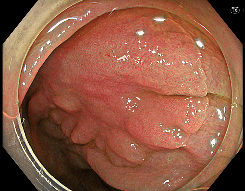
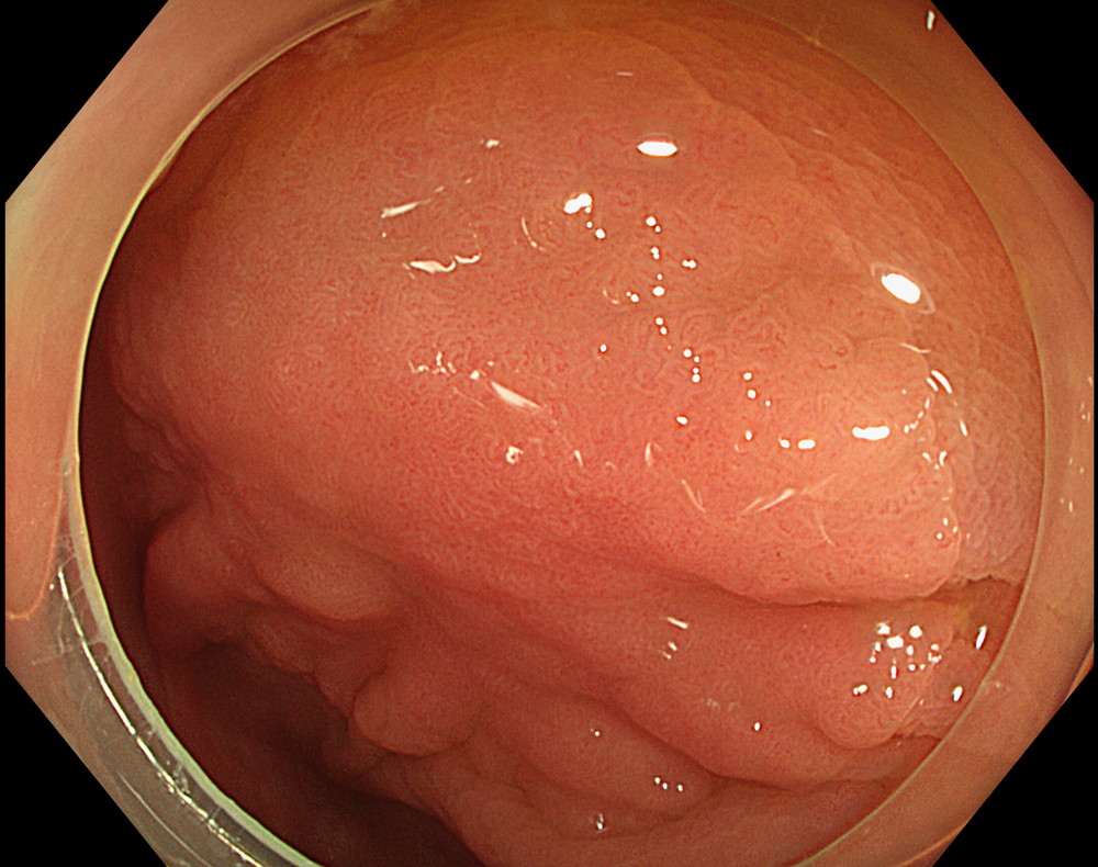
2. LST-NG with TXI (Mode 1)
Enhancement : A8
NBI Mode : NA
TXI Mode : Mode1
RDI Mode : NA
BAI-MAC : On
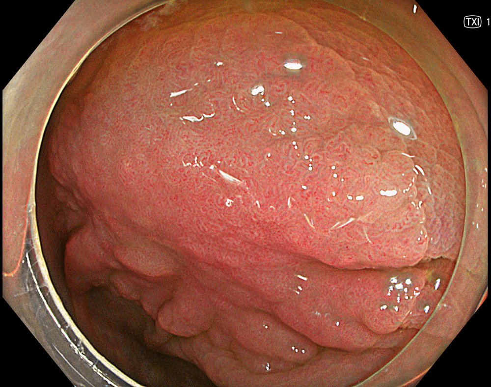
3. LST-NG with TXI (Mode 2)
Enhancement : A8
NBI Mode : NA
TXI Mode : Mode2
RDI Mode : NA
BAI-MAC : On
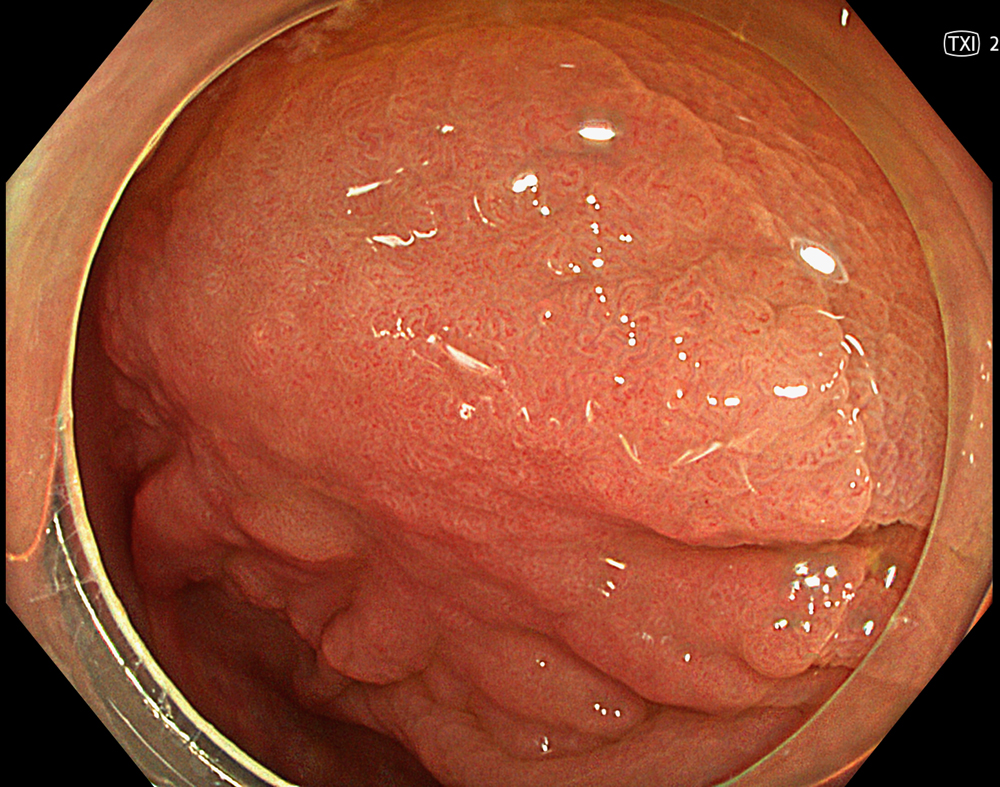
4. LST-NG in WLI
Enhancement : A8
NBI Mode : NA
TXI Mode : NA
RDI Mode : NA
BAI-MAC : On
5. LST-NG with TXI (Mode 1)
Enhancement : A8
NBI Mode : NA
TXI Mode : Mode1
RDI Mode : NA
BAI-MAC : On
6. LST-NG with TXI (Mode 2)
Enhancement : A8
NBI Mode : NA
TXI Mode : Mode2
RDI Mode : NA
BAI-MAC : On
7. LST-NG in NBI
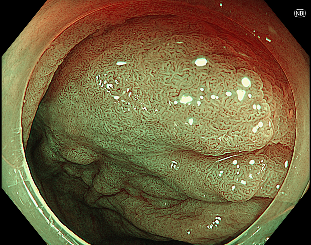
Enhancement : A8
NBI Mode : On
TXI Mode : NA
RDI Mode : NA
BAI-MAC : On
8. NBI with magnification (Near focus)
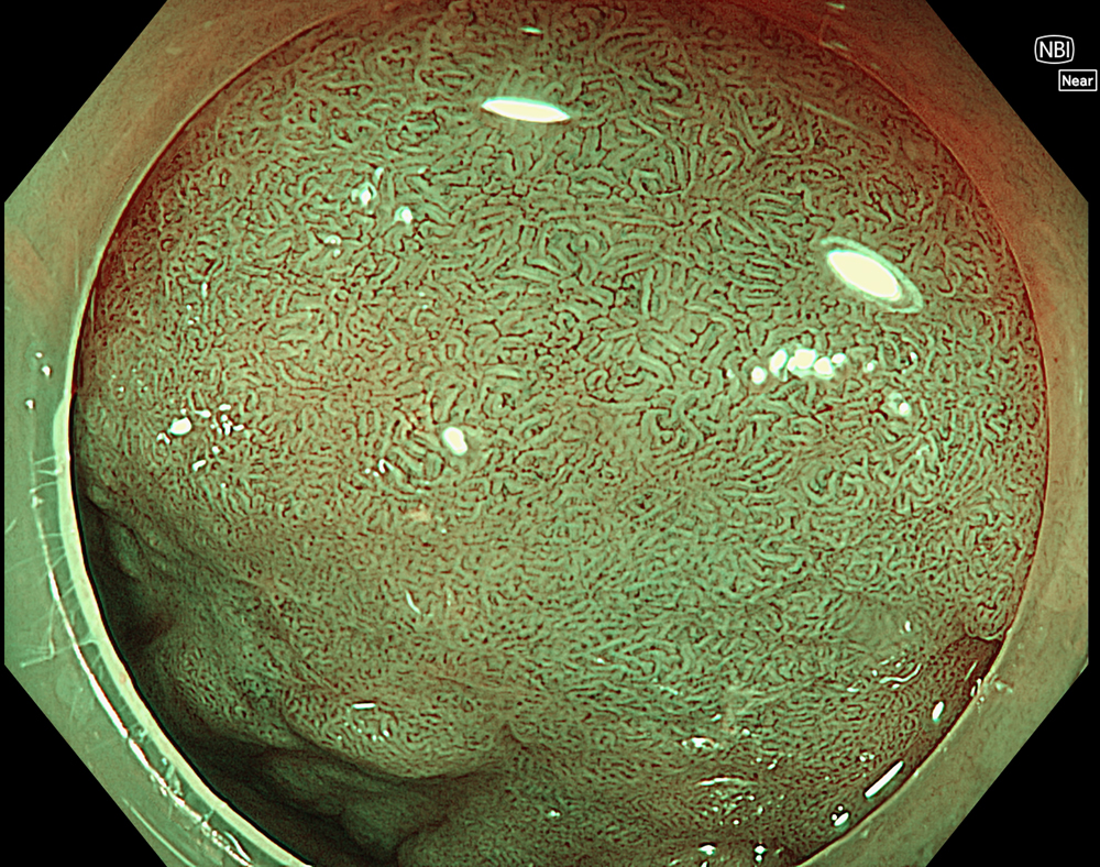
Enhancement : A8
NBI Mode : On (Near focus)
TXI Mode : NA
RDI Mode : NA
BAI-MAC : On
9. Bleeding in WLI
Enhancement : A8
NBI Mode : NA
TXI Mode : NA
RDI Mode : Off
BAI-MAC : On
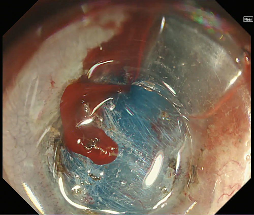
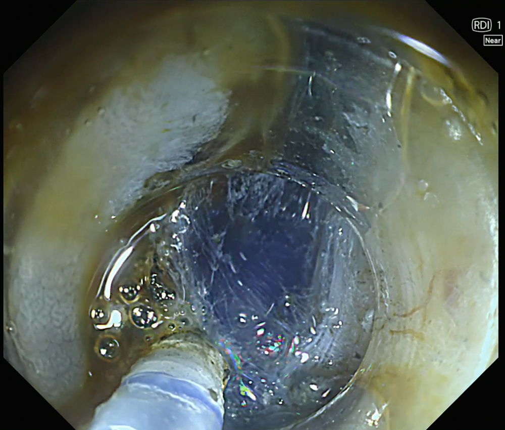
10. Bleeding in RDI Mode 1
Enhancement : A8
NBI Mode : NA
TXI Mode : NA
RDI Mode : Mode 1
BAI-MAC : On
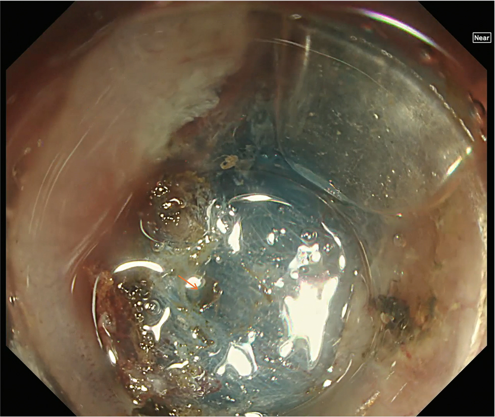
11. Hemostasis in RDI Mode 1
Enhancement : A8
NBI Mode : NA
TXI Mode : NA
RDI Mode : Mode 1
BAI-MAC : On
12. Hemostasis confirmed in WLI
Enhancement : A8
NBI Mode : NA
TXI Mode : NA
RDI Mode : Off
BAI-MAC : On
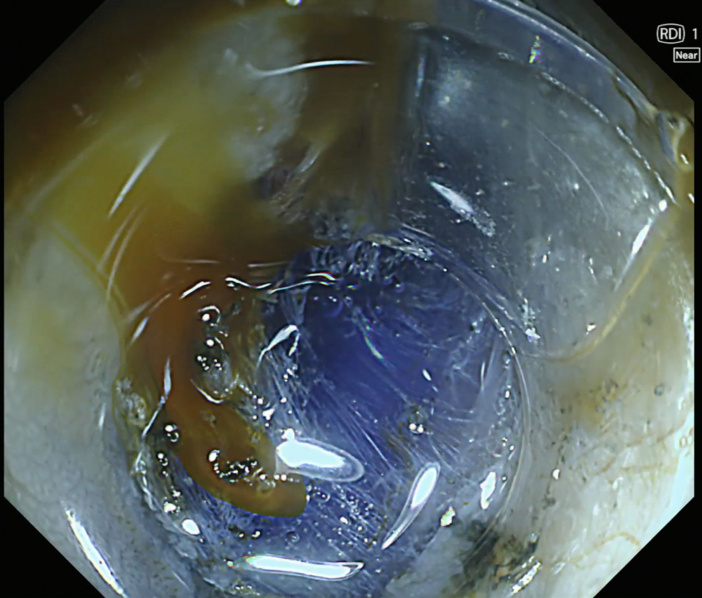
13. Submucosal layer visualization with RDI Mode 1
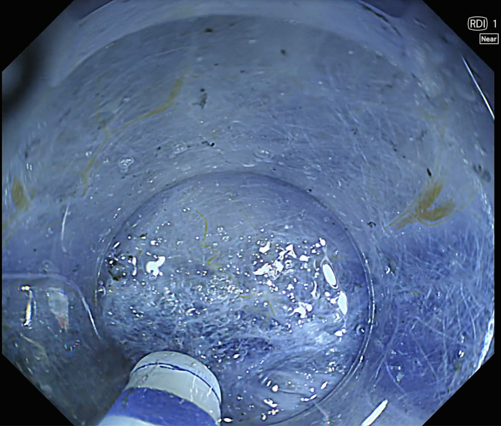
Enhancement : A8
NBI Mode : NA
TXI Mode : NA
RDI Mode : Mode 1
BAI-MAC : On
Case Video
This video demonstrates stepwise lesion evaluation using EVIS X1 imaging modalities. TXI is used to improve visualization of lesion margins and overall extent, followed by NBI and magnified NBI with near-focus observation to assess surface and vascular patterns.
This video shows the use of RDI mode 1 during ESD. After activation of RDI mode 1, the bleeding focus is clearly visualized, allowing contact coagulation using a DualKnife J™. In addition, RDI mode 1 combined with indigo carmine enhances visualization of the submucosal layer, facilitating identification of the cutting line during dissection.
Overall Comment
This case demonstrates the clinical utility of the EVIS X1 system using a CF-EZ1500DI colonoscope for the evaluation and treatment of a large laterally spreading tumor.
TXI improved delineation of the lesion margins and overall extent, while magnified NBI enabled detailed assessment of surface and vascular patterns, suggesting a JNET type IIb pattern.
During ESD, RDI mode 1 enhanced visualization of bleeding points and supported appropriate hemostatic management.
In addition, RDI mode 1 combined with indigo carmine improved visualization of the submucosal layer, allowing clearer identification of the cutting line and more controlled dissection.
* Specifications, design and accessories are subject to change without any notice or obligation on the part of the manufacturer.
- Content Type