Disclaimer
The techniques and clinical opinions presented in this material reflect the personal experience and professional judgment of the healthcare professional and do not necessarily represent the views of Olympus. This material is intended for healthcare professionals only. Users should always refer to the applicable Instructions for Use (IFU) and use Olympus products in accordance with the approved indications and local regulatory requirements. The healthcare professional presenting this material has been engaged by Olympus and compensated at fair market value for their services.
Gastric Case 6

Dr. Ashutosh Mohapatra
Sai Institute of Gastroenterology & Liver Sciences
India
Scope: GIF-EZ1500
Case: Patient endoscopy finding include scattered gastric atrophy in areas of intestinal metaplasia. In areas of Intestinal Metaplasia there are several lesions and out of that one lesion is neoplastic having demarcation line with irregular, heterogeneous, densely distributed micro-vessels suggestive of high grade of dysplasia
Organ: Stomach
Patient Information: M, 50s, belongs to eastern India, non-vegetarian presented with dyspepsia
1. WLI Image
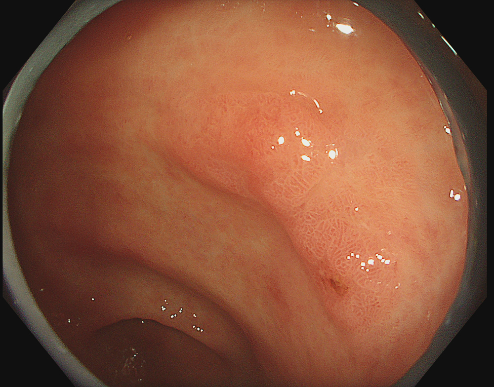
#WL #B5
2. TXI 1 Image
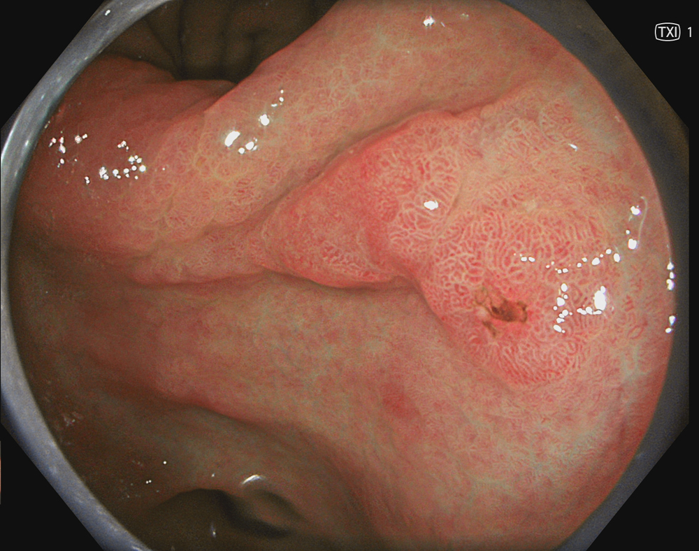
#TXI-1 #Low level enhancement
3. TXI 2 Image
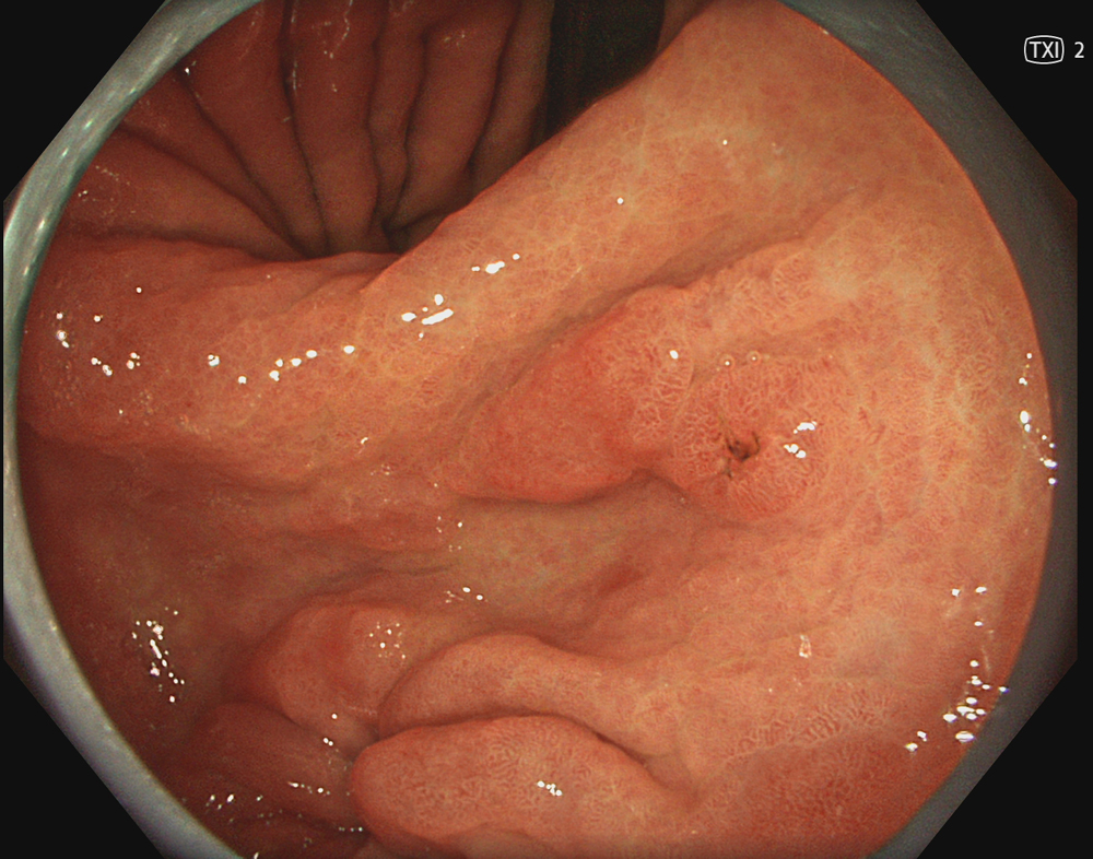
#TXI-1 #low level enhancement
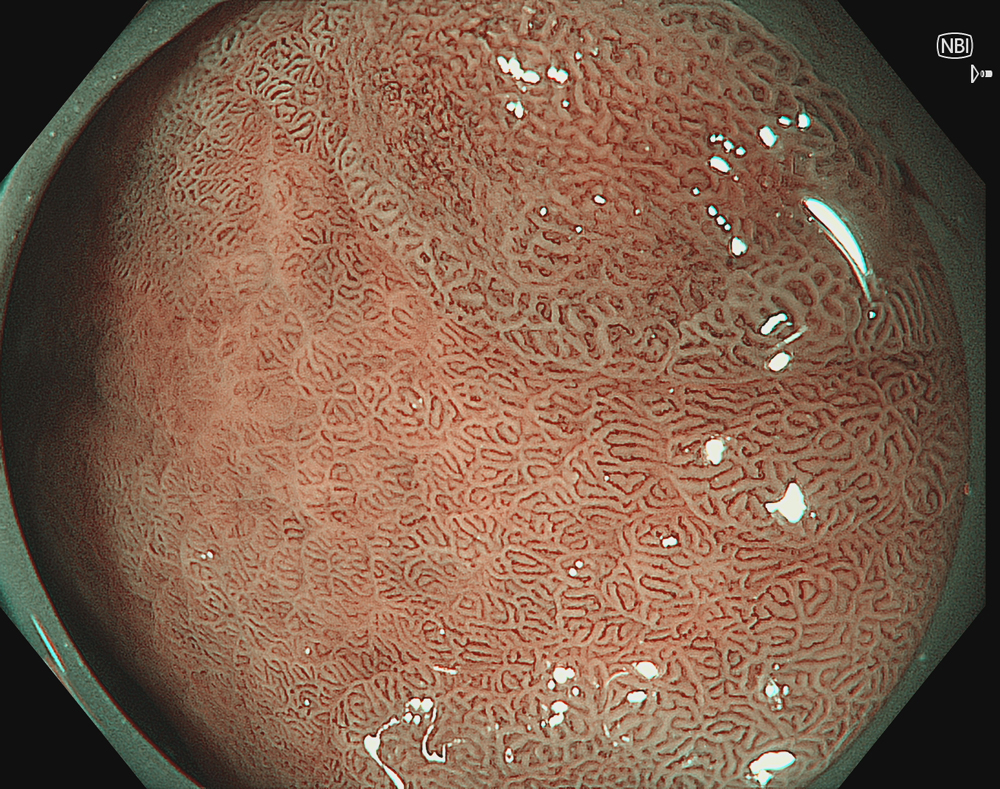
4. NBI
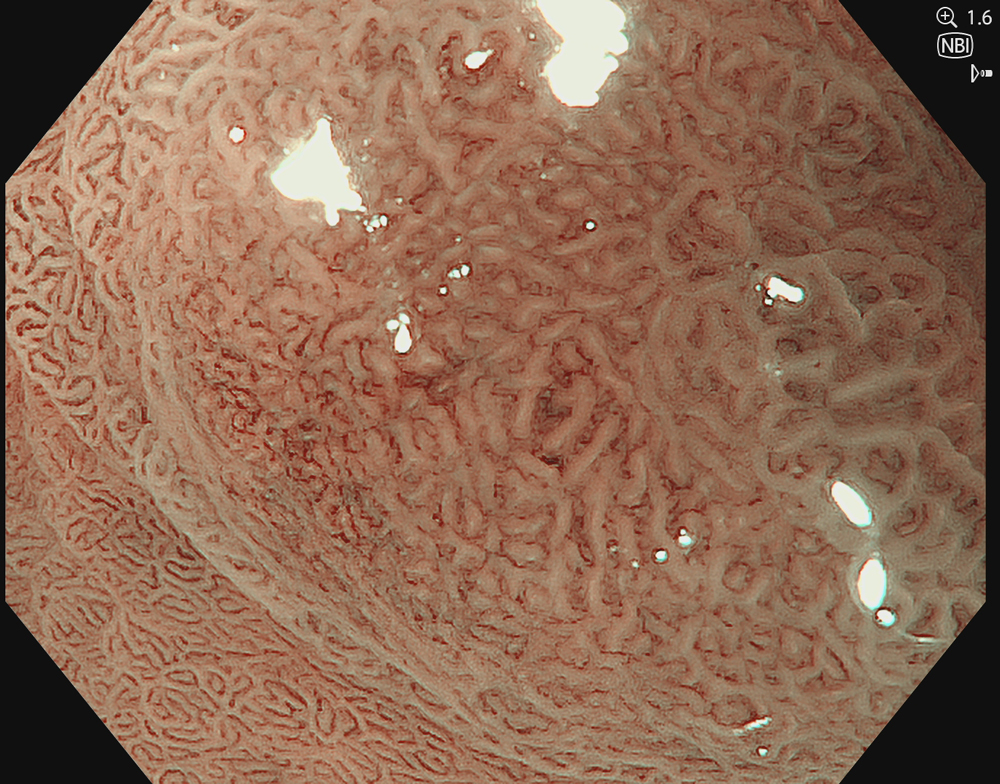
#NBI #B8
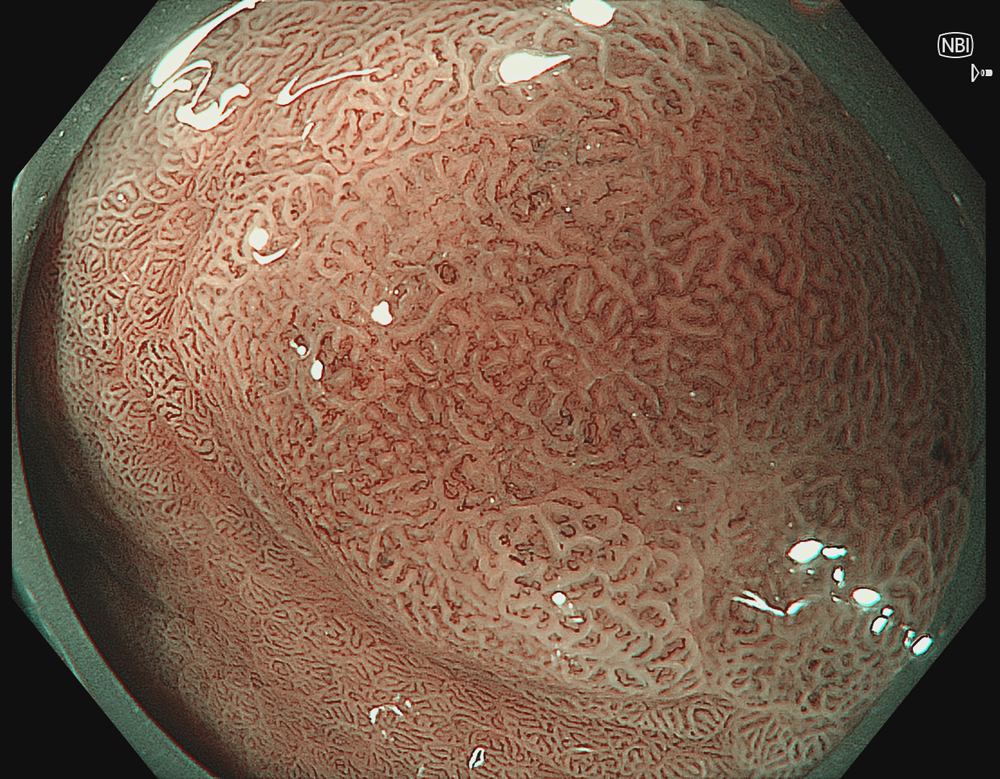
5. NBI in Near Mode
#NBI #B8
6. NBI in Near Mode with 1.6X optical zoom
#NBI #B8
Case Video
Video shows the scattered atrophy with Intestinal Metaplasia which is enhanced in TXI modes ( both 1 and 2) and on NBI image of posterior wall of incisura has a lesion with sharply defined demarcation line and irregular, heterogeneous, densely packed micro-vessels, some of which traversed the crypt epithelium, a pattern highly suggestive of high‑grade dysplasia.
Overall Comment
A male in his 50’s with no significant past medical history presented with dyspepsia and underwent upper GI endoscopy. White-light imaging revealed an approximately 20 mm superficially elevated lesion with a depressed component (Paris IIa+IIc).
NBI of the depressed component revealed a clear demarcation line with irregular, heterogeneous microvascular architecture and an irregular micro-surface pattern, raising concern for a neoplastic lesion.
* Specifications, design and accessories are subject to change without any notice or obligation on the part of the manufacturer.
- Keyword
- Content Type