Pancreatobiliary case 1

Jong Ho Moon, MD, PhD, FASGE, FACG, FJGES
Professor of Medicine
Director, Digestive Disease Center
SoonChunHyang University School of Medicine,
Bucheon/Seoul, Korea

Il Sang Shin, MD, PhD
Assistant Professor
Digestive Disease Center
SoonChunHyang University School of Medicine,
Bucheon/Seoul, Korea
Scope: TJF-Q290V
Organ: Ampulla of Vater
Patient information: F, 70s
Medical history: No past medical history
1. WLI Observation
Enhancement : A7
NBI Mode : NA
TXI Mode : NA
RDI Mode : NA
BAI-MAC : On
2. NBI Observation
Enhancement : A7
NBI Mode : 2
TXI Mode : NA
RDI Mode : NA
BAI-MAC : On
3. TXI Observation
Enhancement : A7
NBI Mode : NA
TXI Mode : 2
RDI Mode : NA
BAI-MAC : On
4. RDI Observation
Enhancement : A7
NBI Mode : NA
TXI Mode : NA
RDI Mode : 1
BAI-MAC : On
Case Video
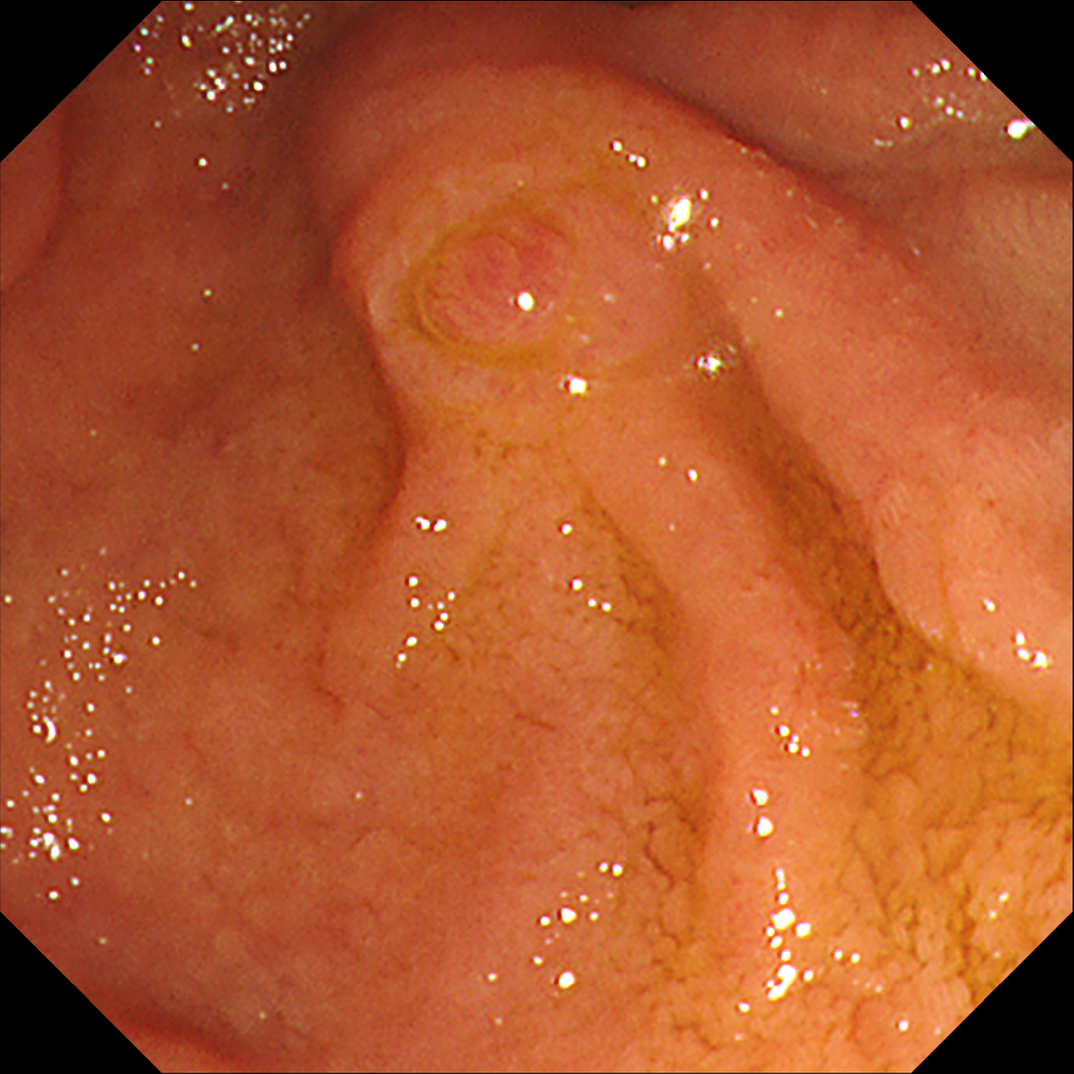
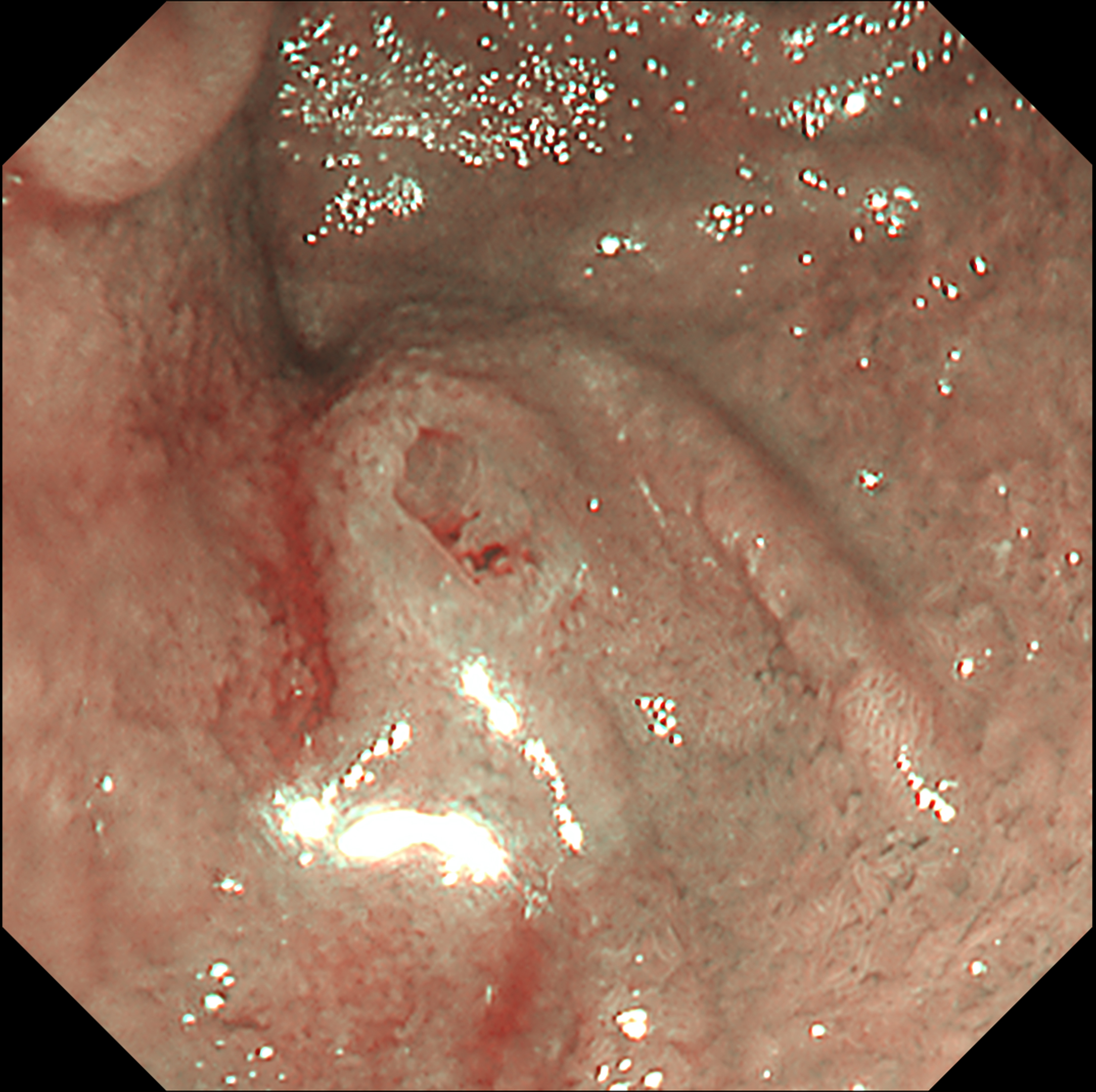
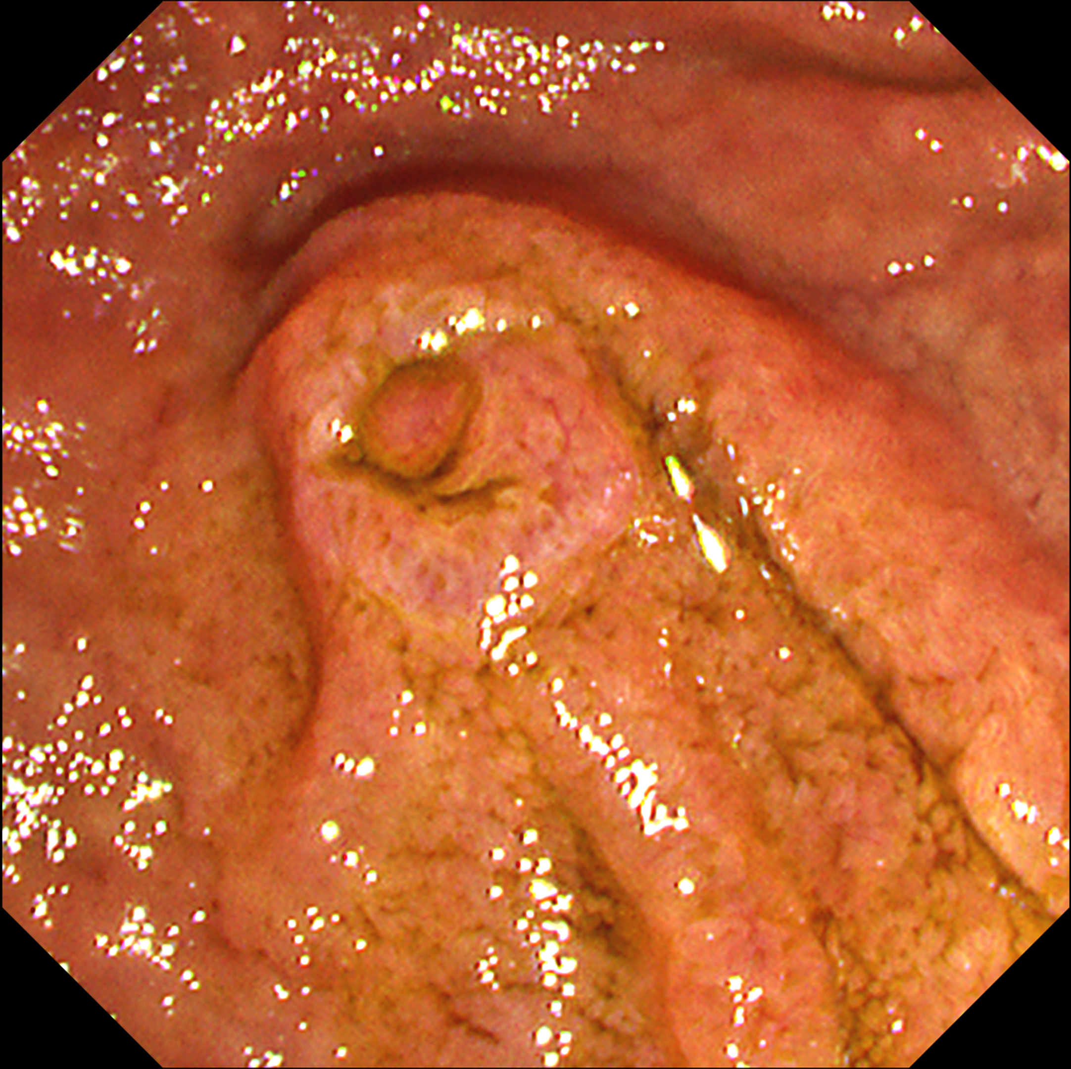
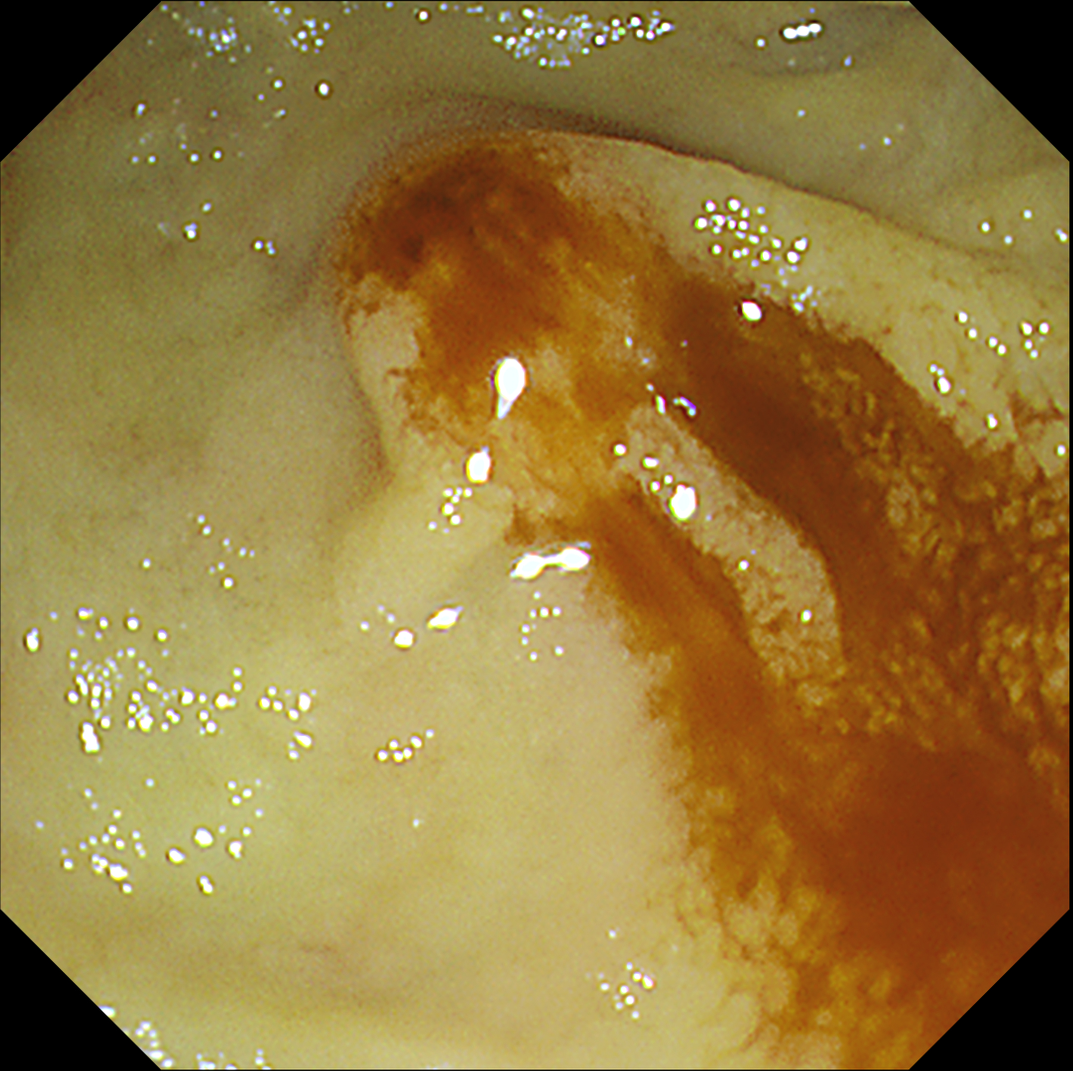
This video shows the usefulness of a new image enhanced endoscopy system in identifying a regular papilla in a patient with suspected ampullary mass on computed tomography (CT) scan. Under white-light imaging (WLI), regular papilla with classic appearance was observed. Under narrow-band imaging (NBI), covering folds and papillary orifices were more apparent. TXI mode 2 shed more light on the anatomical features of the surface than NBI, while maintaining visual naturalness and being less disturbed from bile acids similar to WLI. After forceps biopsy, RDI mode 1 more easily identified the hemostatic condition of ampulla of Vater, making it easier to determine whether there is a bleeding point that requires endoscopic hemostasis. Even if the papilla appears normal, the application of TXI and RDI modes can help to accurately assess the anatomical structure and abnormality of the ampulla of the Vater.
Overall Comment
This case demonstrates that observation using TXI and RDI modes in ampulla of Vater, although which seems to be normal, can be helpful for an effective observation and safe biopsy sampling by keeping the biopsy location with pancreatic duct as far as possible. With less disturbance from bile acid than narrow-band imaging, TXI emphasizes the difference in color tone of the mucosal surface, enhancing the accurate evaluation of the anatomical structure including frenulum, circular fold, and even polypoid nodule of papillary orifice. RDI can effectively identify hemostatic status and possible bleeding points, helping to quickly determine whether endoscopic hemostasis is necessary after tissue sampling.
* Specifications, design and accessories are subject to change without any notice or obligation on the part of the manufacturer
- Content Type