Pancreatobiliary case 2

Jong Ho Moon, MD, PhD, FASGE, FACG, FJGES
Professor of Medicine
Director, Digestive Disease Center
SoonChunHyang University School of Medicine,
Bucheon/Seoul, Korea

Il Sang Shin, MD, PhD
Assistant Professor
Digestive Disease Center
SoonChunHyang University School of Medicine,
Bucheon/Seoul, Korea
Scope: TJF-Q290V
Organ: Ampulla of Vater
Patient information: F, 60s
Medical history: Medication for hyperlipidemia and hypothyroidism
1. WLI Observation
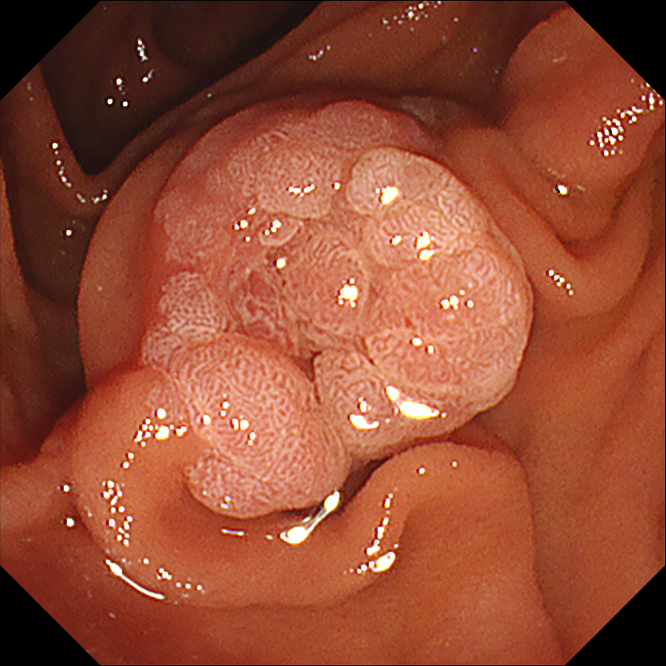
Enhancement : A7
NBI Mode : NA
TXI Mode : NA
RDI Mode : NA
BAI-MAC : On
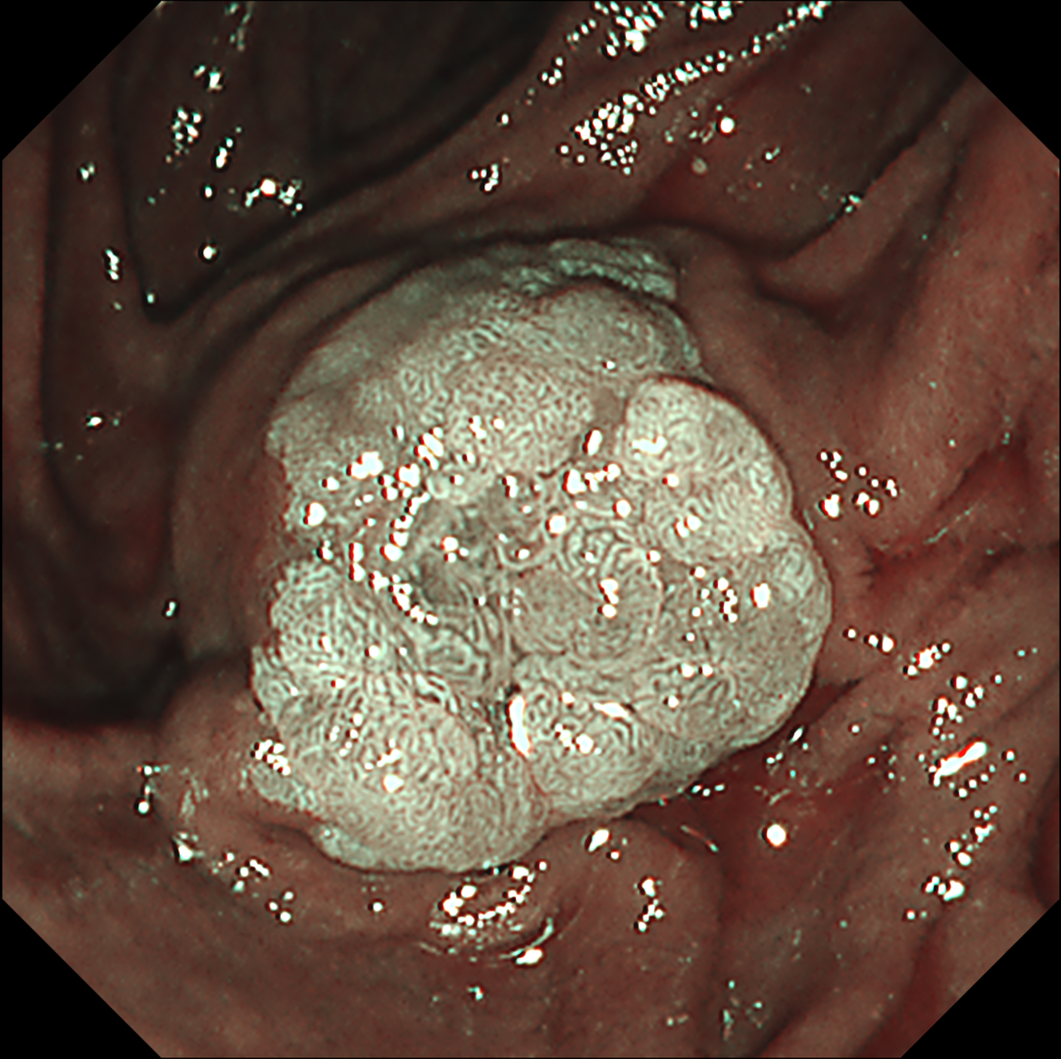
2. NBI Observation
Enhancement : A7
NBI Mode : 2
TXI Mode : NA
RDI Mode : NA
BAI-MAC : On
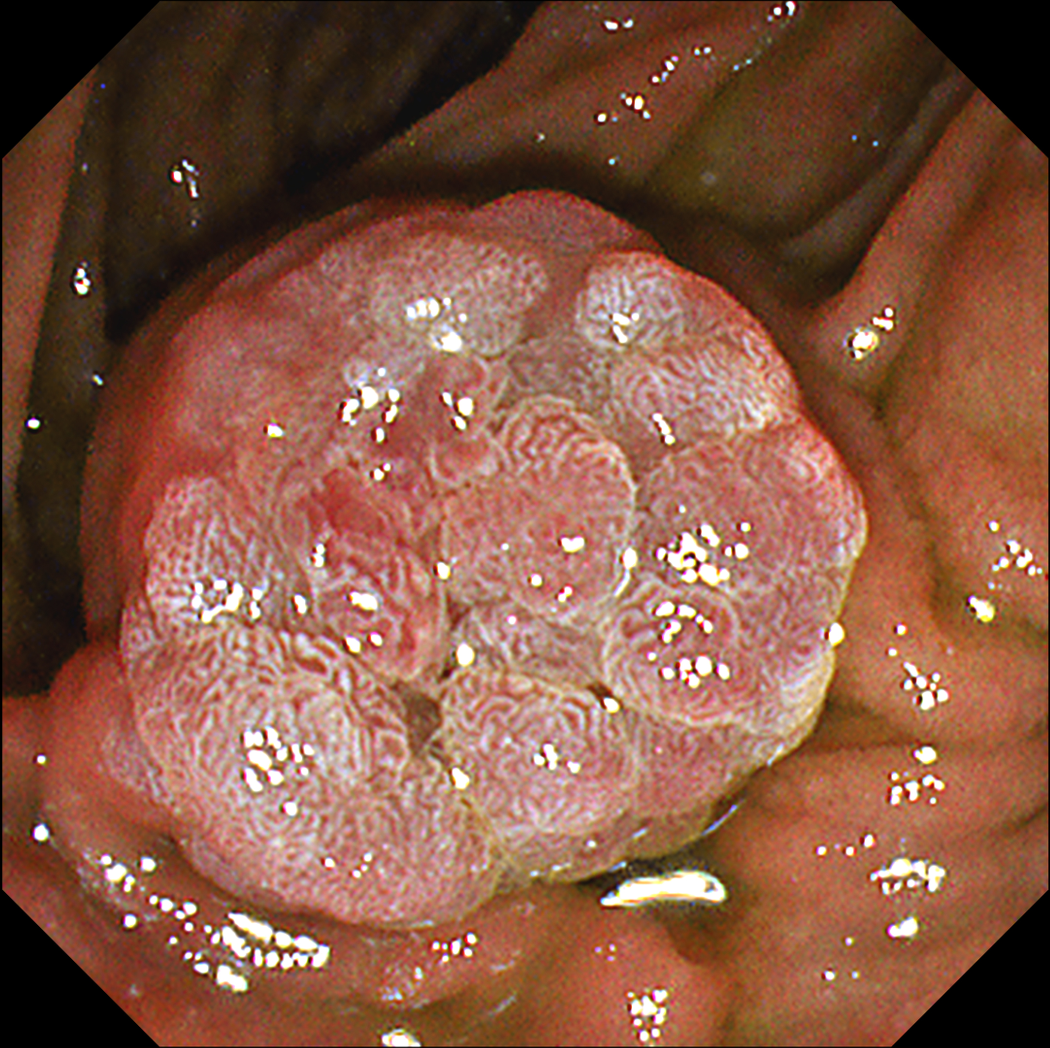
3. TXI Observation
Enhancement : A7
NBI Mode : NA
TXI Mode : 1
RDI Mode : NA
BAI-MAC : On
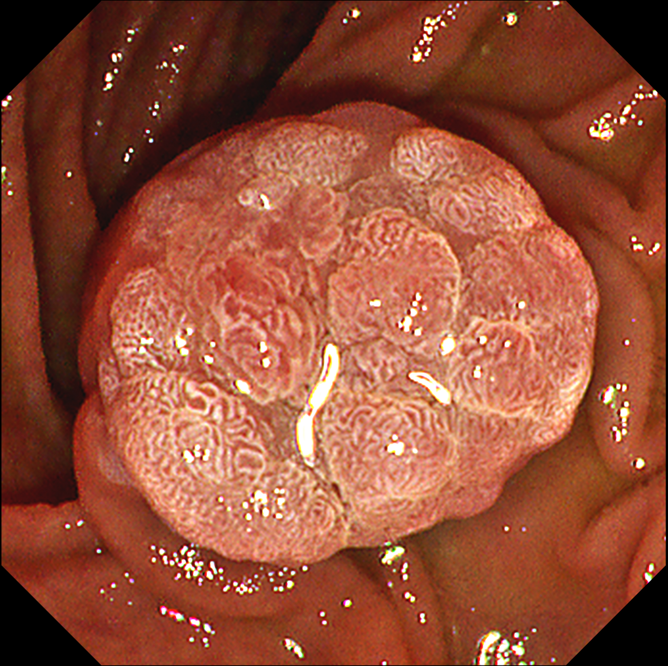
4. TXI Observation
Enhancement : A7
NBI Mode : NA
TXI Mode : 2
RDI Mode : NA
BAI-MAC : On
Case Video
This video demonstrates the usefulness of a new image-enhanced endoscopy system in a patient with suspected ampullary adenoma. On white-light imaging (WLI), a lobular mass-like lesion without ulceration or bleeding was observed. On narrow-band imaging (NBI), the lesional boundaries were clearly observed with strong contrast. Texture and color enhancement imaging (TXI) mode 1 helped to observe the surface meandering microvessels well while further emphasizing the surface cone-like lobularity. TXI mode 2 highlighted the surface irregular nodularity and pit pattern while maintaining visual naturalness. In patients with suspected ampullary mass, applying TXI mode can help to better identify the characteristics of mass-like lesion of ampulla of Vater and make treatment decision.
Overall Comment
This case shows that the observation of the mass-like lesion in ampulla of Vater using TXI mode strongly emphasized the surface lobularity and the irregular meandering surface microvessels. TXI highlights the difference in color tone of the surface mucosae, so the pine-cone like surface lobularity and surface microvessels of suspicious ampullary adenoma could be usefully identified using TXI mode.
* Specifications, design and accessories are subject to change without any notice or obligation on the part of the manufacturer
Jong Ho Moon, MD, PhD, FASGE, FACG, FJGES
Il Sang Shin, MD, PhD Case 3: Regular follow-up exam for discrimination of ampullary adenoma
Jong Ho Moon, MD, PhD, FASGE, FACG, FJGES
Il Sang Shin, MD, PhD
- Keyword
- Content Type


