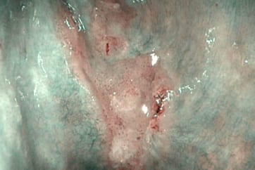
◆ Recurrent superficial cancer in the right piriform sinusafter chemoradiotherapy

Endoscopic Finding:
White light image only demonstrates the small and thin protruding whitish lesion in the right piriform sinus. However, NBI image demonstrates branched and slightly dilated vessels on the surface of the tumor after the endoscope is brought close to the lesion. This finding suggests that the cancer invades into the subepithelial layer. Furthermore NBI finding shows the cancer horizontal margin clearly showing the disappearance of the arborescent vascular network at the borderline of the normal part and cancer lesion.
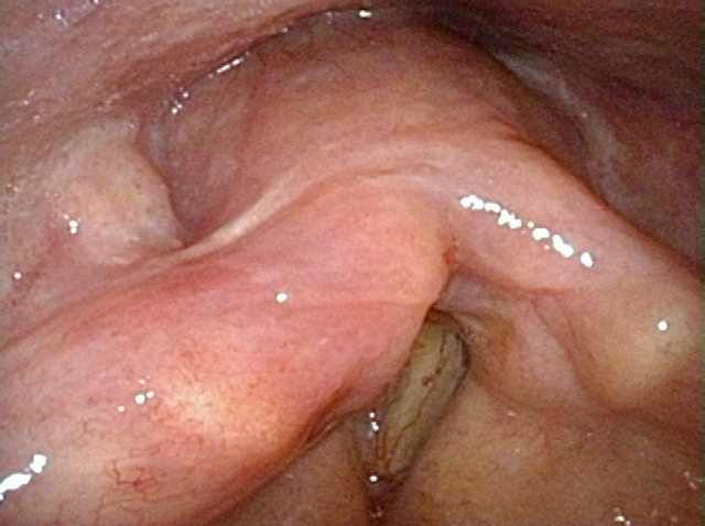
White Light Image
Equipment: OTV-S190, CLV-S190, ENF-VH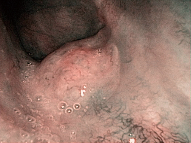
NBI Image
Pathological Finding:
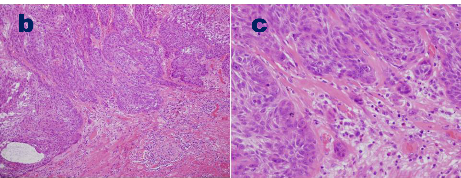
Figure b is the enlarged view of white quadrangle in figure a and figure c is the enlarged view of white quadrangle in figure b.
There was the squamous cell carcinoma making cancer nest and invading into the subepithelial layer.
Tumor size was 10mm x 7mm and tumor thickness was 2mm.
Both horizontal margin and vertical margin were negative.
No lymphatic, no venous and no perineural invasion existed.
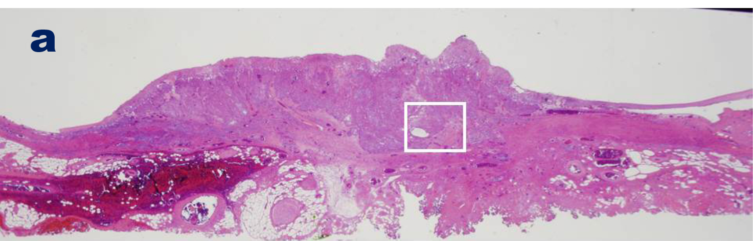
Pathological image (macro)
Pathological image (micro)
Acknowledgment:
Hiroshi Kawachi, Head, Department of Pathology, The Cancer Institute Hospital of JFCR and Yusuke Kiyokawa,
Director, Department of Otolaryngology, Japanese Red Cross Musashino Hospital, contributed to this atlas.
QR code