Author: Ritesh Agarwal
Hospital: Professor, PGIMER Chandigarh Hospital
Disclaimer:
- TXI™ Technology is not intended to replace histopathological sampling as a means of diagnosis
- The positions and statements made herein by Dr. Agarwal are based on Dr. Agarwal’s experiences, thoughts and opinions. As with any product, results may vary, and the techniques, instruments, and settings can vary from facility to facility. The content hereof should not be considered as a substitute for carefully reading all applicable labeling, including the Instructions for Use. Please thoroughly review the relevant user manual(s) for instructions, risks, warnings, and cautions. Techniques, instruments, and setting can vary from facility to facility. It is the clinician’s decision and responsibility in each clinical situation to decide which products, modes, medications, applications, and settings to use.
- The EVIS X1™ endoscopy system is not designed for cardiac applications. Other combinations of equipment may cause ventricular fibrillation or seriously affect the cardiac function of the patient. Improper use of endoscopes may result in patient injury, infection, bleeding, and/or perforation. Complete indications, contraindications, warnings, and cautions are available in the Instructions for Use (IFU)
- Dr Agarwal, the authoring physician(s) of this presentation, are/ is a paid consultant(s) to Olympus Corporation of the Americas.
Procedure Information
Scope: BF-1TH1100
Location: Trachea
Patient information: Male, 57 years old
Medical history: Not Significant
1. WLI in Anterior and lateral wall of Trachea
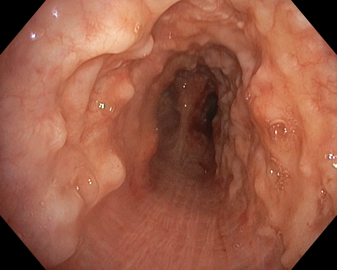
Diffuse nodules seen over anterior and lateral wall of trachea sparing posterior wall.
2. TXI™ Technology in Anterior and lateral wall of Trachea
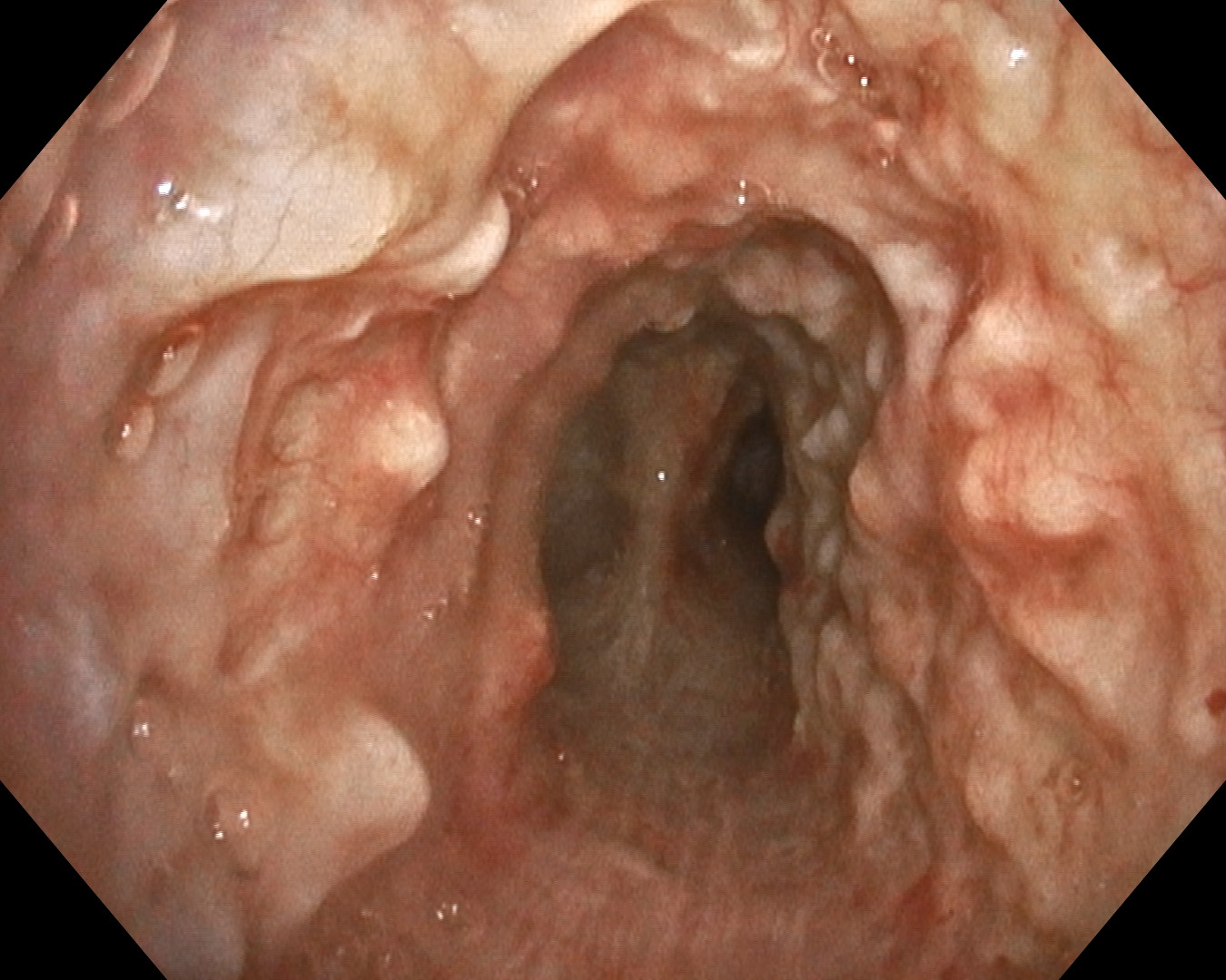
Enhanced characterization of mucosa and nodules over anterior and lateral wall of trachea
Overall Comment
This was a 57-year-old gentleman who presented with hemoptysis. He used to smoke cigarettes occasionally. Flexible bronchoscopy was performed for hemoptysis to exclude airway tumors. On bronchoscopy, we observed nodules in the anterior and lateral wall of the trachea. The nodules spared the membranous portion of the trachea. A diagnosis of tracheobronchopathia osteochondroplastica was made.
- Keyword
- Content Type