Hospital: Thoraxklinik, University of Heidelberg, Germany
Disclaimer:
- TXI™ and RDI™ Technologies are not intended to replace histopathological sampling as a means of diagnosis
- The positions and statements made herein by Dr. Herth’s are based on Dr. Herth’s experiences, thoughts and opinions. As with any product, results may vary, and the techniques, instruments, and settings can vary from facility to facility. The content hereof should not be considered as a substitute for carefully reading all applicable labeling, including the Instructions for Use. Please thoroughly review the relevant user manual(s) for instructions, risks, warnings, and cautions. Techniques, instruments, and setting can vary from facility to facility. It is the clinician’s decision and responsibility in each clinical situation to decide which products, modes, medications, applications, and settings to use.
- The EVIS X1™ endoscopy system is not designed for cardiac applications. Other combinations of equipment may cause ventricular fibrillation or seriously affect the cardiac function of the patient. Improper use of endoscopes may result in patient injury, infection, bleeding, and/or perforation. Complete indications, contraindications, warnings, and cautions are available in the Instructions for Use (IFU)
- Dr Herth, the authoring physician(s) of this presentation, are/ is a paid consultant(s) to Olympus Corporation.
Scope: BF-1TH190
Patient information:
58 years, male
Ex smoker (45 packyears)
Coronary artery disease
Medical history:
Patients reported on chronic coughing,
Lowdose computertomograhy showed no lung abnormalities
Lung function: FEV1 63%
1. WLI
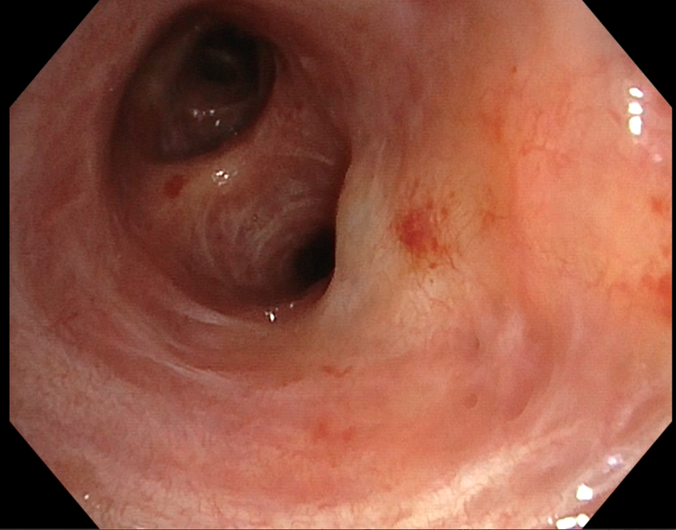
In the white light mode vasculary engorgement was visible.
2. TXI™ Technology
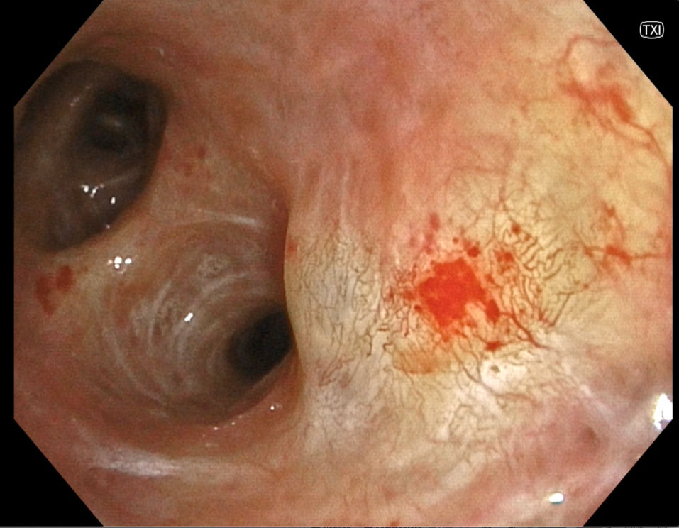
In TXI™ Technology, the vascular pattern was seen more clearly. Vascular slings, as well as dotted vessels, are visible.
3. RDI™ Technology Mode 2
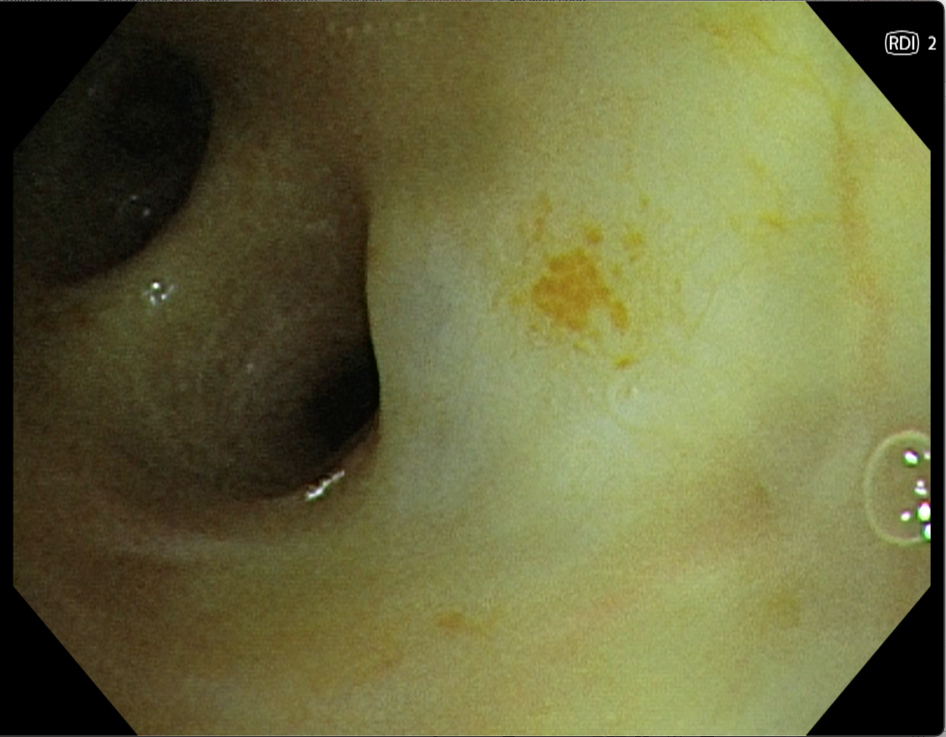
In RDI™ Technology 2 especially the dotted vessels are highlighted.
4. RDI™ Technology Mode 3
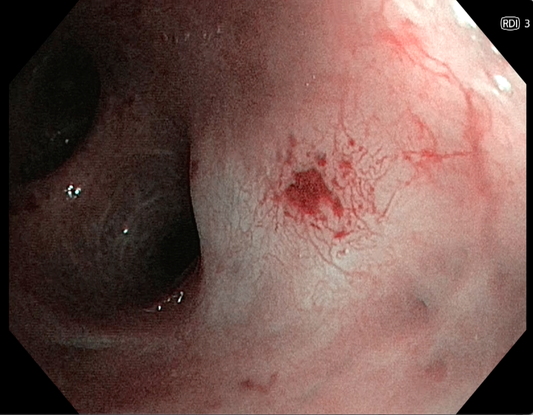
RDI™ Technology 3 offers an alternative mode showing comparable patterns like TXI™ Technology in this case.
Pathological Finding
Vascular changes have been seen at the middle lobe entrance.
With the help of TXI™ and RDI™ Technologies, the vascular pattern was seen nicely and targeted biopsies have been taken, showing a moderate dysplasia.
Overall Comment
With the help of the new features the pathological finding was visibly better and it was possible to distinguish normal and abnormal areas.
The positioning of the forceps for the biopsy was more precise.
* Specifications, design and accessories are subject to change without any notice or obligation on the part of the manufacturer
- Keyword
- Content Type