Junichi Inokuchi, MD. Katsunori Tatsugami, MD. Prof. Seiji Naito, MD. Kyushu University, Japan
Disclaimer:
- NBI™ Technology is not intended to replace histopathological sampling as a means of diagnosis.
- The positions and statements made herein by Dr. Inokuchi, Dr. Tatsugami, and Dr. Naito are based on their collective experiences, thoughts and opinions. As with any product, results may vary, and the techniques, instruments, and settings can vary from facility to facility. The content hereof should not be considered as a substitute for carefully reading all applicable labeling, including the Instructions for Use. Please thoroughly review the relevant user manual(s) for instructions, risks, warnings, and cautions. Techniques, instruments, and setting can vary from facility to facility. It is the clinician’s decision and responsibility in each clinical situation to decide which products, modes, medications, applications, and settings to use.
- Dr. Inokuchi, Dr. Tatsugami, and Dr. Naito are compensated consultants of the Olympus Corporation.
- All images are courtesy of Dr. Inokuchi, Dr. Tatsugami, and Dr. Naito, except where otherwise noted.
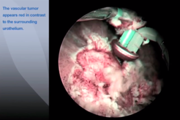
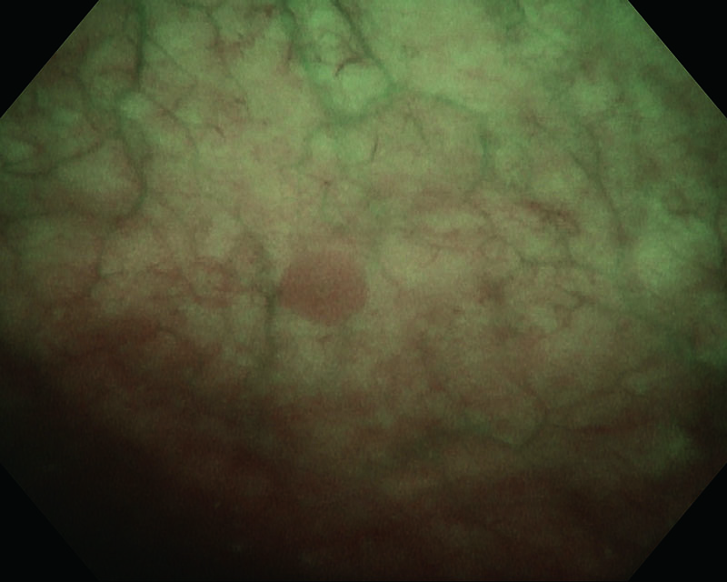
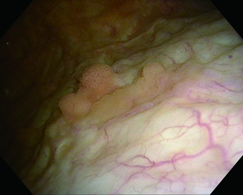
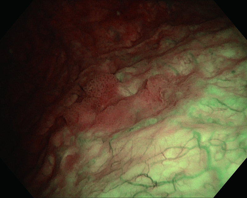
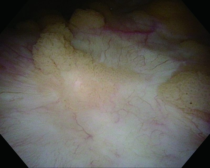
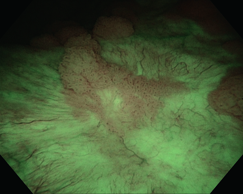
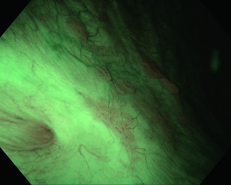
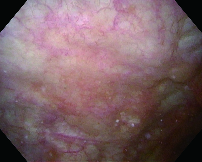
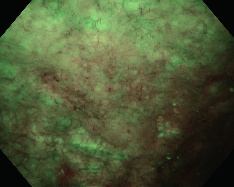
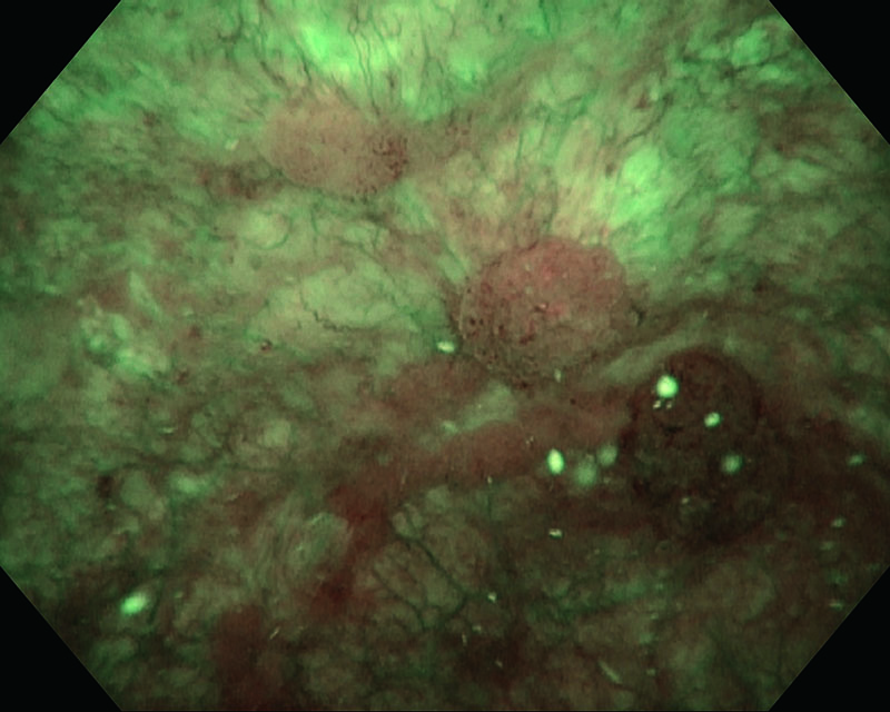
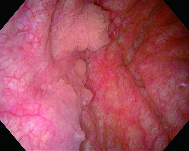
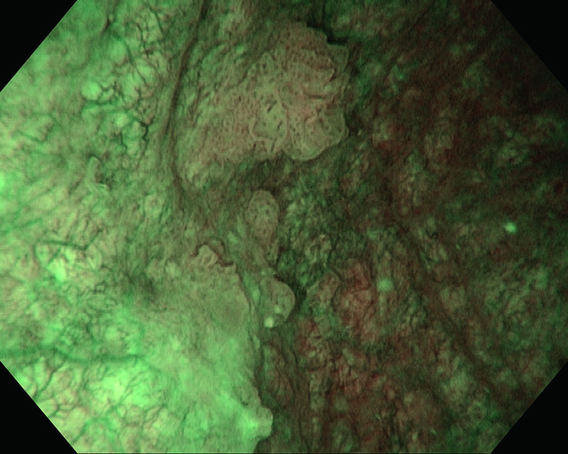
Papillary peduncular tumor/Sessile tumor, age 80, female
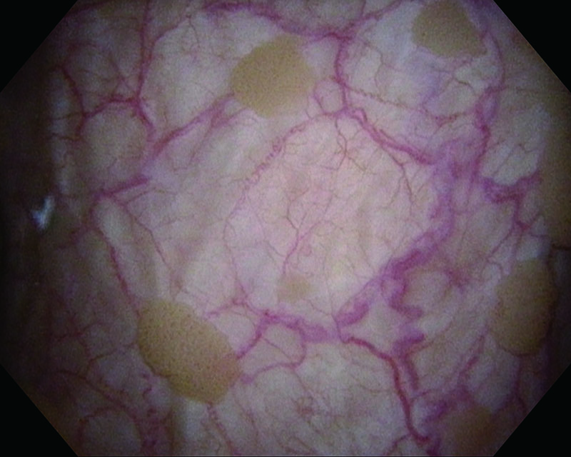
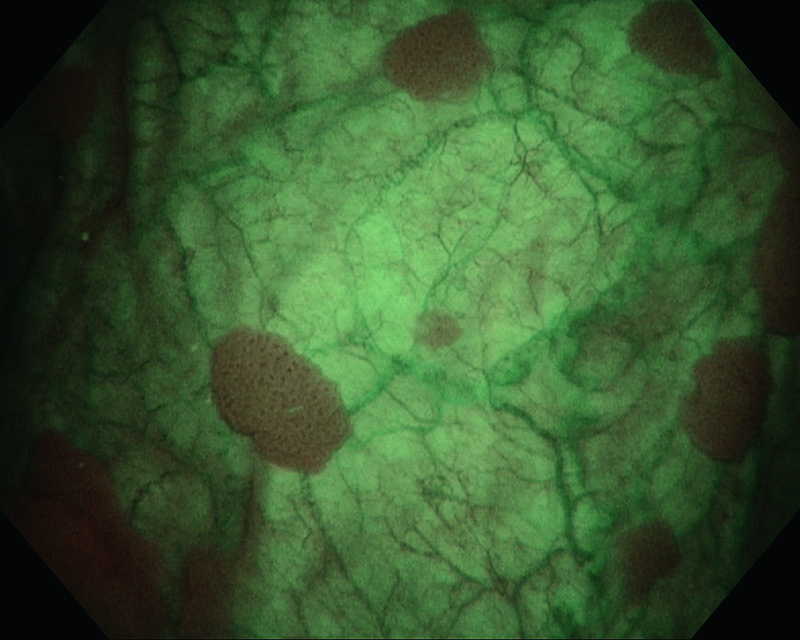
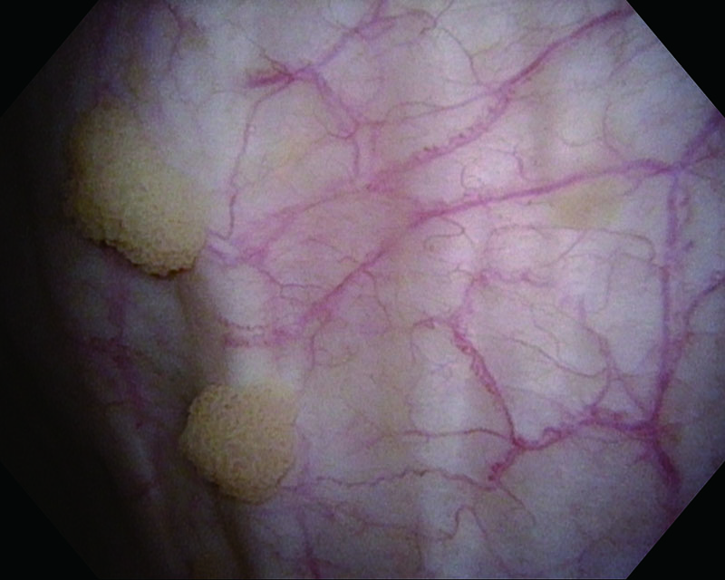
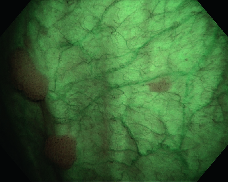
Comments
A small tumor is highlighted under NBI™ Technology which was suspected under WLI.
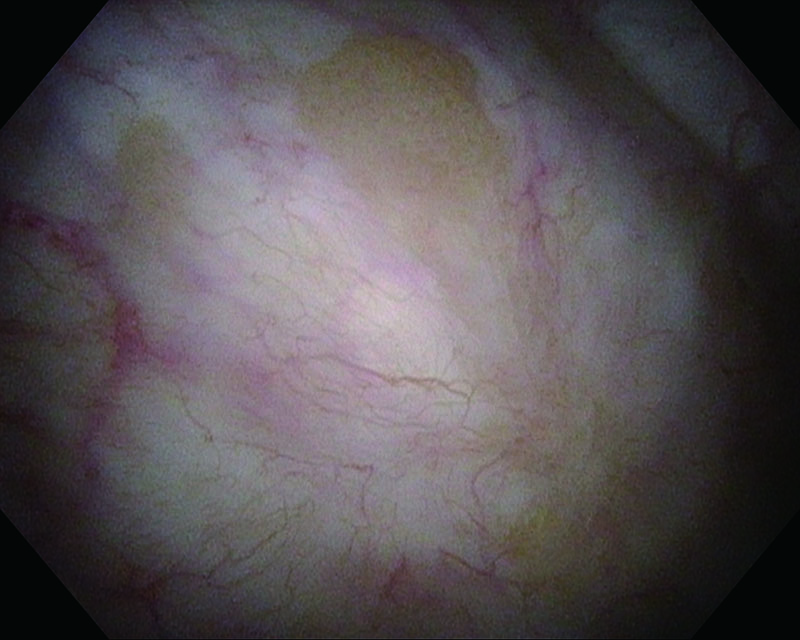
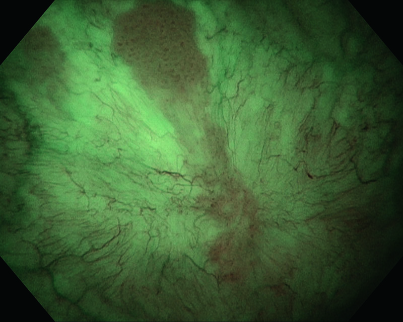
Papillary peduncular tumor, age 61, male
Comments
Utilizing NBI™ Technology enables us to enhance visualization of the marginal region of the tumor.
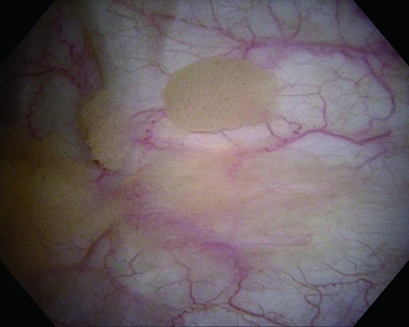
Papillary peduncular tumor, age 82, male
Comments
Utilizing NBI™ Technology enabled us to visualize a marginal region of small tumors which were difficult to visualize under WLI.
Papillary sessile tumor, age 82, female
Comments
Utilizing NBI™ Technology enabled us to enhance visualization of the marginal region of the tumor. NBI™ Technology also enabled us to identify surrounding small tumors which were difficult to identify under WLI.
Papillary peduncular tumor/Sessile tumor, age 80, female
Comments
Recurrent tumor which occurs more often in bladder. Utilizing NBI™ Technology enabled us to enhance visualization of marginal regions of the tumor.
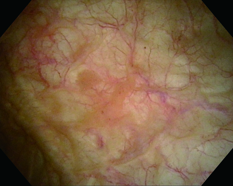
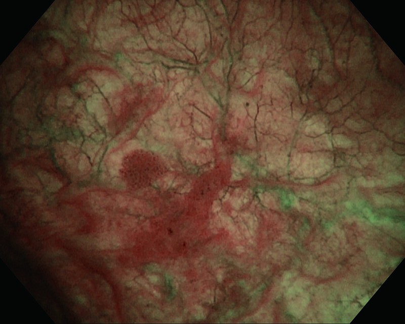
Rubor mucosa, age 81, male
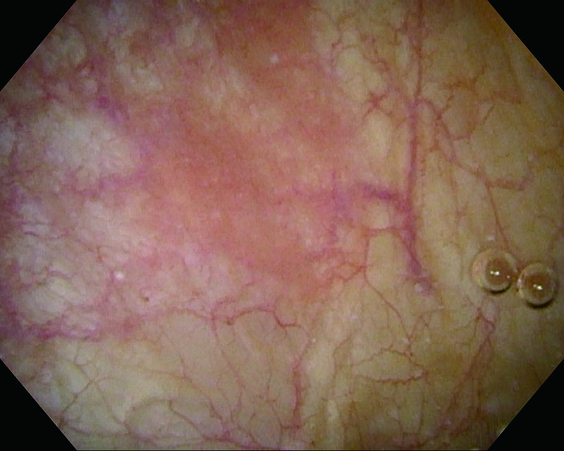
Comments
Bladder CIS. Utilizing NBI™ Technology enabled us to enhance visualization of marginal region.
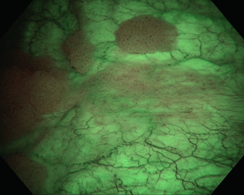
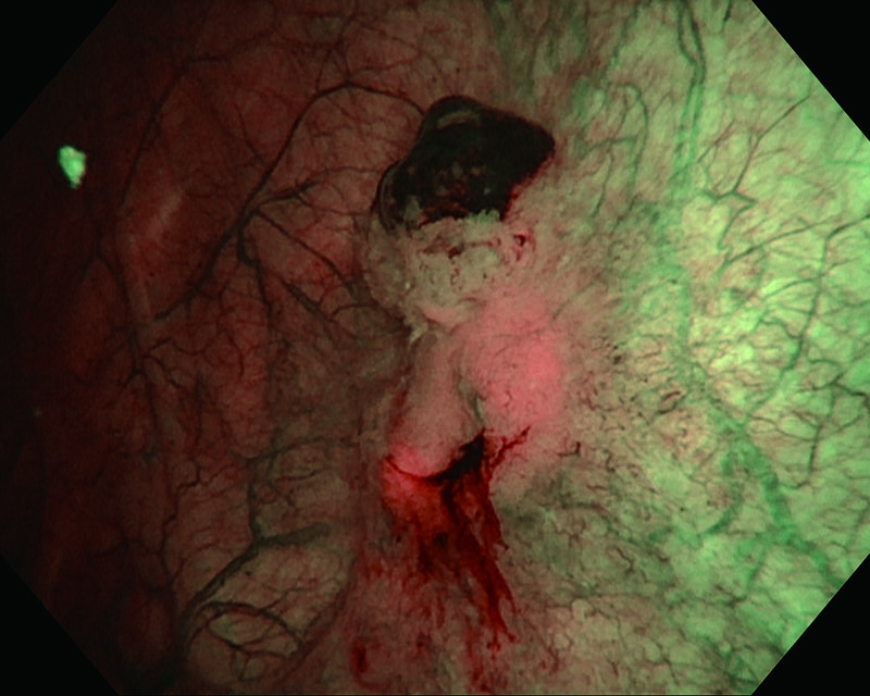
Rubor mucosa, age 81, male
Comments
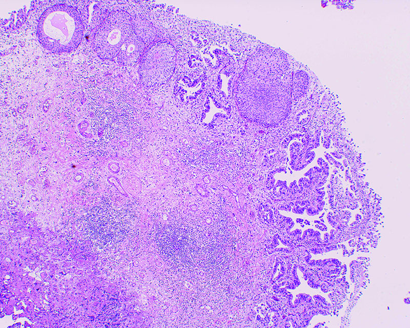
The case suspected carcinoma in situ and identified rubor bladder mucosa. Histopathologic examination revealed CIS.
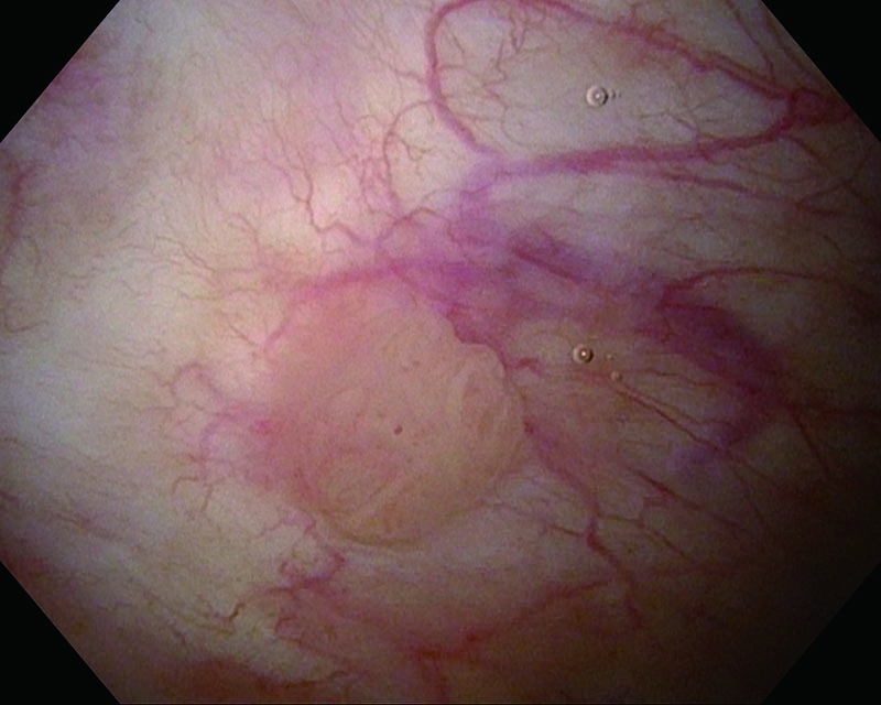
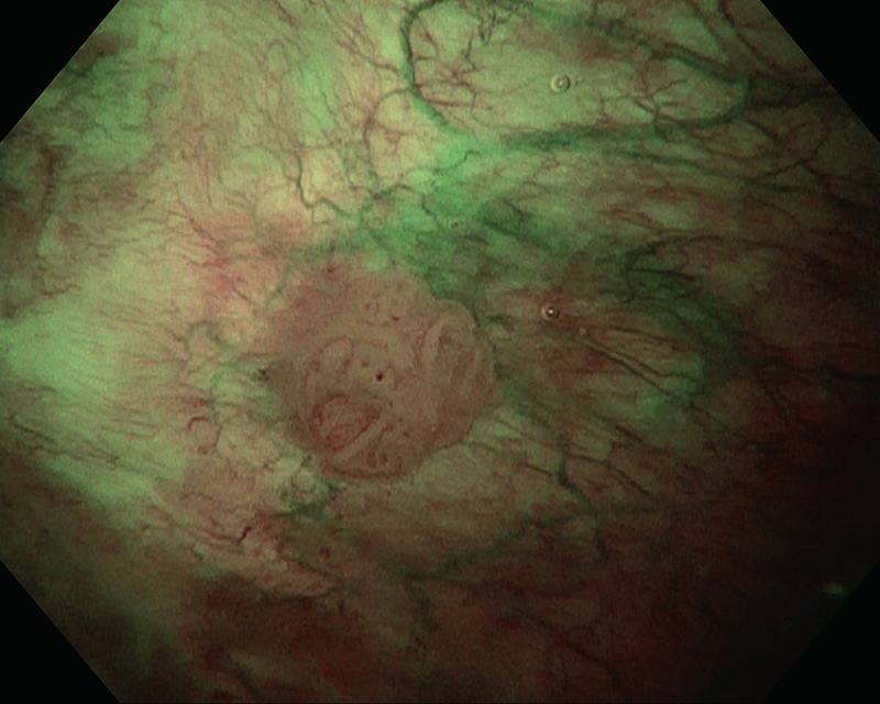
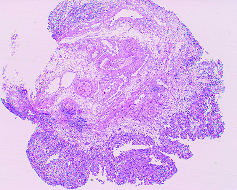
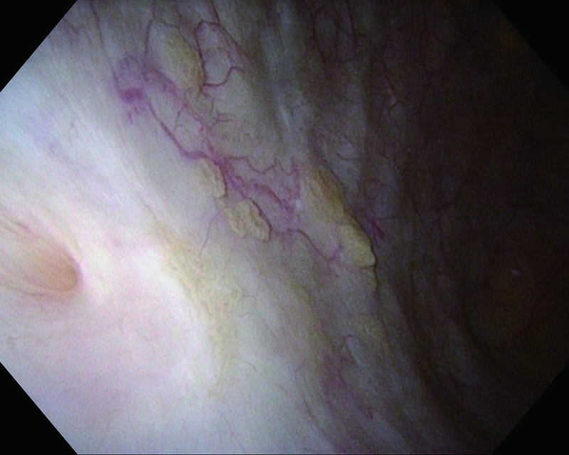
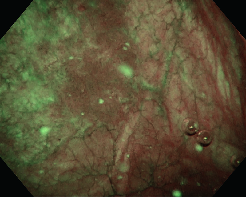
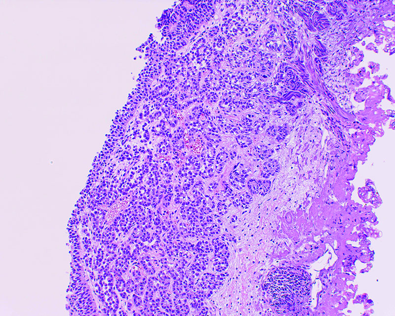
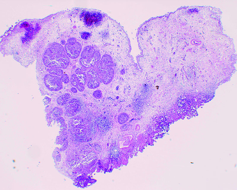
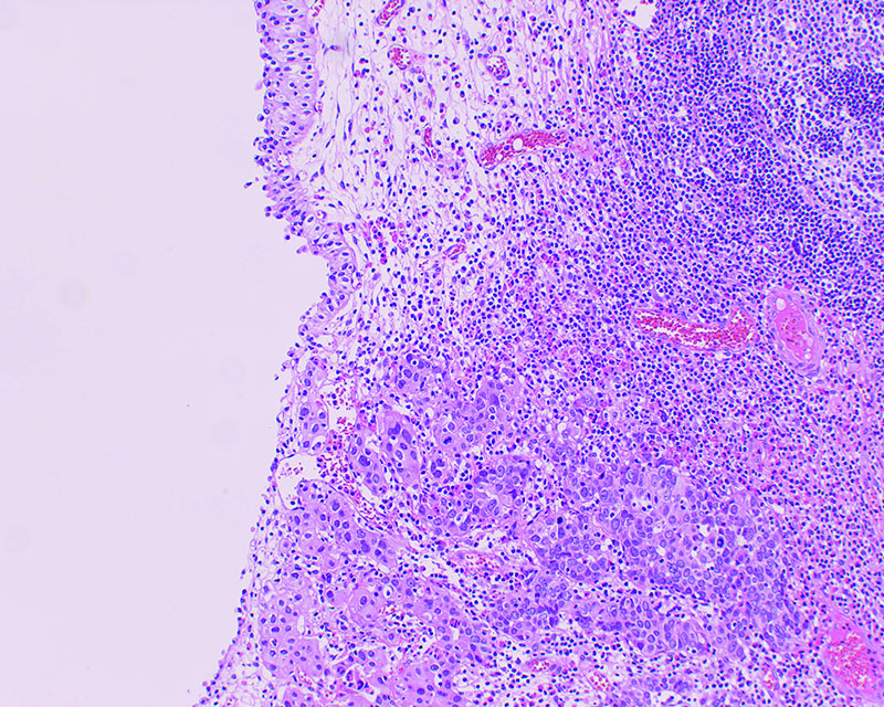
Papillary peduncular tumor/Sessile tumor + rubor, age 81, male
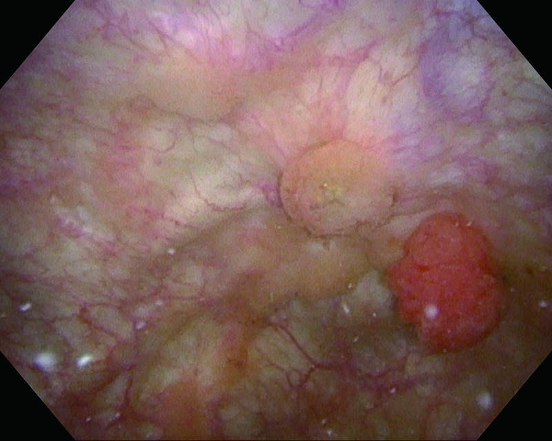
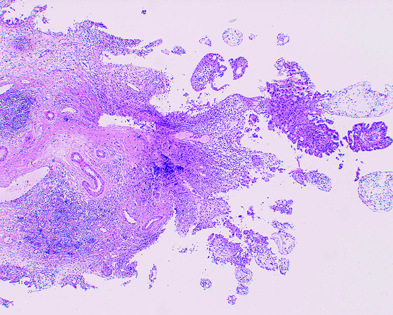
Comments
Utilizing NBI™ Technology enabled us to visualize a papillary tumor and surrounding aberrant mucosa more definitely.
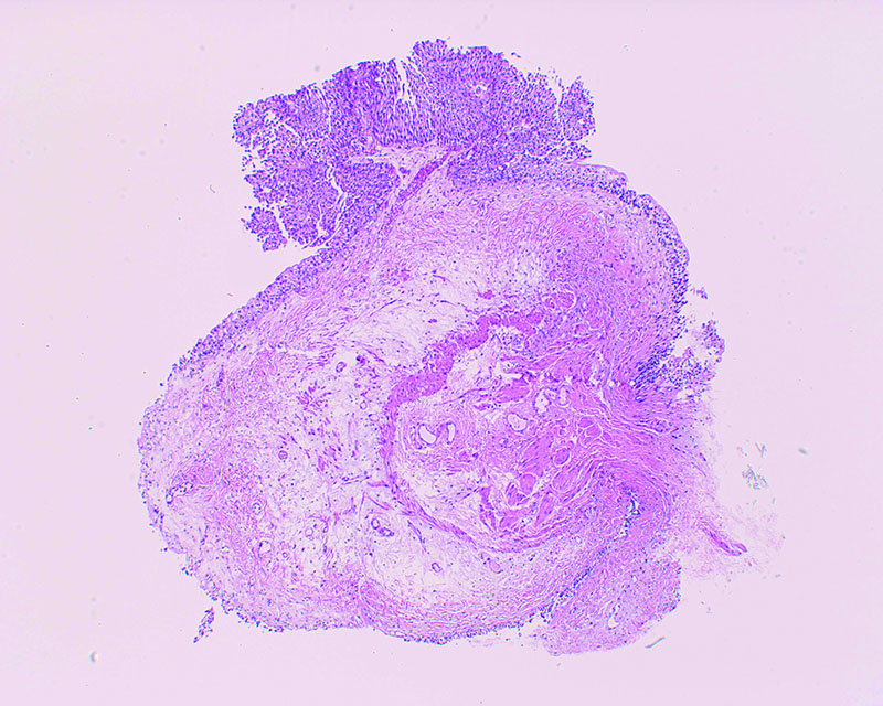
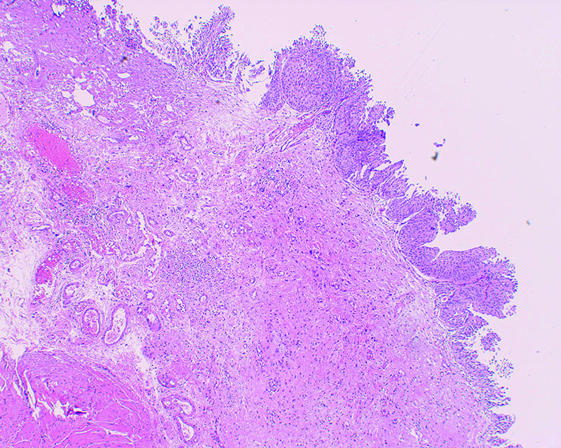
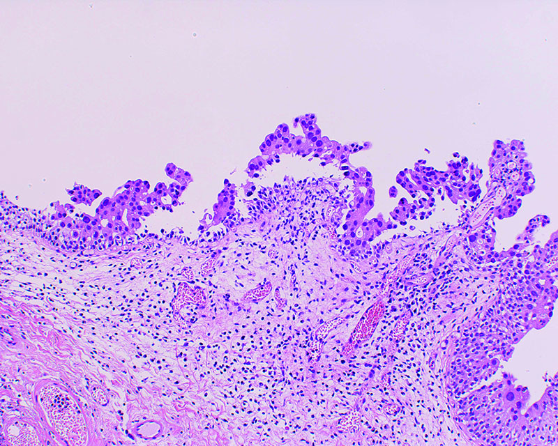
Histopathologic examination revealed T1+is.
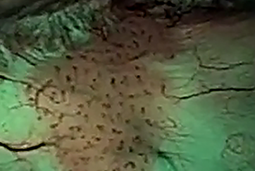
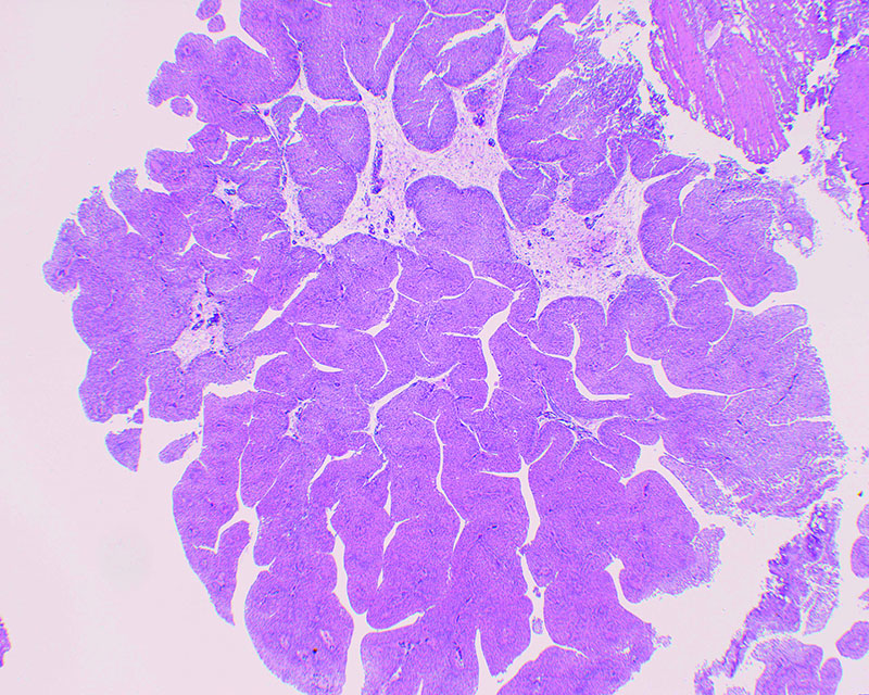
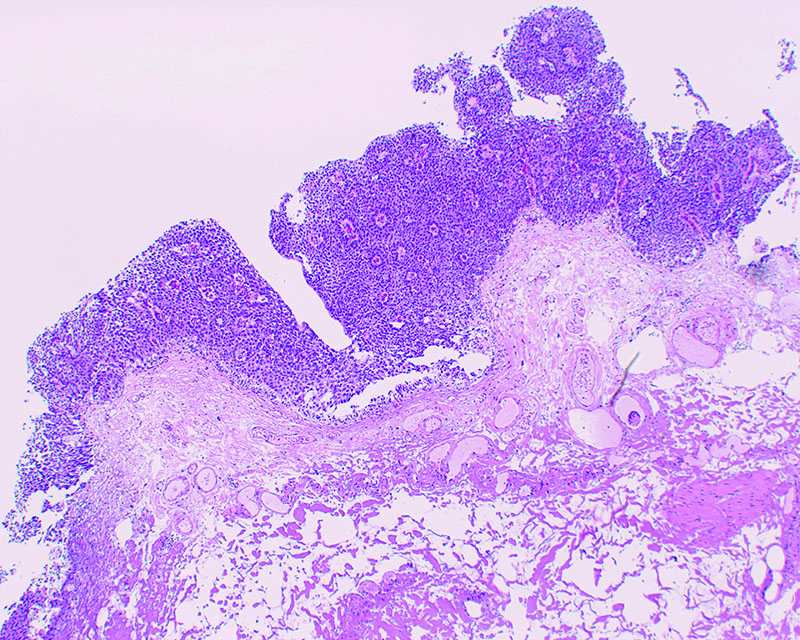
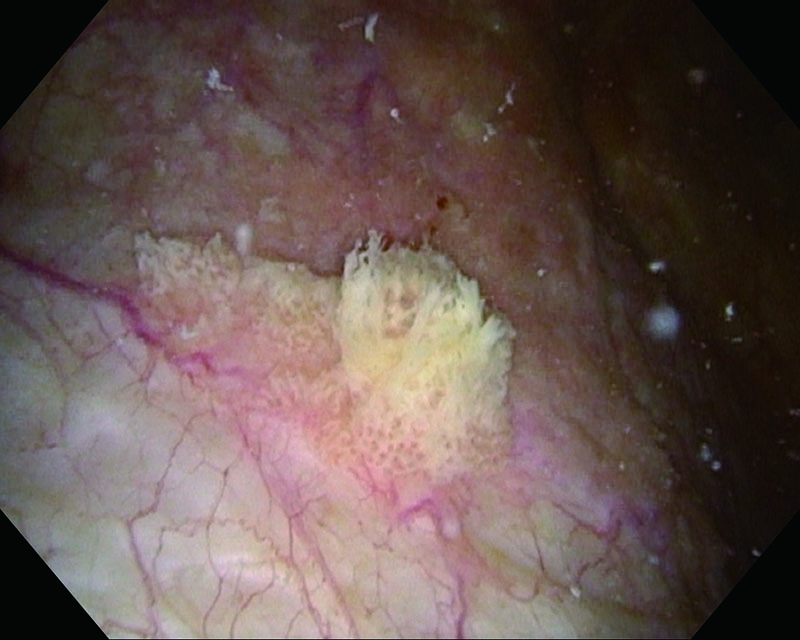
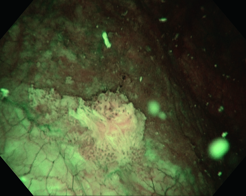
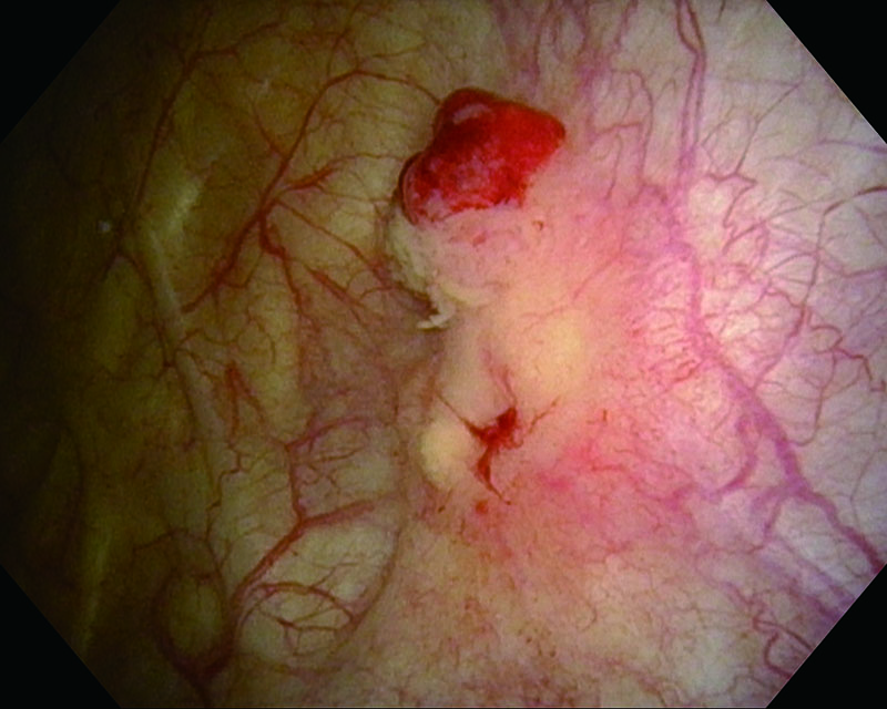
Nodular sessile tumor, age 82, female
Comments
Short tumor which visualization of marginal region was unclear under WLI. Histopathologic examination of resected specimen revealed pT1.
Papillary peduncular tumor/Sessile tumor, age 84, male
Comments
In this case the TUR specimen was T1, high grade. Utilizing NBI™ Technology enabled us to enhance visualization of marginal region.
Nodular sessile tumor, age 78, male
- Content Type