Prof. Jean de la Rosette, MD. Academic Medical Centre, Netherlands
Disclaimer:
- NBI™ Technology is not intended to replace histopathological sampling as a means of diagnosis.
- The positions and statements made herein by Dr. de la Rosette are based on Dr. de la Rosette’s experiences, thoughts and opinions. As with any product, results may vary, and the techniques, instruments, and settings can vary from facility to facility. The content hereof should not be considered as a substitute for carefully reading all applicable labeling, including the Instructions for Use. Please thoroughly review the relevant user manual(s) for instructions, risks, warnings, and cautions. Techniques, instruments, and setting can vary from facility to facility. It is the clinician’s decision and responsibility in each clinical situation to decide which products, modes, medications, applications, and settings to use.
- Dr. de la Rosette is a compensated consultant of the Olympus Corporation.
- All images are courtesy of Dr. de la Rosette, except where otherwise noted.
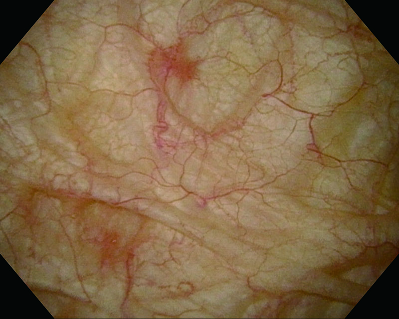
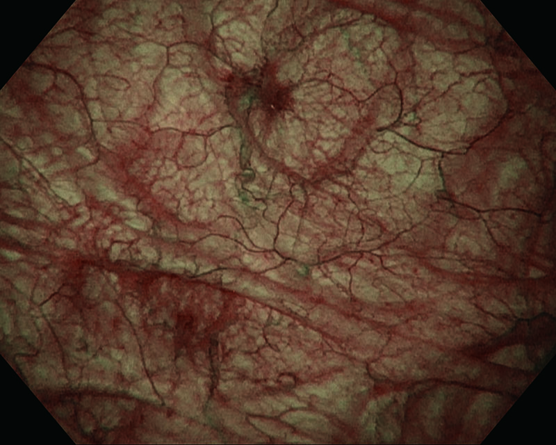
Multiple papillary tumors, age 74, male
White Light
NBI™ Technology
White Light
NBI™ Technology
White Light
NBI™ Technology
White Light
NBI™ Technology
White Light
NBI™ Technology
Comments
Multiple papillary lesions, especially on the bladder trigone, posterior and anterior wall were visible with WLI.
After NBI™ Technology enhancement, additional multiple papillary fields were visualized. Histology showed pTa, Low grade (G1).
Multiple papillary tumors, age 88, male
White Light
NBI™ Technology
Comments
Multiple papillary lesions, especially on the left bladder wall and behind the right ostium,
clearly visible after NBI™ Technology enhancement. Histology showed pTa, Low grade (G1).
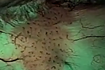
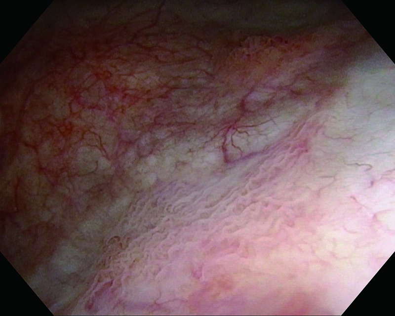
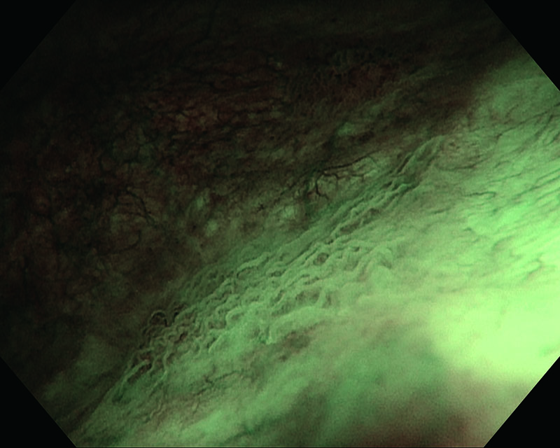
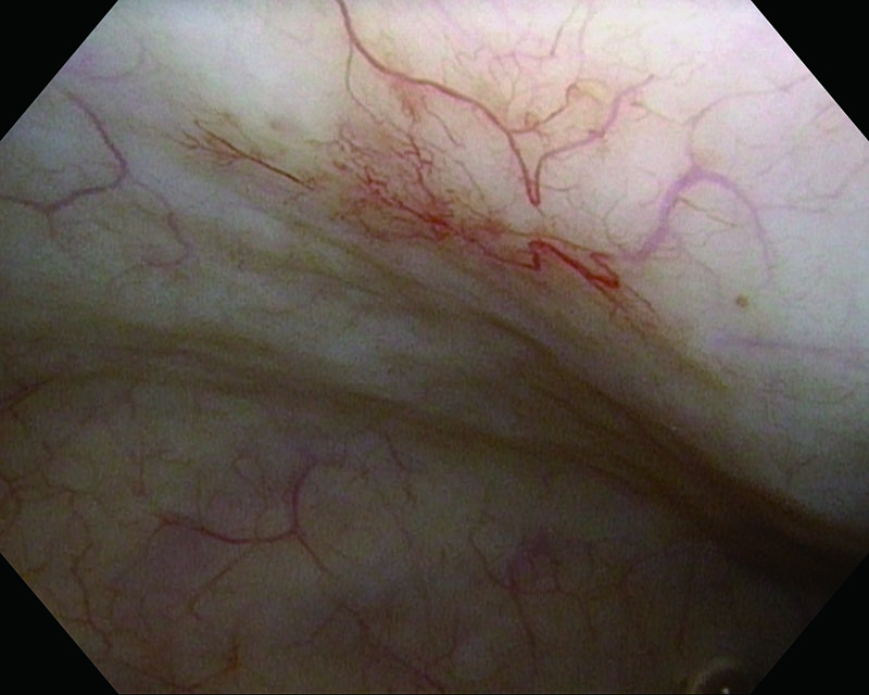
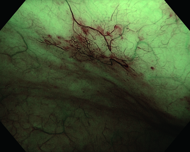
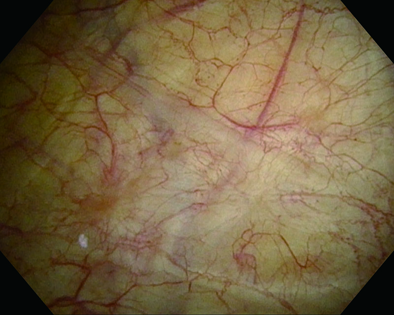
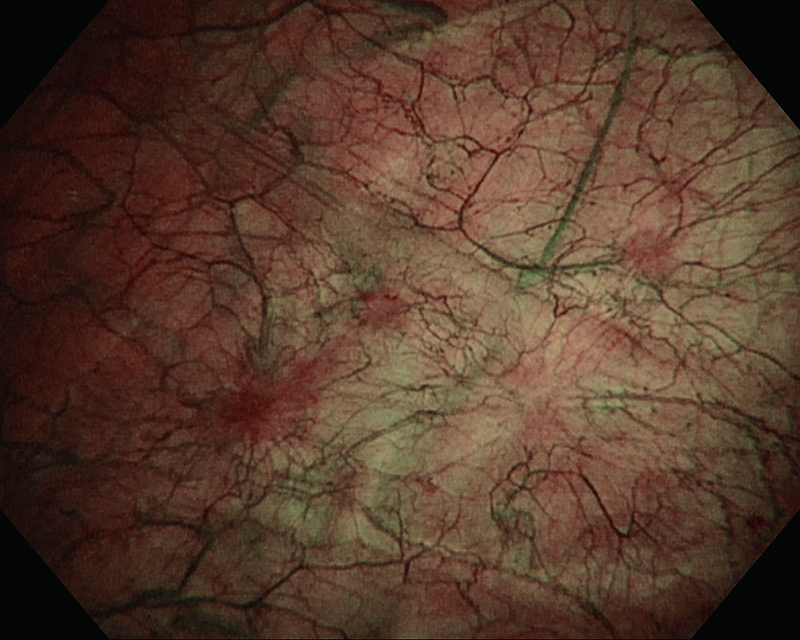
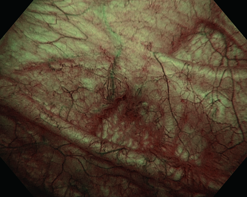
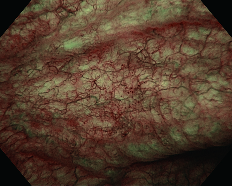
Congestive mucosa of the bladder's trigone, age 28, female
White Light

NBI™ Technology
Comments
Congestive mucosa of the bladder trigone. NBI™ Technology enhances the hyper vascularized area. Histology showed pTa, Low grade (G2).
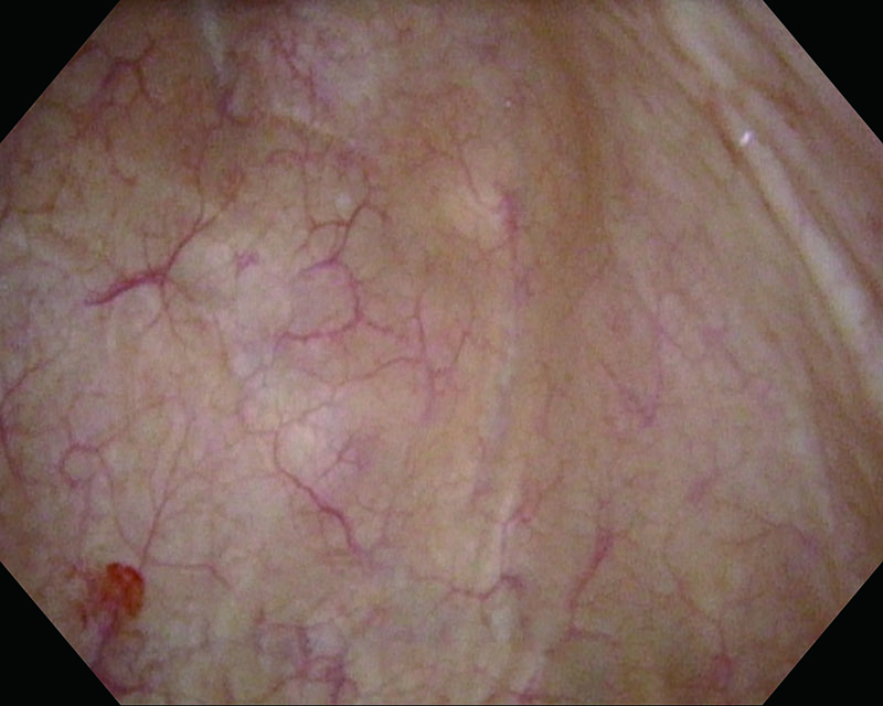
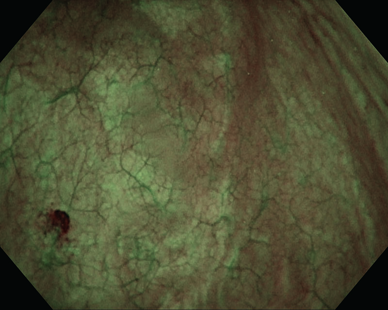
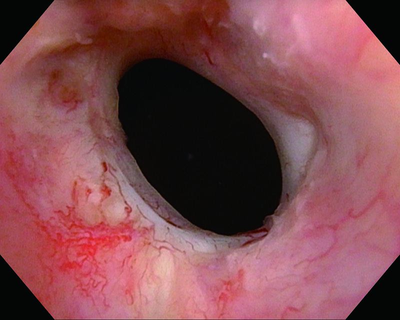
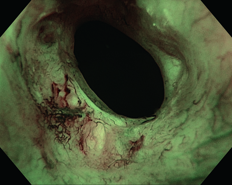
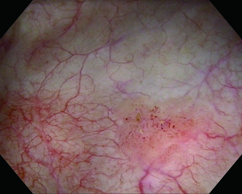
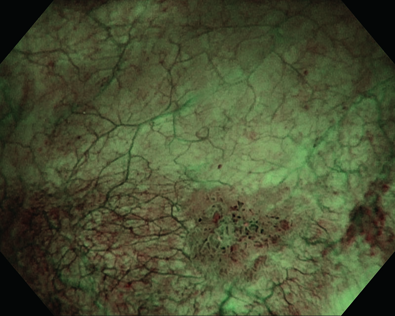
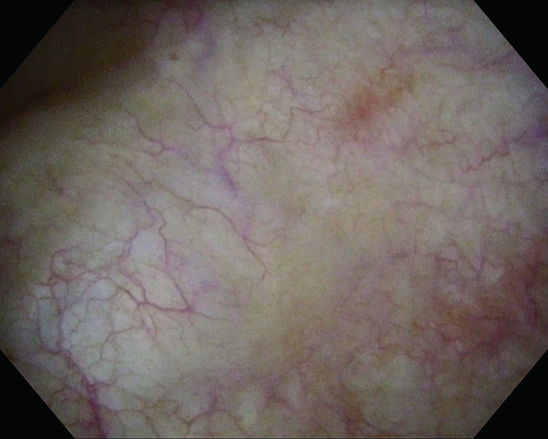
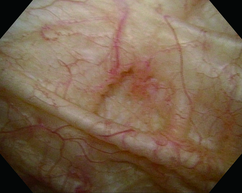
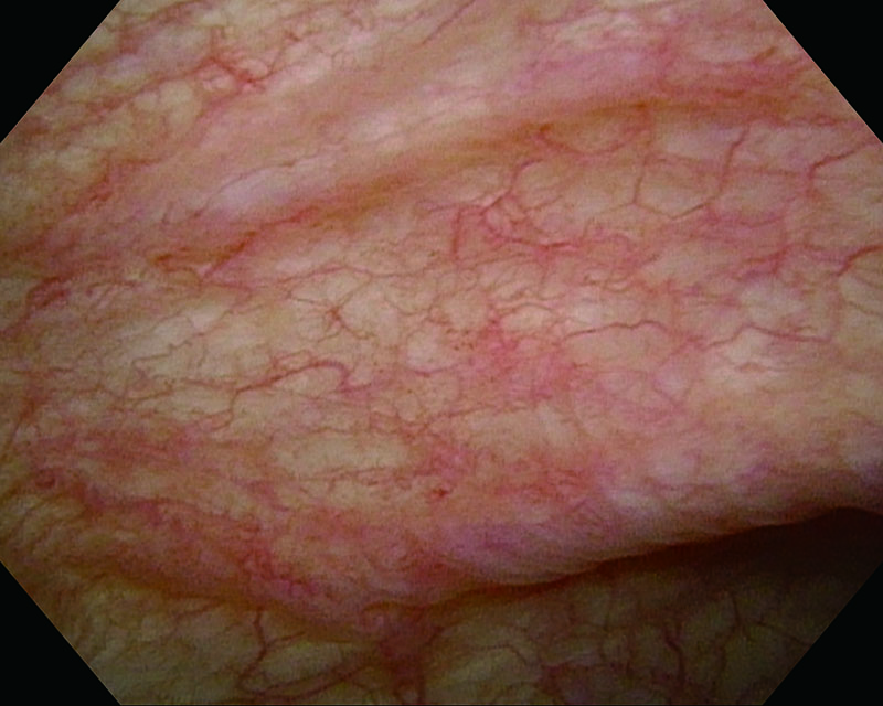
Flat Lesion, age 69, male
White Light
NBI™ Technology
White Light
NBI™ Technology
White Light
NBI™ Technology
Comments
Suspicious superficial lesions adjacent to the right ostium, visible after NBI™ Technology enhancement.
Histology showed pTa, Low grade (G2).
Junichi Inokuchi, MD. Katsunori Tatsugami, MD. Prof. Seiji Naito, MD. Kyushu University, Japan
Angelo Naselli, MD. Prof. Paolo Puppo, MD. Oncological Urology, Istituto Clinico Humanitas Mater Domini, Castellanza, Varese, Italy
- Content Type