Colorectal Case 21

Prof. Yasushi Sano
Kansai Medical University, Osaka, Japan
Sano Hospital, Kobe, JapanScope: CF-EZ1500 DI
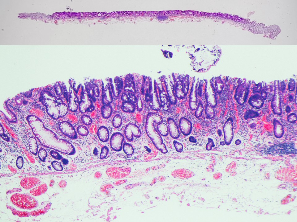
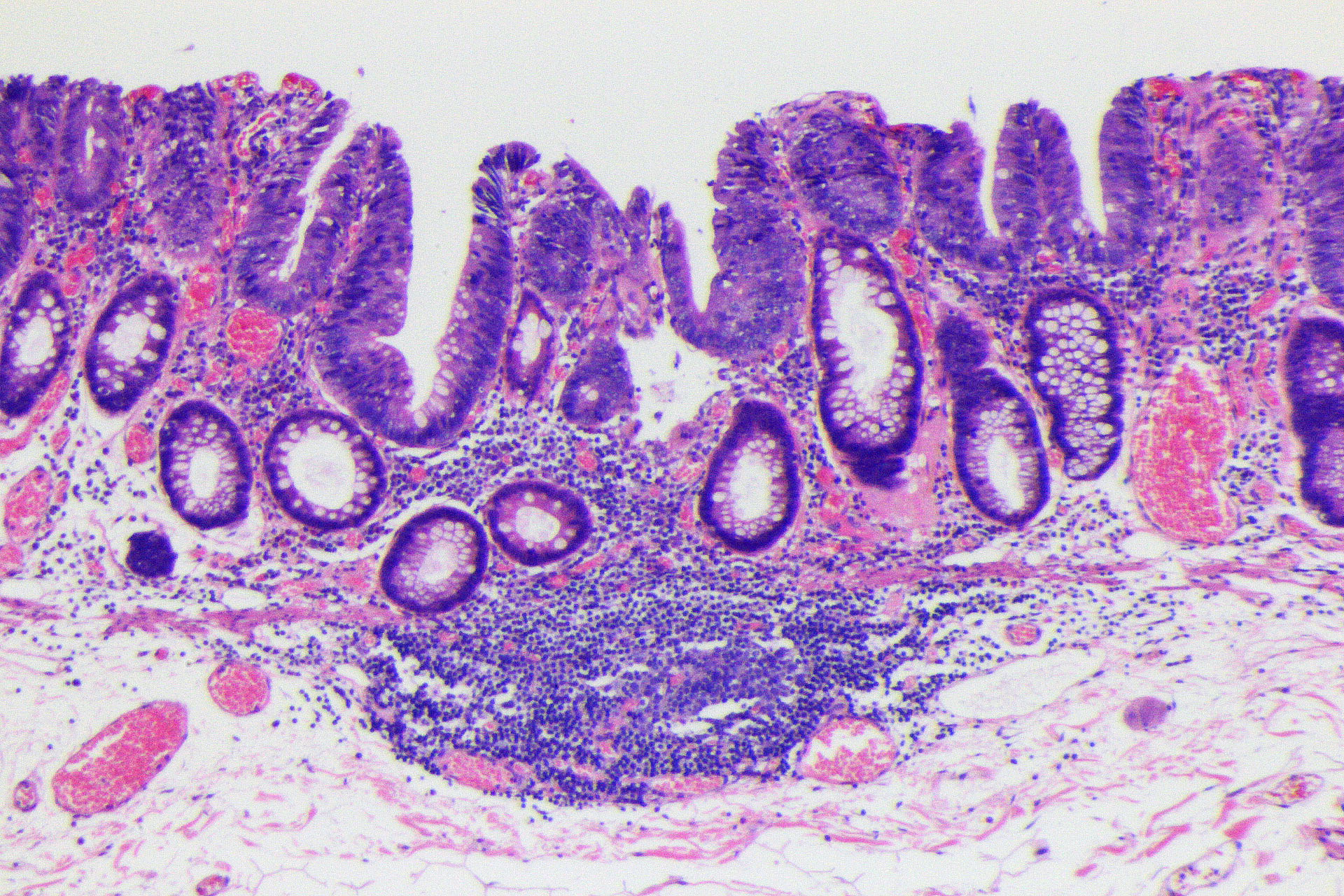
Histology: Tubulovillous adenoma, low-grade dysplasia, including small foci of high-grade dysplasia with lymphoid follicular formation.
Organ: Transverse colon
Patient information: M, 60s
Medical history: Postoperative for pharyngeal cancer, Screening colonoscopy
1. WL

Enhancement : A8
NBI Mode : NA
TXI Mode : NA
RDI Mode : NA
BAI-MAC : On
2. TXI

Enhancement: A8
NBI mode: NA
TXI Mode: NA
RDI Mode: NA
BAI-MAC: On
3. NBI

Enhancement : A8
NBI Mode : On (mode 3)
TXI Mode : NA
RDI Mode : NA
BAI-MAC : On
4. NBI with magnification

Enhancement : A8
NBI Mode : On (mode 3)
TXI Mode : NA
RDI Mode : NA
BAI-MAC : On
5. NBI with magnification

Enhancement : A8
NBI Mode : On (mode 3)
TXI Mode : NA
RDI Mode : NA
BAI-MAC : On
6. Chromoendoscopy

Enhancement : A8
NBI Mode : NA
TXI Mode : NA
RDI Mode : NA
BAI-MAC : On
7. Chromoendoscopy with magnification

Enhancement : A8
NBI Mode : NA
TXI Mode : NA
RDI Mode : NA
BAI-MAC : On
8. Chromoendoscopy with magnification

Enhancement : A8
NBI Mode : NA
TXI Mode : NA
RDI Mode : NA
BAI-MAC : On
9. Crystal violet

Enhancement : A8
NBI Mode : NA
TXI Mode : NA
RDI Mode : NA
BAI-MAC : On
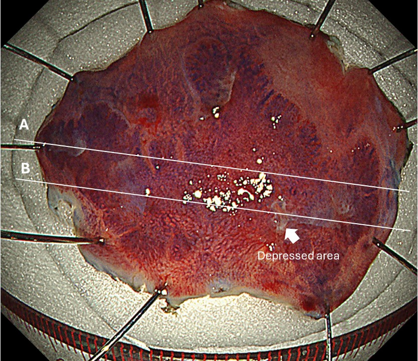
10. Cutting line for histology
11. Histology (low power)
12. Histology (high power)
Case Video
Video 1: Observation by WL, TXI, NBI
Video 2: Observation by chromoendoscopy with Indigo carmine and Crystal violet staining
Overall Comment
LST-NG lesions are considered precursor lesions of PCCRC 1,2) and should not be missed on colonoscopy. In this LST-NG case, the lesion was detected by pale redness. NBI observation was very useful in diagnosing the extent. In general, LST-NG lesions sometimes show a depressed area, and detailed endoscopic observation of this area is important for endoscopic diagnosis. In this case, a JNET 2B region was observed in the depressed area. The pathological diagnosis of the depressed area was adenoma with high-grade dysplasia. In Japan, it is recommended that dye endoscopy be added to the endoscopy to improve sensitivity when a lesion showing JNET 2B is observed.
1. Matsuda T, Fujii T, Sano Y, et al. Randomised comparison of post-polypectomy surveillance intervals following a two-round baseline colonoscopy: the Japan Polyp Study Workgroup. Gut. 2020 Nov 2;70(8):1469–78.
2. Sano Y, Hotta K, Matsuda T, et al. Japan Polyp Study Workgroup. Endoscopic Removal of Premalignant Lesions Reduces Long-Term Colorectal Cancer Risk: Results From the Japan Polyp Study. Clin Gastroenterol Hepatol. 2023 Aug 6:S1542-3565(23)00588-8.
* Specifications, design and accessories are subject to change without any notice or obligation on the part of the manufacturer
Prof. Yasushi Sano Case 22: 0-Is (submucosal invasive carcinoma), 11mm, JNET 2B
Prof. Yasushi Sano
- Keyword
- Content Type