Colorectal Case 10

Disclaimer:
- NBI™ and TXI™ technologies are not intended to replace histopathological sampling as a means of diagnosis
- The positions and statements made herein by Dr. Supakij Khomvilai are based on Dr. Supakij Khomvilai’s experiences, thoughts and opinions. As with any product, results may vary, and the techniques, instruments, and settings can vary from facility to facility. The content hereof should not be considered as a substitute for carefully reading all applicable labeling, including the Instructions for Use. Please thoroughly review the relevant user manual(s) for instructions, risks, warnings, and cautions. Techniques, instruments, and setting can vary from facility to facility. It is the clinician’s decision and responsibility in each clinical situation to decide which products, modes, medications, applications, and settings to use.
- The EVIS X1™ endoscopy system is not designed for cardiac applications. Other combinations of equipment may cause ventricular fibrillation or seriously affect the cardiac function of the patient. Improper use of endoscopes may result in patient injury, infection, bleeding, and/or perforation. Complete indications, contraindications, warnings, and cautions are available in the Instructions for Use (IFU)
1) data on file with Olympus (DC00489968)
2) data on file with Olympus (DC00567392)
Scope: CF-EZ1500DL
Patient information: F, 60s
Medical history: Screening for Colorectal cancer
1. White light observation
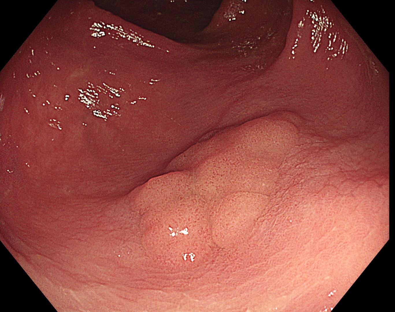
Enhancement : A8
NBI Mode : NA
TXI Mode : NA
RDI Mode : NA
BAI-MAC : On
2. TXI™ technology observation
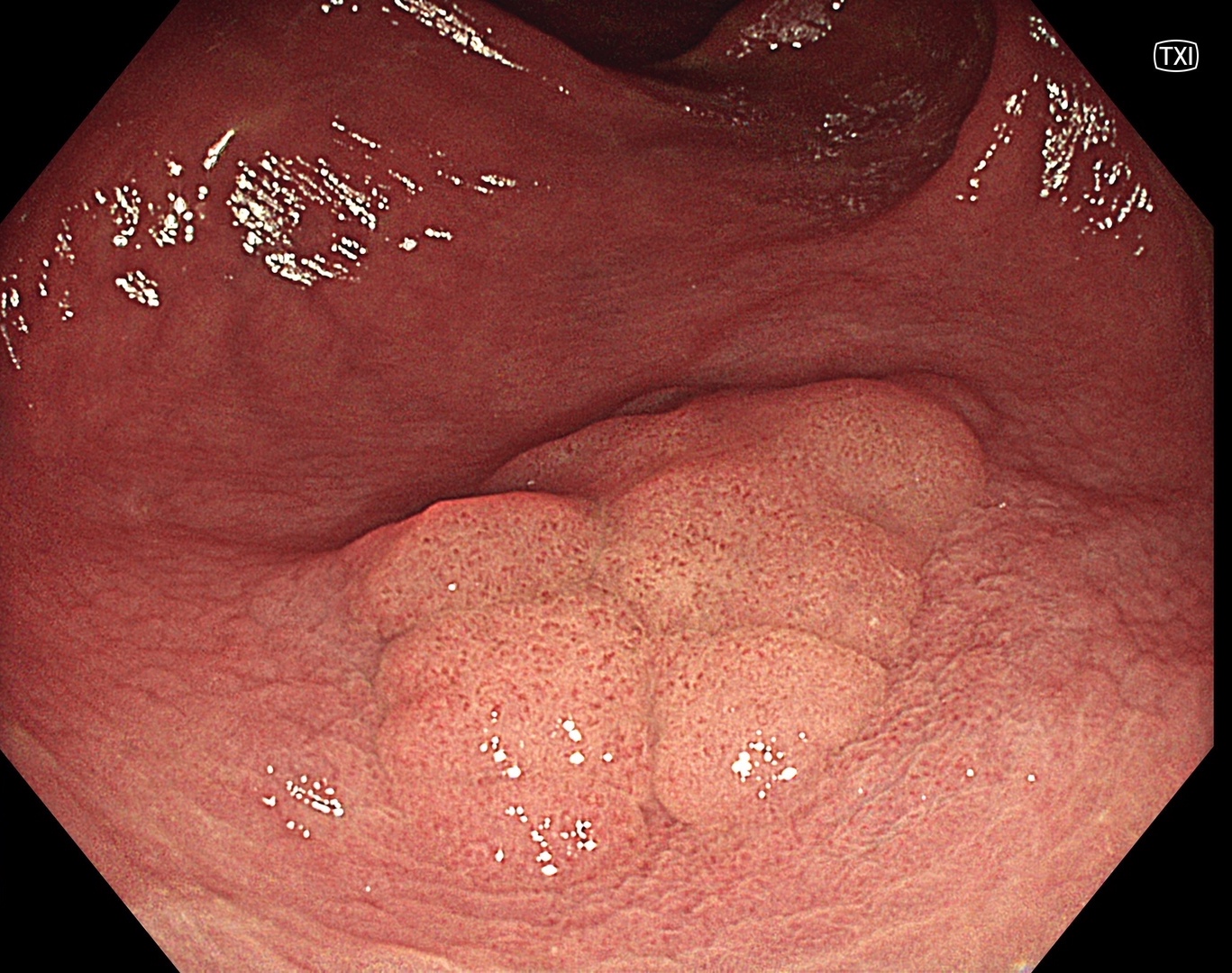
Enhancement : A8
NBI Mode : NA
TXI Mode : 2
RDI Mode : NA
BAI-MAC : On
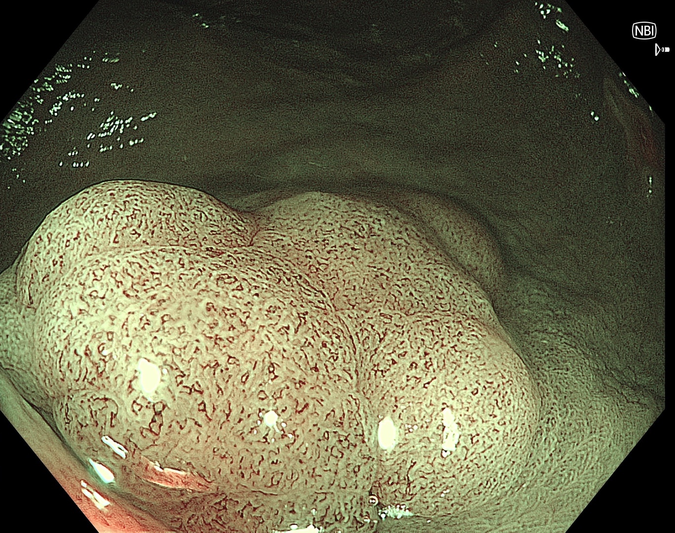
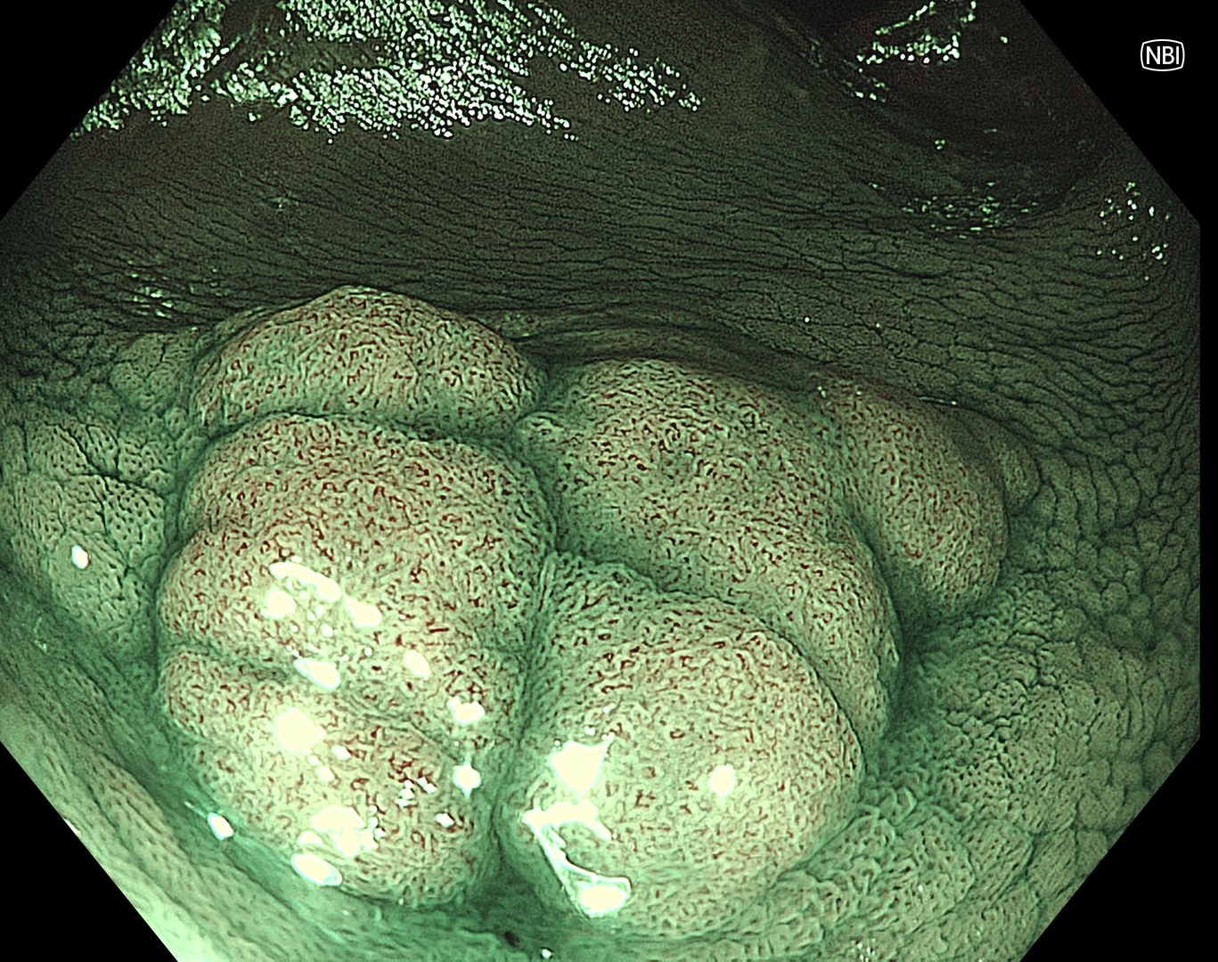
3. NBI™ technology Observation
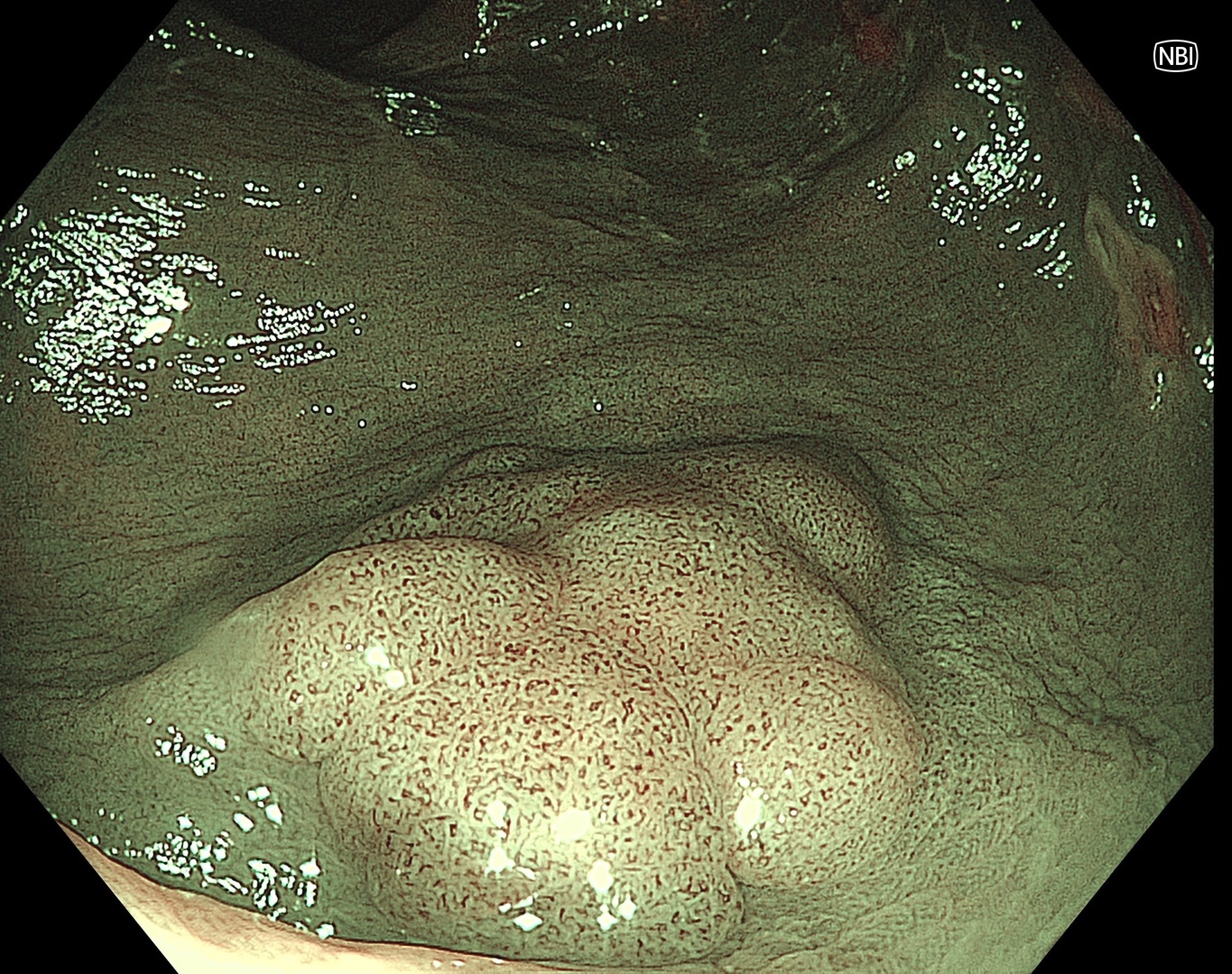
Enhancement : B8
NBI Mode : 3
TXI Mode : NA
RDI Mode : NA
BAI-MAC : On
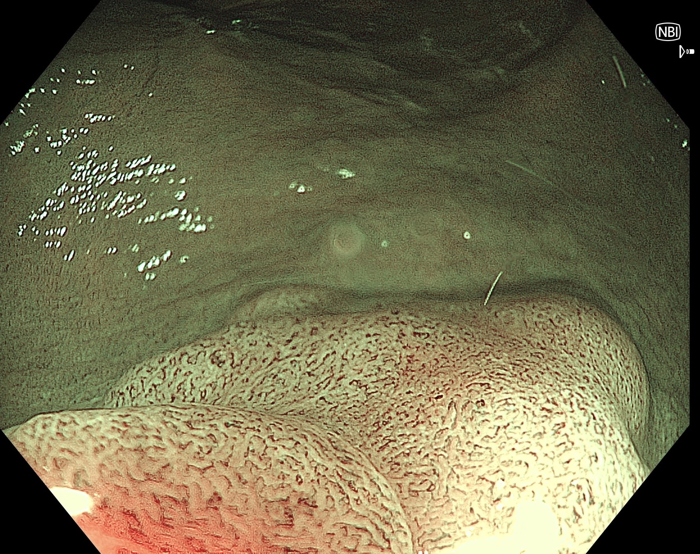
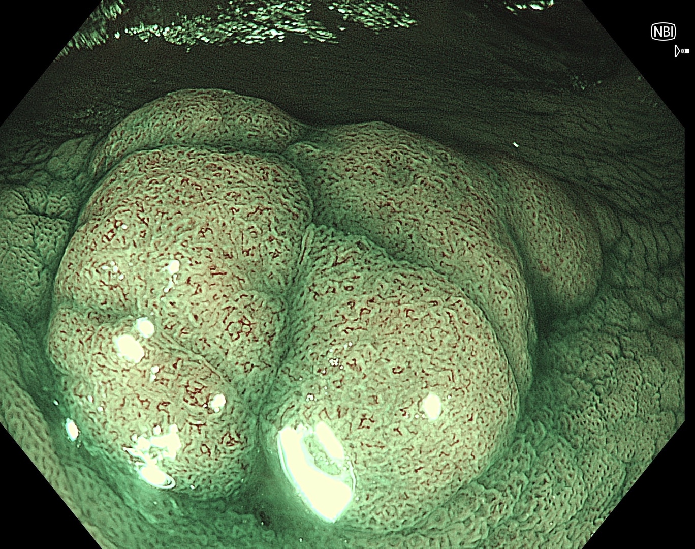
4. NBI™ technology Observation
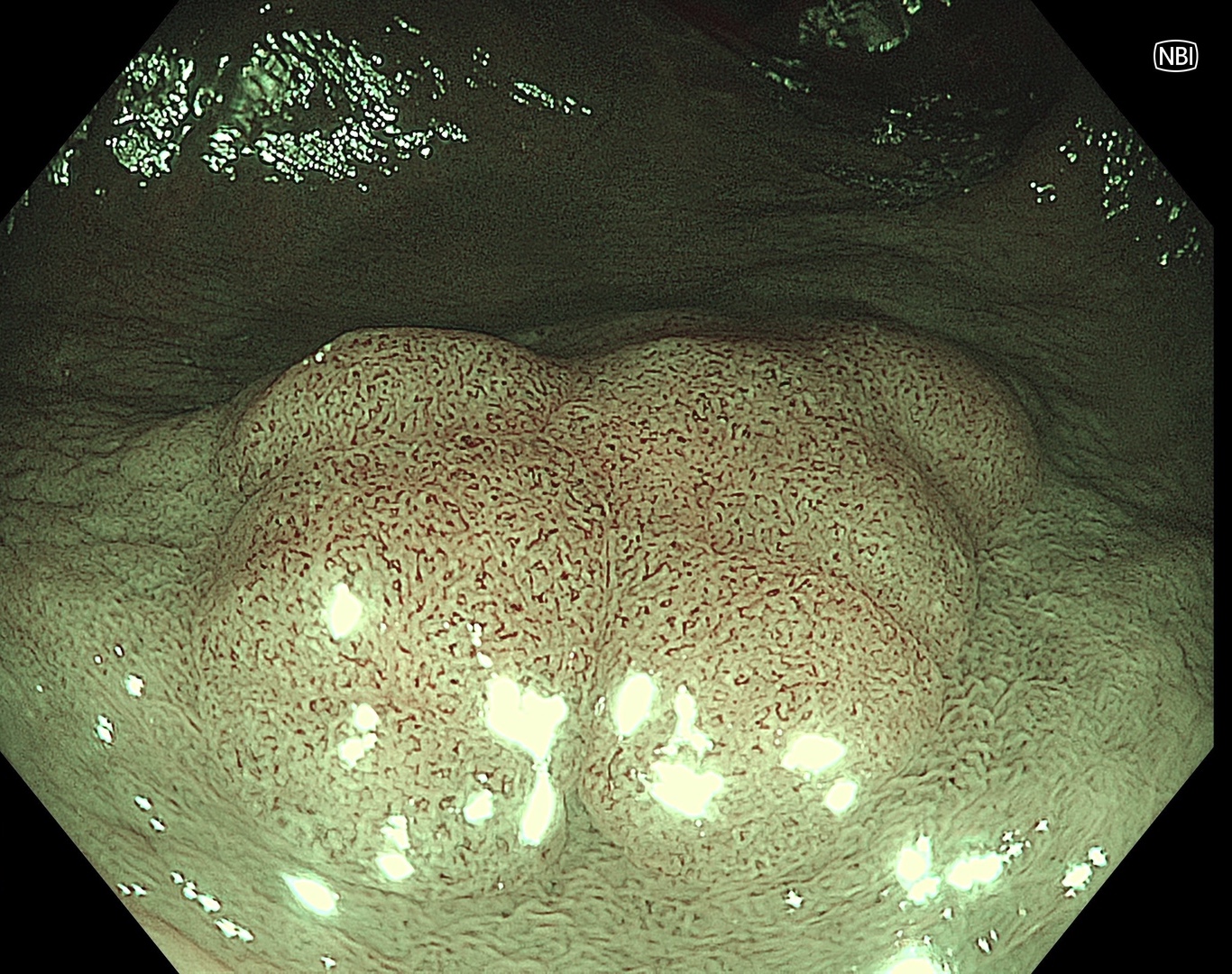
Enhancement : B8
NBI Mode : 3
TXI Mode : NA
RDI Mode : NA
BAI-MAC : On
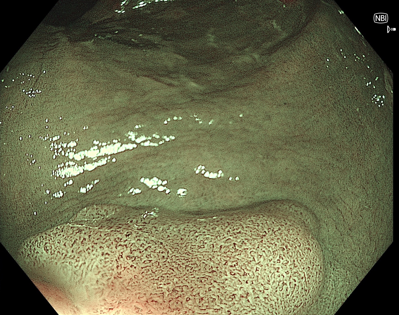
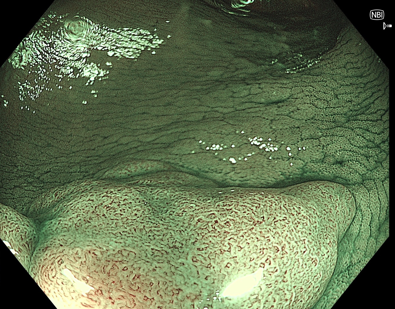
5. NBI™ technology with Near Focus
Enhancement : B8
NBI Mode : 3
TXI Mode : NA
RDI Mode : NA
BAI-MAC : On
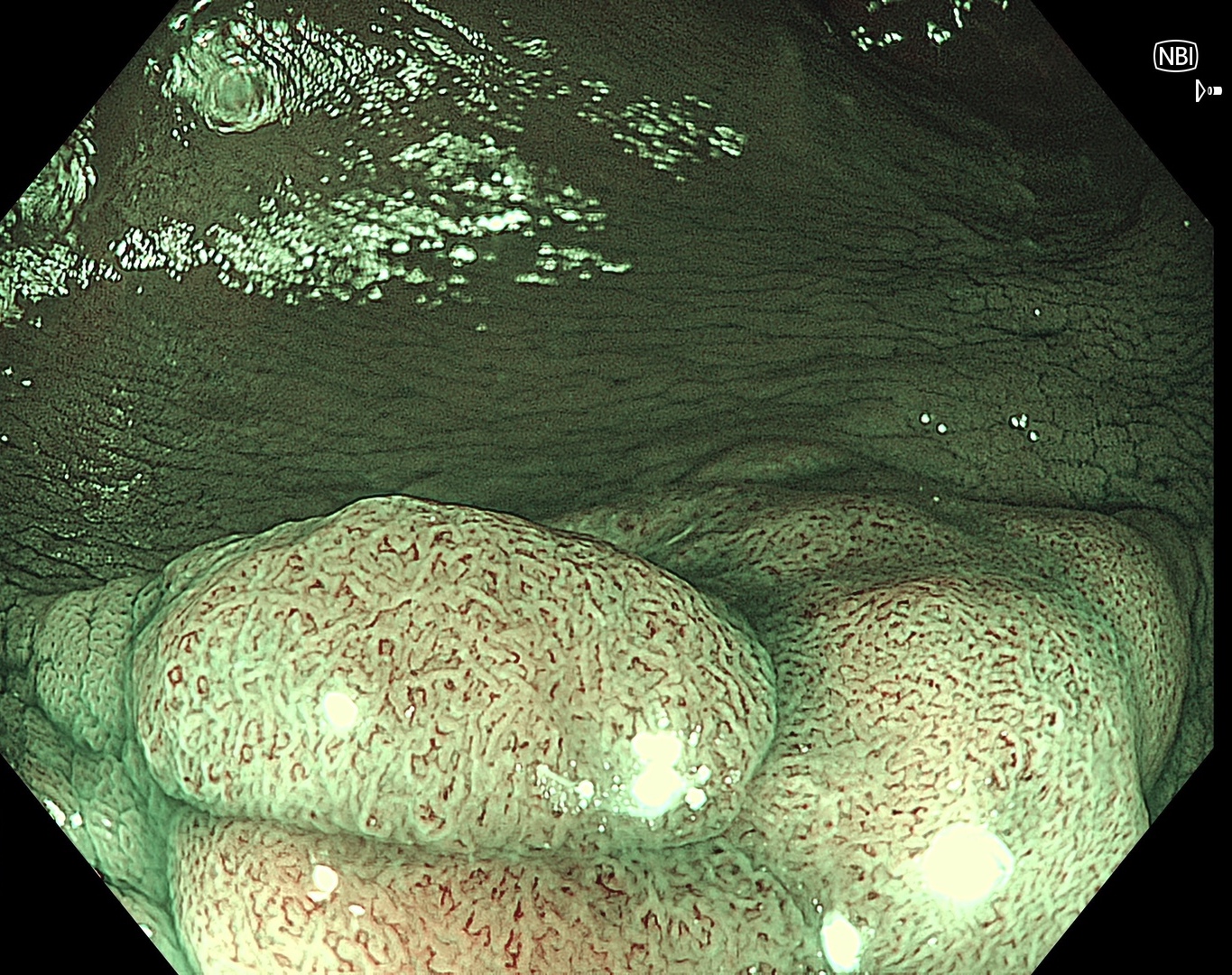
6. NBI™ technology with Near Focus
Enhancement : B8
NBI Mode : 3
TXI Mode : NA
RDI Mode : NA
BAI-MAC : On
7. NBI™ technology with Near Focus
Enhancement : B8
NBI Mode : 3
TXI Mode : NA
RDI Mode : NA
BAI-MAC : On
8. Indigo carmine spray
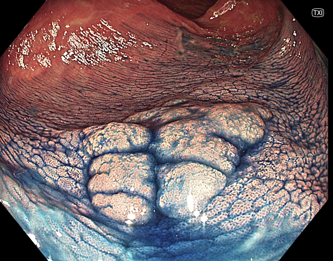
Enhancement : A8
NBI Mode : NA
TXI Mode : 2
RDI Mode : NA
BAI-MAC : On
9. NBI™ technology with Indigo carmine spray
Enhancement : B8
NBI Mode : 3
TXI Mode : NA
RDI Mode : NA
BAI-MAC : On
10. NBI™ technology, Indigo carmine & Near Focus
Enhancement : B8
NBI Mode : 3
TXI Mode : NA
RDI Mode : NA
BAI-MAC : On
11. NBI™ technology, Indigo carmine & Near Focus
Enhancement : B8
NBI Mode : 3
TXI Mode : NA
RDI Mode : NA
BAI-MAC : On
12. NBI™ technology, Indigo carmine & Near Focus
Enhancement : B8
NBI Mode : 3
TXI Mode : NA
RDI Mode : NA
BAI-MAC : On
* Specifications, design and accessories are subject to change without any notice or obligation on the part of the manufacturer
Dr. Supakij Khomvilai Case 11: Colonic polyp (Tubular adenoma)
Dr. Supakij Khomvilai
- Keyword
- Content Type


