03 Anterior Ethmoidectomy
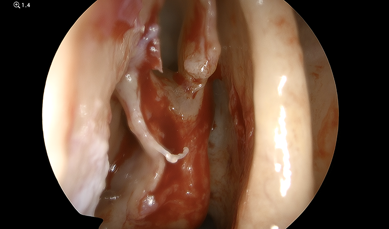
Once the uncinate has been removed and the maxillary sinus identified, it should be possible to completely view the anterior face of the bulla ethmoidalis.
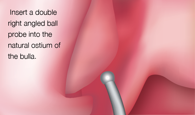
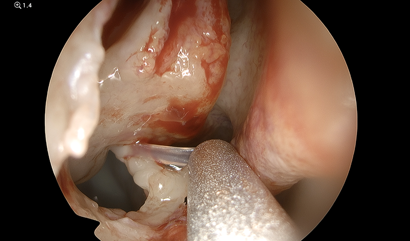
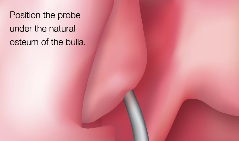
Using the double right angled ball probe, identify the natural ostium of the bulla on its medial side.

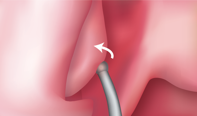
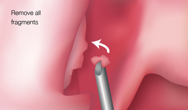
Fracture the anterior face of the bulla forward with the ball probe.
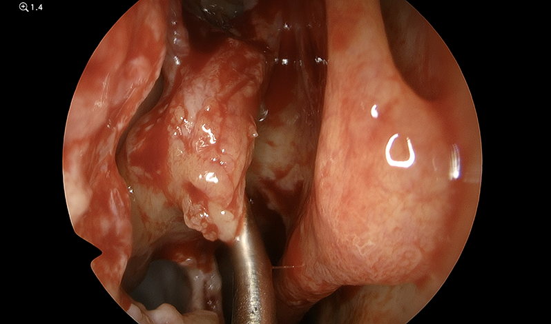
The fragments of the anterior wall of the bulla can then be removed using a combination of thru-cutting instruments and the microdebrider.
Note:
・It is important that the tip of the microdebrider is visible at all times and no pressure whatsoever is exerted on the lateral nasal wall to prevent accidental injury to the lamina papyracia.
・The bulla may be closely applied to the ground lamella of the middle turbinate. If so, the junction between the anterior and posterior ethmoid will be the posterior wall of the bulla. If the bulla sits anterior to the ground lamella (basal lamella), then there will be a cleft behind the posterior wall of the bulla (the retro-bullar recess).
・When removing the anterior face of the bulla, it is important to remember the position of the anterior ethmoidal artery. The artery is usually located one cell posterior to the frontal recess. If the bulla reaches the skull base then the anterior ethmoidal artery may be anterior, at the level of the anterior wall or just posterior to the anterior wall of the bulla ethmoidalis. As such, a surgeon should be prepared for each of these eventualities. The position of the anterior ethmoidal artery should be checked prior to surgery by detailed examination of the patient’s CT scans.
- Content Type