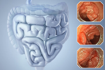
Colorectal Case 20

Prof. Yasushi Sano
Kansai Medical University, Osaka, Japan
Sano Hospital, Kobe, JapanScope: CF-EZ1500 DI
Histology: Tubulovillous adenoma, low-grade dysplasia, including small foci of high-grade dysplasia with traditional serrated adenoma-like appearance.
Organ: Rectum
Patient information: M, 70s
Medical history: FIT positive
6. NBI with magnification

Enhancement : A8
NBI Mode : On (mode 3)
TXI Mode : NA
RDI Mode : NA
BAI-MAC : On
Case Video
Video 1: Observation by WL, TXI, NBI
Video 2: Observation by chromoendoscopy with Indigo carmine and Crystal violet staining
Overall Comment
EVIS X1 with CF-EZ1500 DI colonoscope provides excellent image quality over previous generations of endoscope system. The clarity and resolution of the endoscopic image are excellent, with the help of TXI function this will enhance the identification of lesions during examination. Once a lesion has been identified, the clear focused image contributes accurate detailed examination of morphology of the lesion. The excellent image quality extends to NBI mode, and together with optical magnification accurate characterization of the lesion can be made.
In this case, the parts of tubulovillous adenoma and traditional serrated adenoma (cicada wing-like findings) are mixed, and the differences in endoscopic findings can be well educated 1).
1. Sano Y, Saito Y, et al. Efficacy of magnifying chromoendoscopy for the differential diagnosis of colorectal lesions. Digestive Endoscopy. April 2005 Pages 105-116.
* Specifications, design and accessories are subject to change without any notice or obligation on the part of the manufacturer
Dr. Ho Dang Quy Dung Case 21: 0-IIa (LST-non-granular type), 25mm, JNET 2A+2B
Prof. Yasushi Sano
- Keyword
- Content Type


