Colorectal Case 28
Mr. Peter Borch-Johnsen
RN, M.Sci, PhD- Student
Department of Medicine
Ersta Hospital
Department of Medical Sciences
Uppsala University
Scope: CF-XZ1200
Organ: Anal canal
Patient information: Male, MSM+HIV
Medical history: Good CD4 count
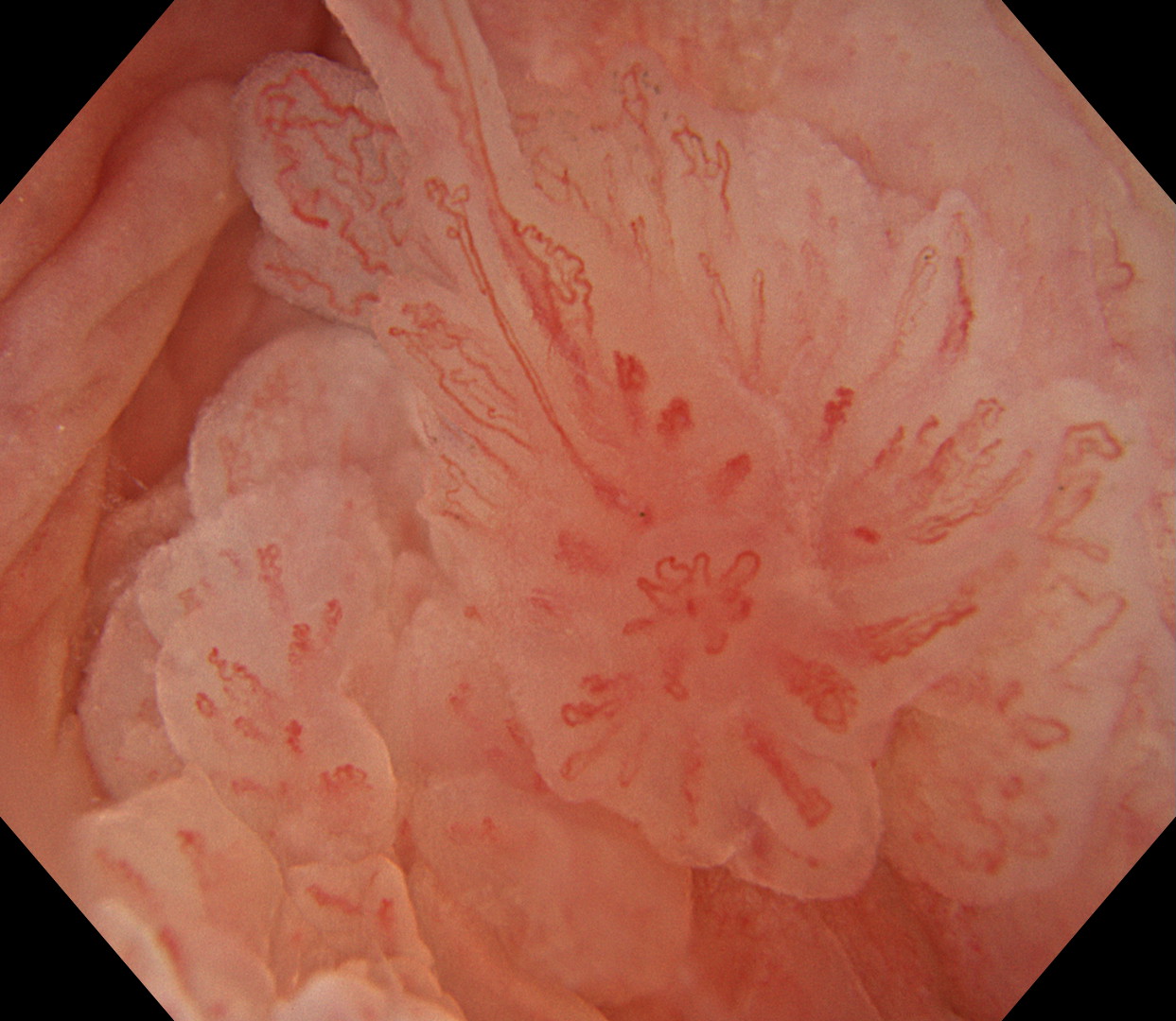
1a. ASIL WLI
1b. ASIL TXI mode 1
1c. ASIL TXI mode 2
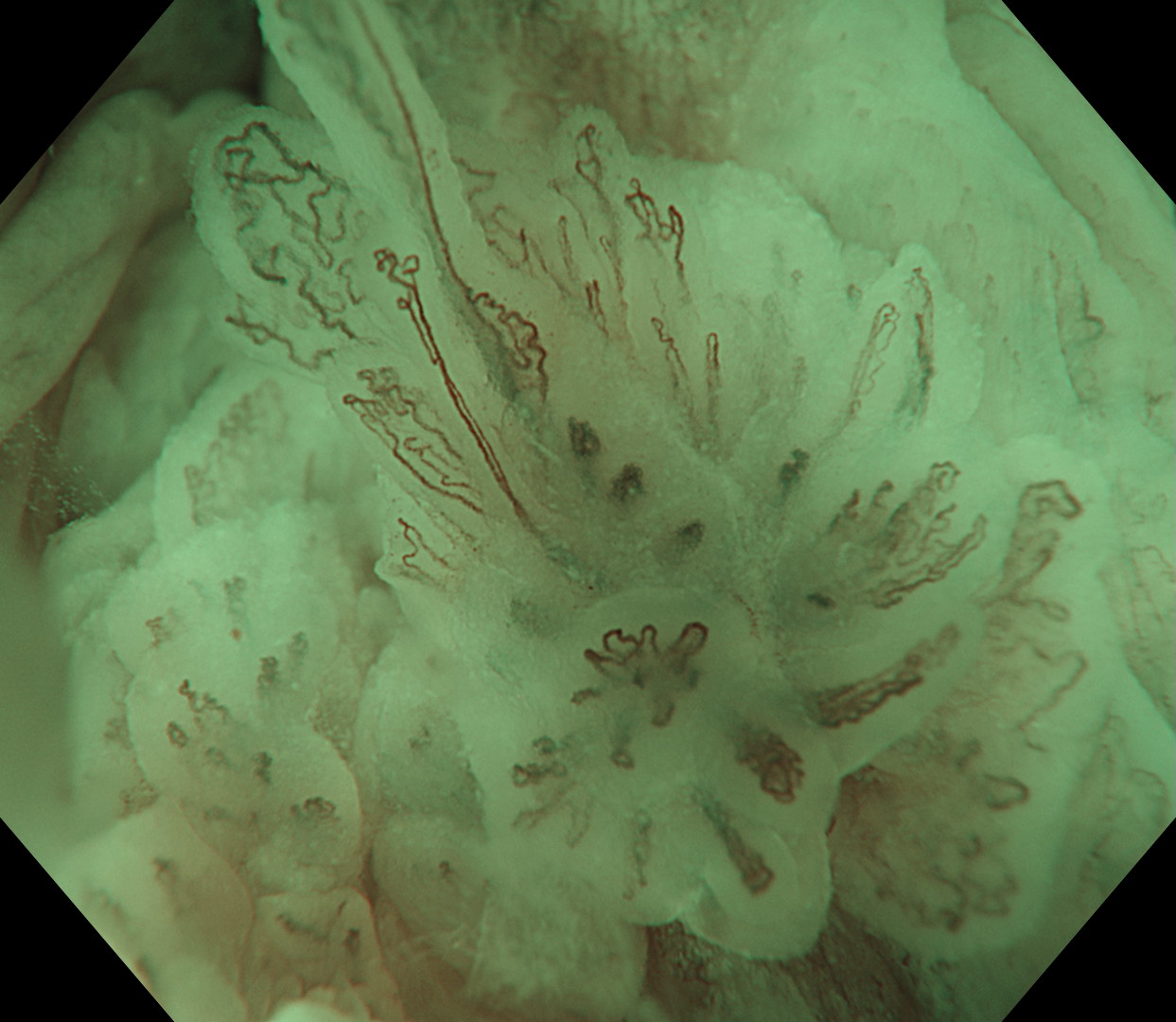
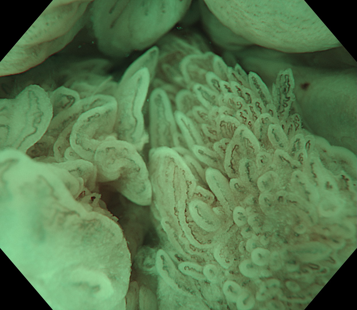
1d. ASIL NBI
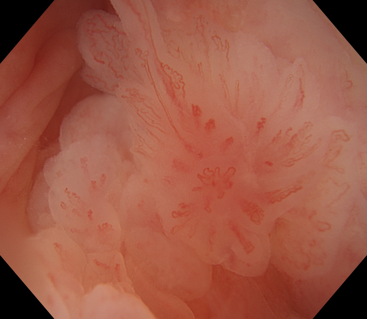
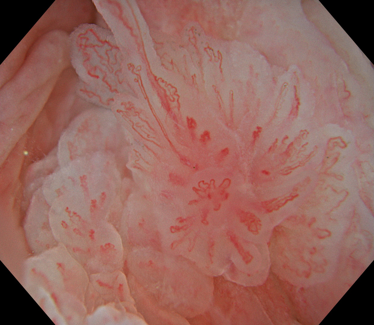
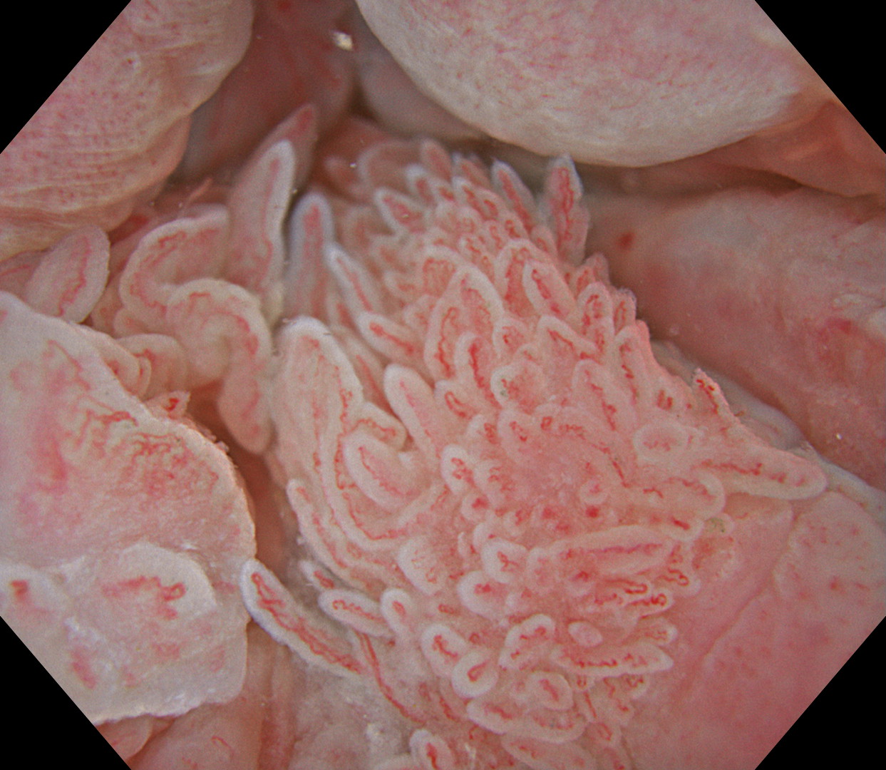
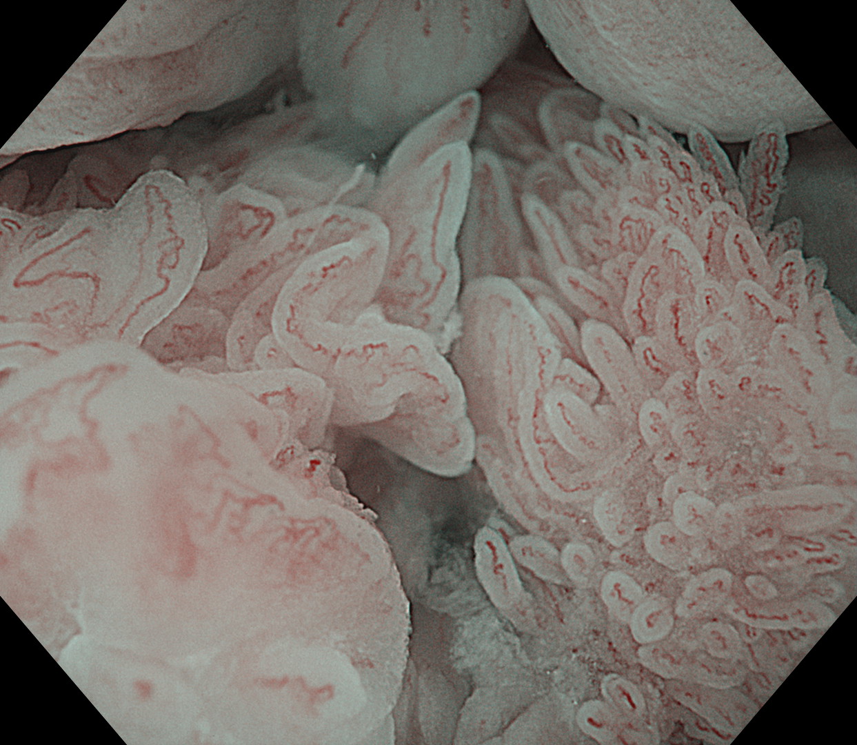
1e. ASIL RDI mode 3
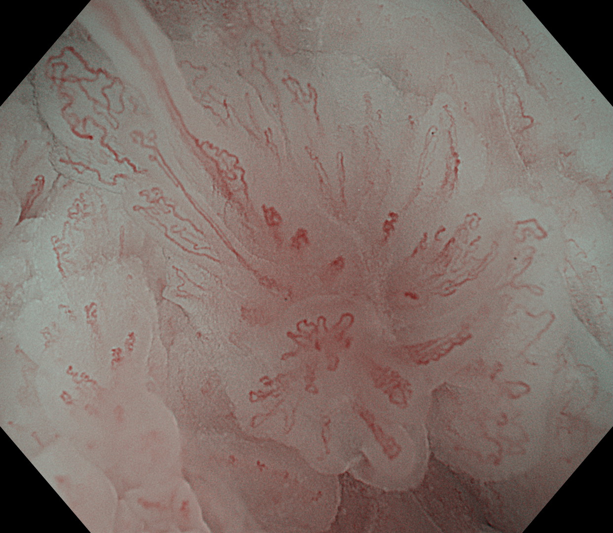
2a. TXI mode 1 ASIL
2b. NBI ASIL
2c. RDI mode 3 ASIL
Case Video
This is a case with multi-focal anal squamous intraepithelial lesion (ASIL) in a high-risk patient for anal cancer (MSM (Men who have sex with men) and HIV+). Referral for colonoscopy due to irregular stools and blood per rectum. Colonoscopy and endoscopic high resolution anoscopy were performed with a zoom colonoscope - CF-XZ1200. A distal attachment was secured leaving approximately 2 mm of free cap at the tip to maintain a suitable distance between the instrument and the mucosa. Co2 is turned off and the anal canal filled with water. TXI mode 1 increases the vascular structure compared with WLI. We usually initiate the examination with TXI mode 1 because it highlights the morphology, vascular structure and colour of the mucosa. NBI and RDI mode 3 are intermittent used and seem to further increase the contrast and resolution making the vessel network even more conspicuous.
Overall Comment
Several studies are ongoing regarding endoscopic detection (optical diagnosis), resection and screening for ASIL in high-risk individuals. The endoscopic method description for detection and resection has previously published.
* Specifications, design and accessories are subject to change without any notice or obligation on the part of the manufacturer
* The product used in this case is sold for limited area. This page is provided as information.
Mr. Peter Borch-Johnsen Cases 32: High quality and high magnification imaging provided by the CF-XZ1200 will advance quality of colonoscopic diagnosis during both screening and detailed examination
- Keyword
- Content Type