Esophageal Case 3

Prof. Stefan Seewald
GastroZentrum Hirslanden, Zurich, Switzerland
Scope: GIF-EZ1500
Case: Squamous Cell Carcinoma
Organ: Esophagus
Patient information: M, 70s
Medical history: Smoker
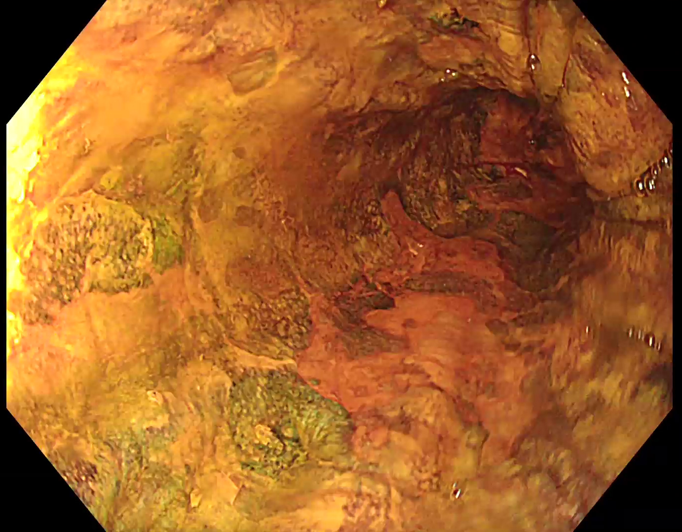
1. WLI
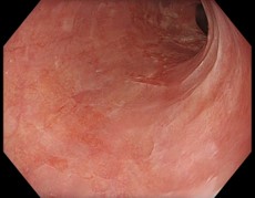
A suspicious reddish lesion can be identified at 6 to 9 o’clock.
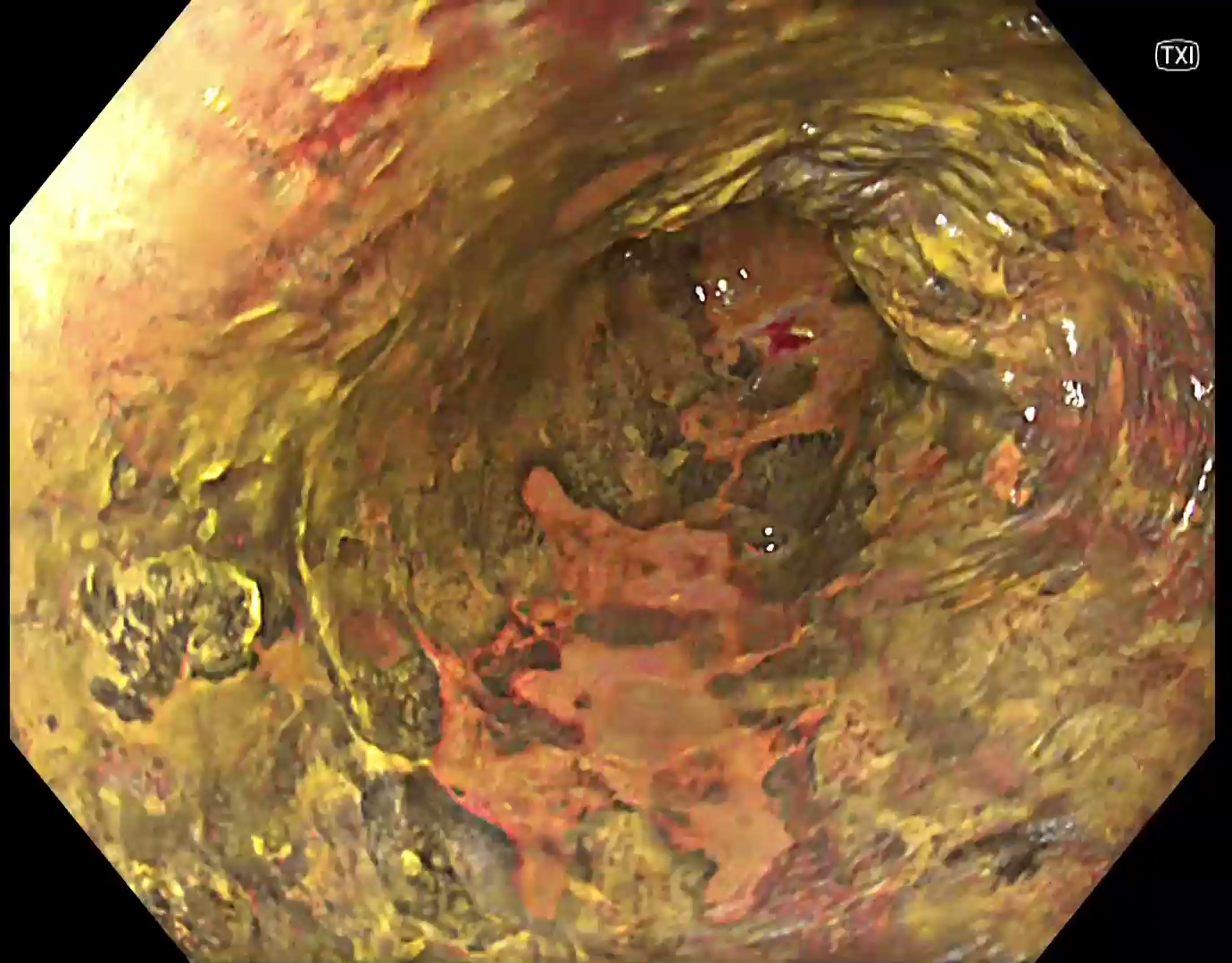
2. TXI
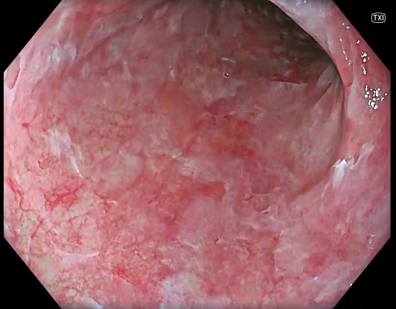
TXI suggests a larger area that may be interpreted as reflux esophagitis at the first glance.
3. NBI
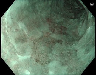
However, with NBI abnormal IPCLs can be identified which require closer examination.
4. Near Focus with NBI
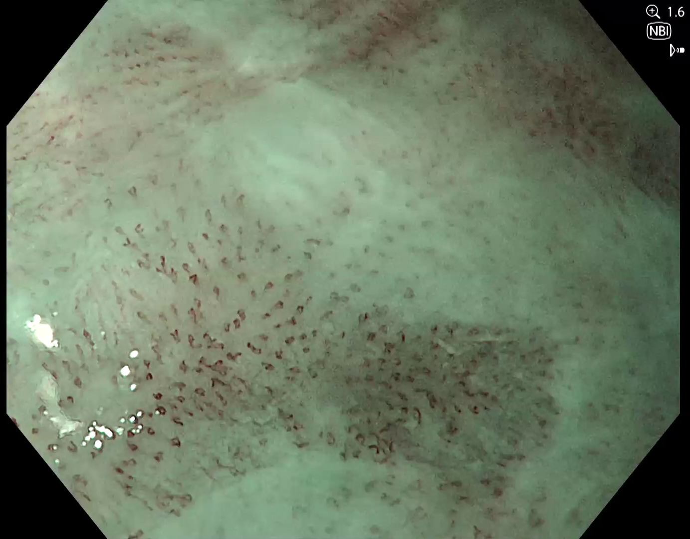
Abnormal IPCLs can be confirmed under near focus with 1.6x electronic magnification, suggesting a squamous cell carcinoma.
5. Lugol staining (WLI)
To confirm the extend of the carcinoma, Lugol staining was applied. Various Lugol-voiding lesions can be identified.
6. Lugol Staining (TXI)
Under TXI 1, the delineation is supported by stronger color contrast of the carcinoma. The pink color sign is more prominent compared to white light.
Case video
Overall Comment
This case presents an incidentally detected squamous cell carcinoma. NBI is considered most beneficial for detection and characterization. TXI in combination with Lugol was helpful to delineate the lesion. TXI improves the delineation with Lugol compared to white light.
* Specifications, design and accessories are subject to change without any notice or obligation on the part of the manufacturer.
Prof. Stefan Seewald Case 4: Esophageal papilloma
Prof. Stefan Seewald
- Keyword
- Content Type


